|
|
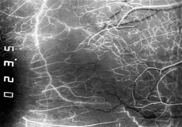
| Fig. 5. Normal temporal angiogram in the same 32-year-old woman as in Figure 3 two seconds later. The superficial episcleral vessels are filling from a superficial branch of the anterior ciliary artery; the deep circle is now difficult to define but can be seen contributing to the iris vascular filling. The limbal arcade and conjunctiva near the limbus are filled from the superficial branches. Both the episcleral plexus and the conjunctival plexuses perfuse late, about 3 mm from the limbus. This watershed zone is important in the etiology of scleral disease. |