|
|
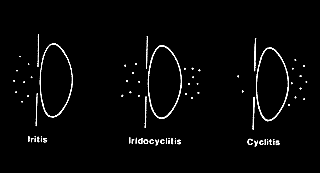
| Fig. 6. Diagram illustrating comparison of density of cells in the anterior chamber and vitreous. Sketching a simple diagram such as this may help to determine the source of the cells. In iritis, most of the cells come from the iris and are present in front of the lens and iris diaphragm. In iridocyclitis, the densities of cells in front of and behind the lens are equal. In cyclitis, most cells are behind the lens in the anterior vitreous. |