|
|
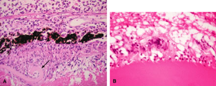
| Fig. 2. A. Enucleated eye of phacoanaphylaxis shows disrupted lens capsule (arrow), iridolenticular adhesion and granulomatous inflammation around the lens. B. Note prominent multinucleated giant cells around disrupted lens cortex in phacoanaphylaxis. |