|
|
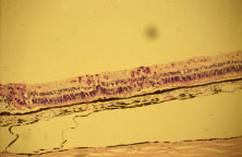
| Fig. 11. Histopathologic specimen showing immunohistochemical staining (red areas) in retinal cells infected with cytomegalovirus (CMV). This is in an area approximately 1 mm posterior to an area of confluent retinal destruction seen at the left of the figure. The small lesions probably correspond to the white dots that are seen just beyond the area of confluent CMV retinitis clinically. These areas of cytomegalic changes are associated with retinal edema and opacification. They are approximately 200 to 300 μm and would be visible as small white areas of opacification. |