|
|
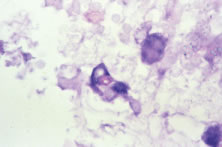
| Fig. 18. Typical cytomegalovirus (CMV) cells with prominent intranuclear inclusions. This retina was double immunostained with an anti-CMV antibody (blue) and an anti-endothelial cell antibody (red factor-VIII associated antigen). Thus, the figure illustrates that CMV can infect vascular endothelial cells. There is abundant retinal necrosis also present. |