|
|
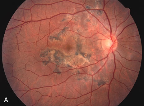
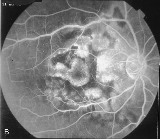
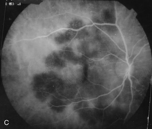
| Fig. 17. A. Classic inactive case of serpiginous choroiditis showing the jigsaw pattern of disease surrounding the fovea. B. Fluorescein angiogram showing staining of the edges of the inactive serpiginous choroiditis. There is blocked fluorescence in the areas of hyperplasia of the retinal pigment epithelium. C. Indocyanine green angiogram showing absence of fluorescence in the area of inactive choroiditis. |