|
|
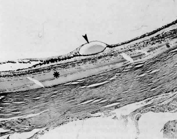
| Fig. 12. A ciliochoroidal effusion in the eye of a 16-year-old boy, who developed sympathetic uveitis after a corneal laceration. A. Scar of the traumatic corneal perforation, with fibrous tissue ingrowth (asterisk) and adherent lens remnants (arrowhead) (H E, × 18). B. The appearance of a ciliochoroidal effusion (asterisk) that was first observed in the eye 2 months after corneal perforation and before the development of uveitis in the fellow eye (H E, × 96). C. The choroid posteriorly is greatly thickened by intense lymphocytic infiltration in which foci of epithelioid cells (arrowheads) display pigment phagocytosis in the absence of necrosis (H E, × 185). |