|
|
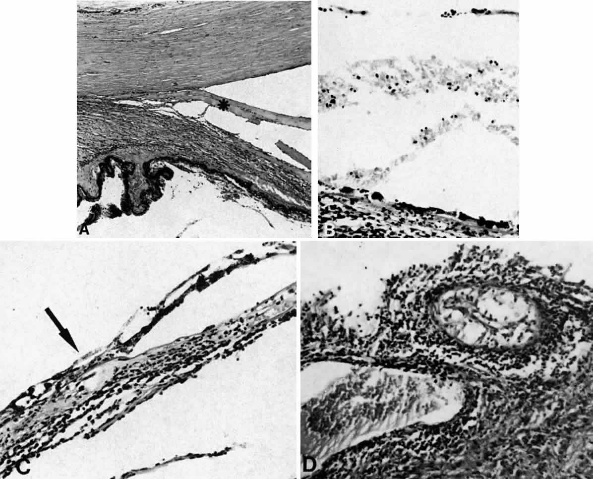
| Fig. 13. A. A ciliochoroidal effusion (asterisk) associated with Toxoplasma retinochoroiditis (H E, × 50). B. The retina temporally is totally necrotic with encysted T. gondii (H E, × 25). C. An old chorioretinal scar (arrow) posterior to the area of necrotic retina (H E, × 160). D. Intense vasculitis and perivascular lymphocytic infiltration of the central vessels in the optic nerve head (H E, × 185). |