|
|
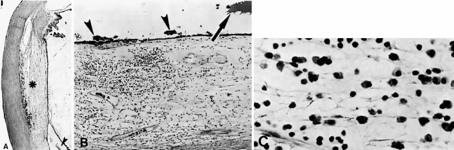
| Fig. 19. A. A ciliochoroidal effusion (asterisk) mistaken for a malignant melanoma and associated with moderately severe panuveitis. A secondary serous retinal detachment (arrowhead) and moderately thickened sclera are evident (H & E, × 15). B. Equatorial choroid with effusion and moderate infiltration of chronic inflammatory cells. Small nodules of proliferated retinal pigment epithelium (arrowheads) and subretinal proteinaceous material (arrow) are shown (H & E, × 80). C. Moderately intense infiltration of plasma cells and some lymphocytes (H & E, × 530). (Courtesy of the Armed Forces Institute of Pathology, Washington, DC) |