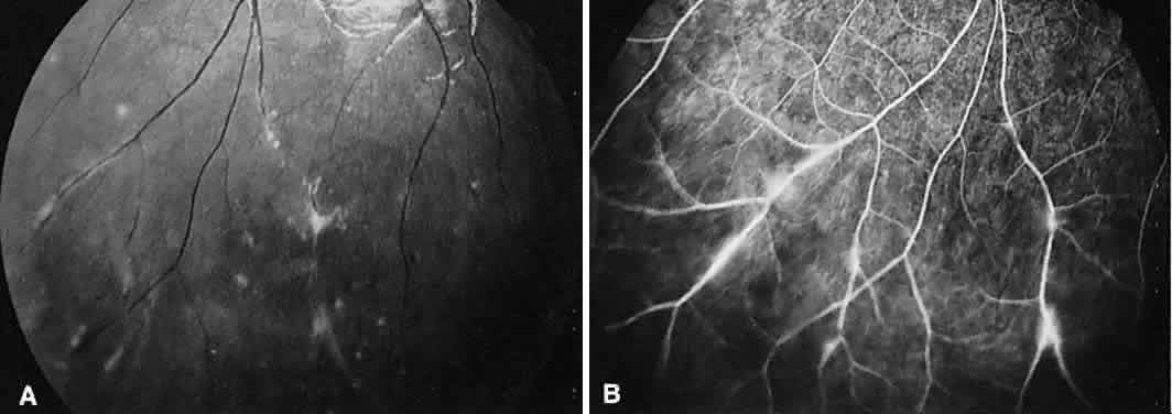
Fig. 9.
Intermediate uveitis.
A.
Red-free photography of the peripheral retina shows sheathing of the retinal venules.
B.
Fluorescein angiography shows staining of the vessel walls with leakage from the peripheral venules.