|
|
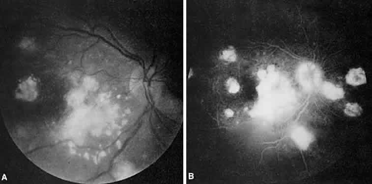
| Fig. 26. Candida endophthalmitis. A. Red-free photograph shows multiple retinal lesions. B. There are multiple areas of hyperfluorescence due to intraretinal and subretinal lesions. Some peripheral atrophic lesions represent age-related macular degeneration. Opacification of the vitreous inferior to the right macular region can be seen. |