|
|
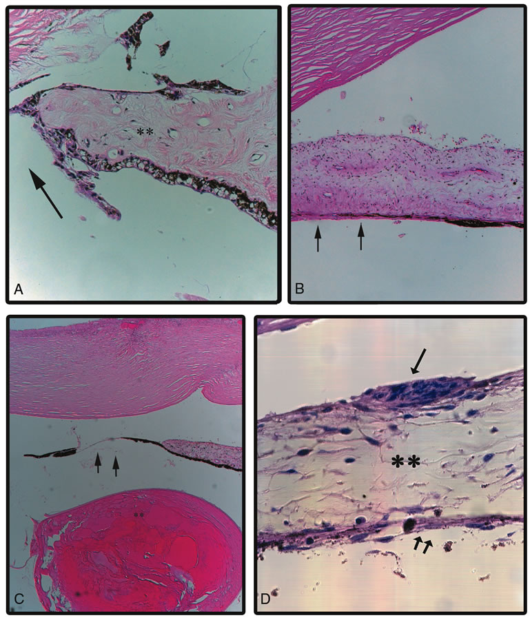
| Fig. 1. Microphotographs. A. Atrophic edge of iris following a peripheral iridectomy (arrow). Note the loss of melanocytes with areas of stromal collagen deposition. Clinically the iris would show hypopigmentation without transillumination defects. B. Iris pigment epithelial atrophy in diabetes mellitus. The iris stroma is preserved with patchy atrophy of the iris pigment epithelium (arrows), which would clinically correspond to a transillumination defect. C. Iris chafing from a sulcus fixated intraocular lens resulting in localized iris atrophy of the stroma and the pigmented epithelium (arrows). Sommerings ring is also seen (**). The intraocular lens is dissolved during processing. D. The iris in Cogan Reese syndrome. Note melanocytic plaque on the iris surface (single arrow), atrophic stroma (*) and pigment epithelium (double arrows). (© University of Illinois at Chicago.) |