|
|
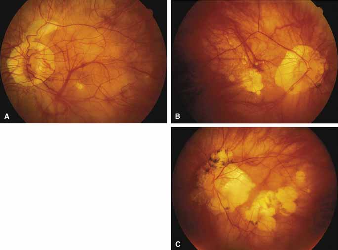
| Fig. 25 Fundus (A) of left eye of a 13-year-old boy with degenerative high myopia (-24.5 +2.50 axis 110 degrees for 20/80-visual acuity) showing marked peripapillary absence of pigment epithelium and a lacquer crack with pigment epithelial and choroidal atrophy extending superior from the disc. Fundi (B, C) of a 50-year-old man with myopic peripapillary and macular choroidal degeneration secondary to high myopia and blue cone monochromatism. Visual acuity was 20/300 OD with -18 D sphere and 20/400 OS with -19 D sphere. Note in all three photographs the myopic “coin” lesions, which for C have enlarged and coalesced. |