Chapter 7 Follicular Conjunctivitis RUBY LAGNADO, AMAR ALWITRY and HARMINDER SINGH DUA Table Of Contents |
DEFINITIONS | |||
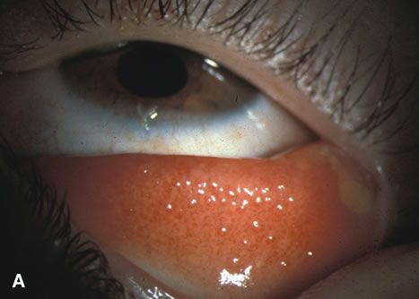
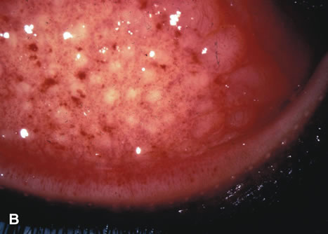
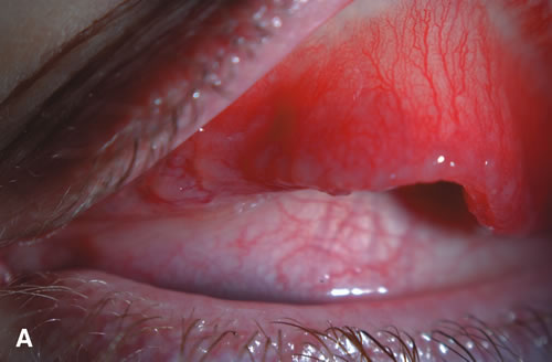
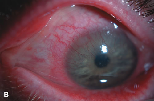
FOLLICLES A follicle is a nonspecific conjunctival response to chronic irritation: mechanical, chemical, or microbial. After the first few weeks of life, lymphoid tissue becomes evident in the human conjunctiva, with a diffuse and indiscriminate distribution. Under the influence of chronic irritation or stimulation, the lymphocytes increase and aggregate into follicles resembling in structure and function lymph nodes or the solitary lymphatic patches of the intestine.1,2 Clinically, follicles are yellowish to grayish white, discrete, round elevations of the conjunctiva. The central part of the follicle is avascular, but dilated blood vessels may surround the base and sweep up from the base over the convexity (Figs. 1 and 2). Follicles are 0.2 to 2 mm in size, although larger follicles may be seen, particularly in chlamydial disease.3
Follicles on the superior tarsal conjunctiva are well defined as a result of the close adherence of the tarsus to the epithelial mucosal tissues. They are also seen in the conjunctival fornices, where they are less regularly spaced and tend to be larger. Conjunctival follicles do not appear in the neonatal period because the lymphoid system is immature, and they are less apparent in children under the age of 2. Follicles histologically consist of lymphoid germinal centers with fibroblasts in the center. T cells occupy the periphery and B cells the center of the infiltrates.4 PAPILLAE Follicles are often clinically confused with conjunctival papillae (Fig. 3). Papillae are vascular changes most prominent on the palpebral conjunctiva, where fibrous septa anchor the conjunctiva to the underlying tarsus. In contrast to follicles, they have a dilated vascular core surrounded by edema and a mixed inflammatory infiltrate producing raised elevations of the conjunctival epithelium. They are nonspecific signs of conjunctival inflammation, most commonly associated with allergic or bacterial conjunctivitis.
FOLLICULAR CONJUNCTIVITIS Follicular conjunctivitis is not a specific disease entity but a connotation for a large number of conditions of varying etiologies that have follicles as a predominant pathologic and clinical manifestation. |
MICROANATOMY OF THE FOLLICLE |
Histologically, the structures of follicles are very similar, regardless of the inciting agent. The characteristic feature is a dense, localized infiltration of the subepithelial tissues by large mononuclear lymphocytes. The larger central mass is surrounded by smaller lymphocytes that frequently form secondary nodules. Other cells, such as plasma cells and polymorphonuclear cells, may appear in certain conditions. The cells of the follicle are supported by a loose and ill-defined reticulum that blends with the surrounding connective tissue. There is no true capsule, but in certain conditions, especially trachoma, a dense, surrounding sheet of connective tissue appears with increasing cicatrization.1,2 Initially, there are prominent lymphatics packed with mononuclear cells and a network of blood vessels surrounding a follicle. Later, endothelial lined spaces may develop and extend toward the center of the follicle. |
FOLLICULOSIS | ||
Folliculosis of childhood is not pathologic, but is a physiologic change
of childhood and adolescence in which follicles are prominent in the
fornix and decrease toward the lid margin (Figs. 4 and 5). There are no associated signs of inflammation, and the condition
is analogous to the lymphoid hyperplasia of tonsils in children. Clinically
insignificant follicles are seen frequently on the upper border
of the tarsus, whereas follicles present in the central portion of the
upper tarsal plate tend to suggest an underlying pathologic process.
|
ADENOVIRUSES |
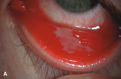
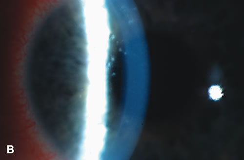
PATHOPHYSIOLOGY AND TAXONOMY Adenoviruses are double-stranded DNA viruses surrounded by a nonenveloped polypeptide icosahedral capsid with glycoprotein projections. They were first detected in 1953 as a latent virus in surgically removed adenoidal tissue from asymptomatic children.5 There are at least 51 serotypes of adenovirus,6 and they have been classified into six species, A to F, on the basis of their hemagglutination properties and biophysical and biochemical criteria.7 Adenoviruses are a common cause of a variety of human illnesses, accounting for approximately 10% of all febrile illnesses in infants and 5% of all such illnesses in children.8 Most of the keratoconjunctivitis and all of the epidemic keratoconjunctivitis types are caused by species D. The nine most recently described serotypes all belong to species D and were first isolated from human immunodeficiency virus (HIV)-infected patients. OCULAR ADENOVIRAL INFECTION Ocular adenoviral infections have a variety of clinical presentations; the two most common forms are epidemic keratoconjunctivitis (EKC) and pharyngoconjunctival fever (PCF). Both are characterized by a marked follicular response. Two less common manifestations are recurrent conjunctivitis and chronic papillary conjunctivitis. The different syndromes tend to be caused by distinct serotypes, although the features do overlap. Ad 3 and Ad 4 are the most common pathogens that cause sporadic keratoconjunctivitis and PCF. Ad 8, 19, and 37 have been implicated in sporadic cases as well as outbreaks of severe EKC.9–11 Although an epidemic due to both types 8 and 19 has been reported, most disease is due to a single serotype. EPIDEMIC KERATOCONJUNCTIVITIS The clinical features of adenoviral keratoconjunctivitis were first described by Fuchs12 in 1889, but it was not until 1955 that adenovirus was identified as the cause of the disease.13 EKC has a wide spectrum of duration and intensity of symptoms. After an incubation period of 2 to 14 days, symptoms begin in one eye; symptoms develop in the second eye 2 to 4 days later. Signs include conjunctival hyperemia and chemosis, intense tearing, and swelling of the conjunctival plica. Ipsilateral preauricular lymphadenopathy is found in many patients. The corneal involvement helps distinguish it from other causes of viral conjunctivitis (Figs. 6 and 7). Corneal subepithelial opacities develop approximately 2 to 4 days after the onset of symptoms. These may stain with fluoroscein and may persist for 2 to 3 weeks. About a week after the onset of symptoms, larger nummular lesions develop. These have irregular, raised borders and a central ulceration that stains with fluorescein. At 2 weeks, the subepithelial infiltrates (SEIs) begin to appear. Focal biopsies have shown these infiltrates to consist of lymphocytes, histiocytes, and fibroblasts accompanied by a disruption of the collagen fiber of Bowman's layer.14,15 The nummular opacities are thought to represent persisting viral replication in subepithelial keratocytes, triggering a host reaction. The immune basis is supported by the clinical observation that the opacities usually resolve with topical steroids but recur when the steroids are discontinued.16 Rarely, these nummular opacities or infiltrates may impair visual function and may persist for months or years. Usually, the conjunctival inflammation and epithelial disease resolve in 10 to 16 days but may last a month. The follicles and lymphadenopathy resolve more gradually in 2 to 4 weeks; however, the SEIs may persist for many months. EKC is also the commonest cause of viral membranous or pseudomembranous conjunctivitis (Fig. 8). Management Currently, there is no specific antiviral therapy available to treat adenoviral keratoconjunctivitis. Cidofovir, or HPMPC, is a broad-spectrum antiviral agent17 that was shown to have significant antiviral activity, reducing viral shedding in rabbits with adenoviral conjunctivitis.18 In humans, cidofovir 1% was shown to lower the frequency of severe corneal opacities, but its clinical use four to 10 times daily is limited by local toxicity.19 The mainstay of treatment is symptomatic in mild cases. In more severe cases, topical corticosteroids are useful where there are SEIs in the visual axis. Topical steroids should otherwise be used with caution in follicular conjunctivitis because herpetic conjunctivitis can occur without corneal involvement. If low-dose steroids are used, they may need to be gradually tapered over months to prevent recurrence. Adenovirus is highly contagious, and measures to limit the spread of infection should be taken. Spread is by person-to-person contact, contaminated ophthalmic instruments, or swimming water. Transmission in ophthalmology clinics has been well reported, most frequently as a result of applanation tonometry; however, slit lamp examination and application of eye drops may also result in spread,20–22 emphasizing the need for adherence to strict hand-washing protocols. Adenoviruses are highly stable and resistant to solvents; however, they are deactivated by hypochlorite solution. The live virus has been found to survive as long as 35 days on plastic surfaces.23 Patients should avoid touching their eyelids and wash their hands frequently. It has been reported that nearly 50% of patients with adenoviral conjunctivitis carry it on their hands.22 Although most patients are culture negative 7 to 10 days after the onset of symptoms,24 positive cultures have been reported for up to 12 months after infection.25 Infected children should be advised to stay at home for 1 week. In the setting of hospital outbreaks, it is essential to implement strict procedures to reduce transmission, and infected personnel should remain off work for 2 weeks. Conjunctival adenovirus infection is highly contagious and occurs worldwide sporadically and epidemically. Although not permanently blinding, adenoviral ocular infection remains the most common ocular viral infection worldwide. The social and economic cost of community epidemics is high.26 |
PHARYNGOCONJUNCTIVAL FEVER |
Adenoviruses also produce the classic triad of fever, pharyngitis, and acute follicular conjunctivitis in PCF. PCF occurs more commonly in children, is caused by serotypes 3 and 7, and is spread by respiratory secretions. The symptoms are similar to EKC, with tearing and foreign body sensation that is initially unilateral. The follicular conjunctivitis produces a watery discharge with associated preauricular or submandibular lymphadenopathy and occasional petechial hemorrhages. Although corneal involvement is not a key feature, there is occasionally a punctate keratitis; however, SEIs are rare. PCF is a self-limiting condition that varies in severity and may last from 4 days to 2 weeks. The virus remains in the conjunctiva for 2 weeks, but fecal excretion can last a month.27 Treatment is symptomatic, and topical steroids are rarely needed. |
RNA VIRUSES |
PARAMYXOVIRUSES Measles—Rubeola The measles (rubeola) virus causes photophobia, an acute catarrhal papillary conjunctivitis, superficial punctate keratitis, superficial pannus, and occasionally Koplick's spots on the conjunctiva and caruncle.28 Rarely, an immune interstitial keratitis may occur. In a healthy individual, the disease usually resolves with no ocular sequelae. However, in debilitated individuals, particularly those who are vitamin A deficient, it can progress to severe keratitis and keratomalacia.29,30 Herpes simplex virus (HSV) infection also may affect children with measles and may be responsible for one-fifth of their corneal ulcers.30 Mumps Conjunctivitis is a common manifestation of mumps characterized by dilatation of the conjunctival and episcleral vessels without marked secretion, and sometimes by chemosis of the bulbar conjunctiva.31 It is self-limiting and resolves spontaneously. A follicular conjunctivitis is not typical but was reported once in the literature.32 The other common ocular features are dacryoadenitis and punctate keratitis followed by interstitial keratitis. Newcastle Disease Newcastle disease virus causes respiratory infections in poultry; in humans, the virus and the vaccine cause a limited infection. It is unique in causing a unilateral follicular conjunctivitis with mild tearing and preauricular adenopathy. Rarely, there is a transient, fine, superficial keratitis in the first week.33 Newcastle disease normally occurs by direct inoculation during processing of infected birds by poultry workers and veterinarians. The disease is self-limiting and does not require therapy. |
TOGAVIRIDAE |
Childhood rubella is often accompanied by a catarrhal conjunctivitis and sometimes a mild follicular conjunctivitis. Rarely, there may be a mild superficial punctate keratitis that does not lead to permanent corneal disease.34 |
PICORNOVIRIDAE |
ACUTE HEMORRHAGIC CONJUNCTIVITIS Acute hemorrhagic conjunctivitis (AHC) has affected millions of people since it first appeared in Ghana in 1969. Large epidemics have occurred in Africa and Asia, and it was first reported in the Western Hemisphere in 1981 in Brazil.35 The major causative agents are enterovirus 70 (EV-70) and coxsackievirus type 24 (CA-24). Adenovirus types 8, 19, and 37 have also recently been recognized as causes of hemorrhagic conjunctivitis, and this may herald a new stage in the evolution of these viruses.36 The clinical features of AHC caused by EV-70 have, in more recent epidemics, been reported to be milder, with a maintained high transmission rate that may be due to biological transformation of EV-70.37Apart from conjunctival secretions, respiratory and oral transmission of coxsackievirus CA-24 variant would explain the rapid and extensive spread of AHC during an outbreak.38 The disease is characterized by a short incubation period and sudden onset of lacrimation, lid swelling, itching, foreign body sensation, and periorbital pain, which rapidly become bilateral. The common signs are papillary conjunctivitis with a minor follicular response; subconjunctival hemorrhages, which are more pronounced temporally; a fine punctate epithelial keratitis; and preauricular adenopathy. The hemorrhages are petechial at first but rapidly become confluent. Subepithelial opacities and erosions are rare, and the conjunctivitis resolves within 4 to 6 days, although the hemorrhages may take longer. Respiratory and gastrointestinal involvement may occur.39 More rarely, neurologic complications, including Guillain-Barré syndrome and Bell's palsy, may develop.40 Corneal superinfection may occur after AHC, and use of topical steroids may predispose an already compromised cornea to develop microbial keratitis. Folk remedies, such as the use of human urine on the eye, have also resulted in gonococcal ophthalmia.41 |
POXVIRIDAE |
PARAPOXVIRUS Orf virus is a member of the paravaccinia group of DNA viruses that cause a contagious pustular dermatitis, predominantly in sheep and goats.42 It occasionally affects humans, commonly causing lesions of the hand. Ocular infection is unusual; however, it was reported in a farmwife who developed a raised ulcerated lesion at the inner canthus, follicular conjunctivitis, and lymphadenopathy after handling sheep.43 This may have been as a result of direct finger inoculation occurring while she carried an infected lamb. Treatment in this case was with idoxuridine 0.5% for the eye, with resolution in 3 weeks. Blindness has been reported in human orf infection, possibly because of secondary bacterial infection.44 |
ORTHOMYXOVIRIDAE |
The orthomyxoviruses include the influenza virus, which can cause an acute follicular conjunctivitis with mucopurulent discharge. |
HERPES SIMPLEX |
Primary herpes simplex infection is frequently associated with a vesicular
lesion on the lid margin, a clinical feature that aids diagnosis. It
typically occurs in children and may have associated upper respiratory
symptoms and stomatitis. Half of all primary HSV infections involving
the lid margin will develop corneal epithelial manifestations ranging
from fine punctate epithelial staining to dendritic ulceration.45 In primary herpetic eye disease, the ulceration is more commonly fine
microdendrites involving the cornea and conjunctiva, unlike the prominent
dendrites in recurrent disease. It has been reported that the incidence of HSV ocular infection presenting as acute follicular conjunctivitis in the absence of corneal or lid signs in cases of clinically diagnosed epidemic keratoconjunctivitis is 1.4% to 7%.46 There is no significant difference in clinical features between primary and recurrent HSV conjunctivitis.47 The clinical features of HSV conjunctivitis are those of a moderate follicular conjunctivitis, sometimes with preauricular node swelling and upper respiratory tract signs. There are rarely corneal changes, and it is bilateral in 13% of cases.47 It has a mean duration of 8 days. The low rate of bilateral disease and the short duration may help discriminate HSV from adenoviral conjunctivitis as clinically differentiating the two disease entities is extremely difficult, particularly in the early stages. Most herpetic infections of the eye are caused by human herpesvirus type 1 (oral); however, type 2 (genital) may occur in the newborn and adults after oral–genital inoculation. Neonates who develop herpes simplex primary conjunctivitis should receive intravenous treatment with acyclovir in addition to topical treatment. The Herpetic Eye Disease Study (HEDS) assessed whether long-term prophylactic treatment with acyclovir would prevent recurrence of ocular herpetic disease.48 This was a prospective randomized, multicenter trial, and although it did not primarily address herpetic conjunctivitis, its findings did suggest that the use of 800 mg (or 400 mg b.i.d) oral acyclovir daily over a year reduced the recurrence of ocular herpetic disease. |
EPSTEIN-BARR |
Epstein-Barr virus (EBV), a ubiquitous DNA herpesvirus, is the most common causative agent of infectious mononucleosis syndrome. Ocular manifestations include a follicular or membranous conjunctivitis with or without subconjunctival hemorrhages. Discrete granular subepithelial infiltrates may be seen, with overlying punctate epithelial keratitis.49 The treatment of EBV-associated ocular disease is not defined because of the small number of documented cases. Other ocular complications include uveitis, choroiditis, retinitis, papillitis, and ophthalmoplegia. |
ADULT INCLUSION CONJUNCTIVITIS |
Chlamydia trachomatis is an obligate intracellular bacterium that can cause several different
conjunctivitis syndromes, varying in nature according to which serotype
is involved. Serotypes A to C are responsible for trachoma and are
discussed in the section on chronic conjunctivitis. Serotypes D to K of C. trachomatis cause an acute follicular conjunctivitis with a mucopurulent discharge. Historically, the diagnosis was made after visualization of chlamydial inclusion bodies on the conjunctival smears, thus giving the disease its name. These trachomata are transmitted sexually, may be asymptomatic, and may involve multiple sites simultaneously. Transmission is through direct contact of the eye with infected genital or urinary secretions.50,51 Indirect transmission can occur in poorly chlorinated swimming pools.52 Eye-to-eye transmission is probably more common than previously thought.53 Adult inclusion conjunctivitis (AIC) is more common in developed countries in the young and sexually active; it occurs within 2 to 3 months after the patient has a new sexual partner. The serious nonocular complications of chlamydial infection, such as epididymitis and salpingitis, make it an important diagnosis to establish and treat. Chlamydia has been reported to be the cause of a follicular conjunctivitis in adults in 5% to 18% of conjunctivitis.54 Approximately 40% to 50% of men with nongonococcal urethritis and 70% of their partners are infected with C. trachomatis,49 although 25% of the females are asymptomatic.55 Serotypes D to K produce a syndrome clinically distinct from trachoma and do not cause permanent visual loss. It is not clear why AIC is more benign, but reasons may include a lack of re-exposure to the agent, the patient's age at initial infection, and a lower innate pathogenicity of the organism. CLINICAL FEATURES The disease affects initially one eye, 1 to 2 weeks after exposure to the organism. The symptoms may be acute or subacute, with redness, irritation, and a mucopurulent discharge. A palpable, nontender preauricular node may develop, but there are no systemic symptoms. Most of the conjunctival findings are located inferiorly, with a predominantly follicular reaction also associated with some degree of papillary reaction (Fig. 9). Limbal swelling is also often present. Pseudomembranes do not occur, and conjunctival scarring, if it does occur, is minimal. In the second week, the cornea may have a mild or moderate superficial punctate keratitis that may be fine or coarse. Later in the infection, peripheral epithelial or subepithelial infiltrates may occur, which have an intact epithelium. Occasionally, iritis will occur in the later stages and, rarely, Reiter's syndrome. A superficial micropannus may develop in those who had subepithelial infiltrates, but there is no frank corneal scarring. DIAGNOSIS After a detailed history and clinical examination a fluorescent monoclonal antibody test is the most sensitive method of rapid diagnosis.56 Giemsa-stained conjunctival smears looking for perinuclear inclusion bodies have a high false-negative rate57 (Fig. 10). In recent years, polymerase chain reaction (PCR) assay has become more widely used as an effective diagnostic tool to accurately detect the presence of chlamydial material in ocular specimens (see section on laboratory diagnosis). TREATMENT Patients and their partners should be screened for nonocular chlamydial infection and for other venereal diseases. Systemic treatment is mandatory. The recommended treatment for chlamydial infection is azithromycin 1 g as a single daily dose for 7 days, or doxycycline 100 mg twice daily for 7 days. Alternative regimes are erythromycin 500 mg four times a day for 7 days, or ofloxacin 300 mg twice a day for 7 days.58 |
NEONATAL INCLUSION CONJUNCTIVITIS |
Neonatal inclusion conjunctivitis (NIC) is caused by Chlamydia serovars D to K and is the most common cause of infectious neonatal conjunctivitis
worldwide.57 It is transmitted through infected genital secretions during passage through
the birth canal. The frequency of genital chlamydial infection
in women from developed countries may be 2% to 8%,59 increasing with lower socioeconomic status. Thirty percent to 40% of
untreated women will have babies affected with NIC, and 10% to 20% will
develop chlamydial pneumonitis.60 Acute conjunctivitis with purulent discharge develops 5 to 12 days after birth. This may manifest earlier if there is premature rupture of membranes. The infection may initially be unilateral, with a papillary reaction, as the immune system is too immature to cause a follicular reaction, and there is no preauricular lymphadenopathy. Pseudomembranes occur in more severe cases as does corneal scarring and neovascularization. Diagnosis is based on laboratory tests. Giemsa-stained inclusion bodies may often be seen, unlike in AIC. Monoclonal fluorescent antibody staining should also be done to assist in the rapid diagnosis. Although McCoy cell culture may be used as a diagnostic tool, the advent of PCR techniques has enabled rapid and accurate diagnosis.61 About half of neonates with chlamydial pneumonitis have had NIC.62 Treatment is oral erythromycin syrup 50 mg/kg per day in two to four divided doses for 14 days. The mother also requires systemic treatment. Tetracycline is contraindicated for the baby or for the mother if she is breast-feeding. Neonatal ocular prophylaxis with silver nitrate or antibiotic ointments does not prevent perinatal transmission of C. trachomatisfrom mother to infant; however, these agents do prevent gonococcal ophthalmia. |
CHRONIC FOLLICULAR CONJUNCTIVITIS | ||||||||||||||||||||||||||||||||||||||||||||||||
Follicular conjunctivitis that lasts for more than 16 days is considered
chronic and may persist for months to years. The causes of chronic follicular
conjunctivitis are listed in Table 1.
Table 1. Diagnostic Features of Chronic Follicular Conjunctivitis
|
CHLAMYDIA TRACHOMATIS | ||||
Trachoma is an ancient disease and remains the most common cause of chronic
follicular conjunctivitis, and the foremost cause of blindness worldwide
after cataract.63 It is the most common cause of preventable blindness. Trachoma was described
in the Egyptian Ebers papyrus in 1900 BC and remains endemic to
that country. It was endemic in Europe into the 20th century. Would-be
immigrants to the United States were screened for trachoma on
arrival at Ellis Island, and those with the characteristic follicles
were turned away. Trachoma has disappeared from Western Europe and North America as a result of improved living conditions. However, trachoma continues to affect 150 million people in 48 countries, of which 6 million are blind.63 In areas where trachoma is constantly present at high prevalence, active disease is found in more than 50% of preschool children. As many as 75% of women and 50% of men over the age of 45 may show signs of scarring disease.64 Infected children are the major reservoir for trachomatous infection in a community.65,66 The prevalence of active trachoma decreases with increasing age, with less than 5% of adults showing signs of active disease. Although similar rates of active disease are observed in male and female children, the later sequelae of trichiasis, entropion and corneal opacification are more common in women than men.67 This is probably related to the close contact of women with children who are suffering from the active disease. In endemic areas, trachoma is directly transmitted from person to person through ocular secretions.68 Flies also act as vectors by feeding on these ocular secretions.65,66 Children with trachoma in endemic areas have C. trachomatisin the upper respiratory tract and gastrointestinal tract, so it may also be transmitted by droplet or fecal contamination. PATHOGENESIS C. trachomatisis an obligate intracellular organism that more closely resembles bacteria than viruses. They contain DNA and RNA, possess a cell wall, and are treatable with tetracycline, erythromycin, and sulfonamides. Acute infection with C. trachomatisimmunotypes A, B, Ba, or C is usually self-limiting with few long-term sequelae. Repeated exposure leads to conjunctival cicatrization and corneal scarring.69 Both humoral and cell-mediated immune responses to infection occur.70 The early formation of follicles that regress is followed by prolonged papillary hypertrophy of the conjunctival epithelium and deposition of subepithelial connective tissue, which contains a diffuse infiltration of lymphocytes and plasma cells. Antichlamydial antibodies can neutralize Chlamydiae, block attachment and internalization of the organism, and produce partial immunity. It may be that the T lymphocytes and cell-mediated immunity are responsible for the conjunctival scarring. Alterations in the extracellular matrix components and collagen metabolism occur in the conjunctival tissue, with new collagen type V formation in active trachoma and scarred trachoma. The conjunctival tissue from patients with active trachoma contains an increased amount of collagen types I, II, and IV. Scarred trachoma is characterized by a marked increase in basement membrane collagen IV and a marked decrease in types I and III. CLINICAL FEATURES The onset and severity of symptoms vary, and in young children, the condition may be entirely subclinical. Acute infectious trachoma causes bilateral, follicular, nonpurulent conjunctivitis 5 days after inoculation52 that may be associated with a tender preauricular lymph node. After 2 to 3 weeks, follicles appear on the superior tarsal conjunctiva and superior limbus. It is not known how long individuals are infectious. There are usually no associated systemic symptoms, although it may sometimes be accompanied by rhinitis, upper respiratory tract infection, or preauricular adenopathy.71 Repeated infection results in conjunctival scarring, cicatricial entropion, and trichiasis, with risk of blinding corneal ulceration. The dry eyes, scarred tear ducts, keratinization of the conjunctiva, and distortion of the lid margin also contribute to the cornea being susceptible to damage. Trichiasis carries an eightfold risk for corneal opacity.72 In countries where the prevalence of active trachomatous infection is higher, such as Tanzania, the progression rate from conjunctival scarring to trichiasis and then corneal opacity is more rapid than in countries, such as the Gambia, where active trachoma is declining.73–75 WORLD HEALTH ORGANIZATION CLASSIFICATION FOR ACUTE TRACHOMA In the World Health Organization (WHO) grading system for acute trachoma, grade TF (trachoma follicles) represents mild trachoma with the presence of five or more follicles in the upper tarsal conjunctiva of at least 0.5 mm diameter (Fig. 11). Grade TI (trachoma inflammation) describes a more severe trachoma with pronounced inflammatory thickening of the upper tarsal conjunctiva that obscures more than half of the normal deep vessels (Fig. 12). Repeated infections cause scarring trachoma (grade TS) in which the upper eyelid is shortened and distorted causing entropion and trichiasis (grade TT), which abrades the eye. Blindness results from progressive corneal opacification (grade CO), which is related to the degree of entropion or trichiasis (Fig. 13).
According to the WHO, the diagnosis of trachoma requires at least two of the following signs: superior tarsal follicles, limbal follicles or Herbert's pits, typical conjunctival scarring, or vascular pannus. Herbert's pits are necrosed limbal follicles; they are the only sign unique to trachoma, but they do not occur in every case (Fig. 14). Early pannus formation is a good predictor for severe conjunctival scarring later in the disease.76
Corneal vascularization occurs in response to the marginal corneal infiltration and is more extensive at the superior limbus. It should be remembered that other corneal diseases, such as Salzmann nodular degeneration or bacterial corneal ulcers may occur in patients with trachoma. The risk of these conditions and subsequent complications, such as perforation or scarring, is increased due to the presence of trichiasis, entropion, and the chronic dry eye state. Patients with vernal catarrh who contract trachoma may develop severe corneal complications. The diagnosis of trachoma can be confirmed by conjunctival cytology77; however, Giemsa staining has been superseded by fluorescent monoclonal antibody staining. PCR analysis is accurate but expensive for diagnosis. A previous history of trachoma does not confer immunity to recurrent disease and whole-organism vaccines are ineffective, so other strategies for trachoma control are required.78 The WHO is promoting the Global Elimination of Trachoma (GET) by the year 2020 and has adopted the SAFE strategy—Surgery for entropion and trichiasis, Antibiotic treatment for active infection, and the promotion of both Facial cleanliness and Environmental improvement to reduce transmission—to achieve this goal. RISK FACTORS Poverty, excessive distance to water, small amounts of water used by the household, the presence of flies, and poor hygiene are risk factors for trachoma.79,80 Active trachoma is associated with young age and close contact between people. Discharge from the eyes and nose may be a source of reinfection.81 It is likely that host factors also play a role in a person's susceptibility to C. trachomatis disease.82 HEALTH EDUCATION Interventions to prevent scarring trachoma by reducing active infection include promotion of facial washing. It has been shown that it is possible to increase facial cleanliness through education even where there is a water shortage.83 Trachoma transmission by flies could be reduced by encouraging the use of latrines and also by insecticides. MEDICAL TREATMENT Historically, sulfonamides were found to be effective in controlling trachoma; however, they caused unacceptable side effects in a few cases and thus had to be discontinued.84 Topical tetracycline ointment was shown to be effective in individual cases when applied once daily for 6 weeks or for 5 days a month for 6 months. However, poor compliance and rapid reinfection when only individual cases were treated meant that this was not successful at the community level.85,86 Treatment with azithromycin as a single oral dose (20 mg/kg) is safe and has been shown to be at least as effective as 6 weeks of supervised tetracycline ointment.87 This has been a major advance, and in 1998, a new international initiative was launched by the Edna McConnell Clark Foundation and Pfizer Inc, in collaboration with the WHO, in which the drug is provided free to national trachoma control programs in selected countries. Azithromycin is a key component of SAFE and is more successful in curing chlamydia and improving clinical outcome, with the added advantage of ease of treatment. It has been recommended that in areas where trachoma is moderately prevalent (more than 35% of children affected), treatment should be undertaken annually; but in hyperendemic areas (more than 50% of children affected), it should be given biannually.88 However, azithromycin-resistant pneumococci have been isolated from the nasopharynx of children after two mass treatments, suggesting the need to monitor resistance.89 SURGICAL TREATMENT Preventing blindness from trachoma, even when transmission is stopped, still requires surgery for the irreversible scarring and trichiasis affecting large numbers of the already exposed population. The operation of choice for trachomatous trichiasis is bilamellar tarsal rotation.90 It is important that this surgery is done before corneal scarring has started to develop, and earlier uptake of surgery requires health education.91 To prevent blinding trachoma, it will not be necessary to eradicate ocular chlamydial infection, but only to reduce the frequency of reinfection in endemic populations to such an extent that severe conjunctival scarring, leading to trichiasis and corneal scarring with subsequent visual loss, is prevented.92 |
PSITTACOSIS |
It had been previously thought that only the chlamydial agents causing
trachoma and inclusion conjunctivitis could produce follicular conjunctivitis
in man. However, in 1968, there was a report of a laboratory technician
involved in research with Bedsonia who developed follicular conjunctivitis proven by conjunctival scrapings
to be caused by Chlamydia psittaci.93 Since then, it has been shown that ocular infections with C. psittaci and C. pneumoniae may be more common than previously recognized. PCR species-specific tests are used to further identify chlamydial antigens in patients with chronic conjunctivitis in whom direct fluorescent antibody tests were positive.94 In patients with nontrachomatous chlamydial infection, a short course of oral antibiotics is inadequate. |
MORAXELLA |
Moraxella lacunata usually produces angular blepharitis but can also cause chronic follicular conjunctivitis with follicles on the upper tarsal plate, and keratitis. It may be associated with preauricular lymphadenopathy. M. lacunata may be incorrectly diagnosed as epidemic keratoconjunctivitis, herpes simplex, or chlamydial infection.95 It can be identified on scraping as large diplobacilli or by culture on blood agar. It is treated with topical tetracycline or erythromycin. |
PARINAUD'S OCULOGLANDULAR SYNDROME | |
Cat-scratch disease is caused by Bartonella species and mainly affects children and young adults. Most patients develop
regional lymphadenopathy preceded by an erythematous papule at the
site of inoculation. Bartonella henselae lives on cats and their fleas and may be transmitted to humans by a scratch
or bite. Bartonellainfection of the eye was first described by Henri Parinaud in 1889, when he reported three patients with chronic fever, regional lymphadenopathy and follicular conjunctivitis (Fig. 15).96 Subsequent reports noted the history of cat exposure in some patients, and the syndrome became known as Parinaud's oculoglandular syndrome.
Parinaud's oculoglandular syndrome is the most common ocular complication of cat-scratch disease, affecting approximately 5% of symptomatic patients.97 It is characterized by unilateral granulomatous follicular conjunctivitis with tender regional lymphadenopathy affecting the preauricular, submandibular, or cervical regions. Conjunctival lesions may ulcerate and usually resolve within several weeks, but the lymphadenopathy may take several months to clear. Other ocular manifestations of cat-scratch disease include neuroretinitis with macular star formation, multifocal retinochoroiditis with panuveitis, branch retinal artery occlusion, serous macular detachment, and papillitis with peripapillary retinal detachment.98 DIAGNOSIS The classic clinical diagnosis of cat-scratch disease requires that at least three of four criteria be met: a history of traumatic cat exposure, a positive skin test in response to cat-scratch disease antigen, characteristic lymph node lesions, and negative laboratory investigations for unexplained lymphadenopathy. The immunosuppressed are more vulnerable to cat-scratch disease from their pets, and systemic dissemination is more likely.99 INVESTIGATION Investigation now depends predominantly on serology testing, but these results can be inconclusive because of several different serotypes leading to antigenic variability.100 Conjunctival biopsies and PCR molecular techniques can be valuable diagnostic tools for lymph node aspirate. TREATMENT Mild disease is self-limiting and does not require antibiotic treatment. Severe disease is treated with rifampicin, ciprofloxacin, trimethoprim, sulfamethoxazole, and gentamicin.101 |
LYME DISEASE |
Ocular findings in Lyme disease were first noted in the original observations
of Steere and colleagues102 in 1977. Animal studies have shown that the Lyme spirochete invades the
eye early and may remain dormant. Nonspecific follicular conjunctivitis
may occur in up to 10% of patients during the flulike illness
of Lyme disease.102,103 The conjunctivitis is usually self-resolving and does not lead
to long-term sequelae. Associated episcleritis, photophobia, and
subconjunctival hemorrhages have been noted.103 Other ocular manifestations of the disease include keratitis, which is
most common within a few months of the onset of disease. This may be
nummular with nonstaining opacities, or it may be interstitial with keratouveitis, neovascularization, scarring, and corneal edema. The corneal
effects are likely to be secondary to a delayed hypersensitivity response
to the Borrelia burgdorferi antigen.104 Inflammatory syndromes, such as vitritis and uveitis, may occur. Neuro-ophthalmic manifestations include neuroretinitis, multiple cranial nerve palsies, optic atrophy, and disc edema. In endemic areas, Lyme disease may be responsible for 25% of new-onset seventh nerve palsy. |
OCULAR MEDICAMENTOSA/TOXIC FOLLICULAR CONJUNCTIVITIS | |
Follicular conjunctivitis may occur as a response of the ocular surface
to the toxicity of compounds, drugs, or their preservatives, that are
frequently instilled into the conjunctival sac (Fig. 16). The response is mediated via destabilization of the tear film or
a direct toxic effect on the surface epithelium. Toxicity is defined as damage to structures or disturbance of function over and
above an agent's therapeutic effect.
Several agents are capable of producing signs and symptoms of ocular medicamentosa. The agents may be prescribed (iatrogenic) or self-instilled (inflicted). The effect may be caused by the drug, its solvent or vehicle, or the preservative used. Toxic follicular reactions with or without inflammation have been described with atropine, miotics, epinephrine, some antiglaucoma medications, and antivirals. Such a reaction is probably the result of the mitogenic effect of the agents.105 Chronic disease, such as glaucoma and dry eye states are associated with high risk for toxicity. Brimonidine was reported to have an incidence of follicular conjunctivitis of 9.6% in treated patients.106 Other conjunctival manifestations of toxicity include cicatrization and papillary reaction. Associated corneal involvement may be significant. Features include punctate keratopathy; coarse, focal keratopathy; pseudodentrites; filamentary keratitis; persistent epithelial defects; and band keratopathy. The diagnosis is based on clinical features, a high index of suspicion, multiple drug usage, and frequent instillation. Increased symptoms of stinging on instillation of the drug may indicate toxicity, as may an initial improvement of symptoms of the underlying condition followed by worsening of symptoms on continued usage. Rose bengal staining helps in identifying early signs of corneal and conjunctival toxicity. A favorable response to withdrawal of drug(s) or substitution with preservative-free preparations is also a strong indicator of toxicity. Treatment includes cessation of drug use where possible or substitution with an alternative or preservative-free preparation. The period to resolution of symptoms and clinical signs after cessation or substitution of the offending drug may vary from a few days to a few months.107 |
MOLLUSCUM CONTAGIOSUM | |
Molluscum contagiosum is the most common poxviral pathogen in humans since
the eradication of smallpox. It causes umbilicated warty growths 2 to 3 mm
in diameter on the lid margins (Fig. 17). The shedding of live virus into the conjunctival fornix causes
follicular conjunctivitis with punctate keratitis; if left untreated, corneal
pannus may develop.108 These responses are most likely a toxic reaction to the viral particles. The
disease is spread by close personal contact and sexual activity. Treatment
is by excision or cryotherapy.
|
ADENOVIRUS |
Although predominantly related to acute disease, rarely adenovirus conjunctivitis runs a chronic course109 or can be intermittent and remitting, with no symptoms between episodes.110 Chronic disease follows acute follicular conjunctivitis when the typical conjunctival follicles are replaced with a papillary reaction. |
AXENFELD'S CHRONIC FOLLICULAR CONJUNCTIVITIS |
Axenfeld's chronic follicular conjunctivitis, or orphan's conjunctivitis, was
originally described in orphanages in France and has
occurred from time to time in boarding schools. This is a mild asymptomatic
disease without significant keratitis. Axenfeld's assistant, Victor
Morax, inoculated himself with scrapings from an infected child
and developed follicular conjunctivitis after an incubation period
of 10 days; his disease was relatively severe at onset, and persisted
for 2 years. Because this syndrome is similar to the mild trachoma once
seen in American Indian boarding schools, it may have been mild trachoma. Axenfeld's conjunctivitis has an insidious onset and is accompanied by a scanty discharge in which mononuclear cells (lymphocytes) predominate. The follicles involve the entire conjunctiva, including the upper tarsal plate, but are firm and do not break up when pressure is applied. The cornea is not involved, and probably for this reason, the disease is asymptomatic. Because Axenfeld's chronic follicular conjunctivitis is not associated with florid corneal involvement and does not produce conjunctival scarring, it has been classified as a disease entity separate from trachoma. Most of the observations on Axenfeld's follicular conjunctivitis were made before the slit lamp was widely used, and it is likely that small degrees of keratitis and pannus may have been overlooked. Moreover, in mild trachoma, a chlamydial agent is rarely demonstrated and the disease produces little, if any, scarring. Therefore, it is possible that Axenfeld's conjunctivitis is a mild form of trachoma that runs a self-limiting course under the relatively hygienic conditions of a children's institution. In such cases, a regular course of trachoma therapy with oral tetracycline or erythromycin should be administered. |
CHRONIC FOLLICULAR CONJUNCTIVITIS OF THYGESON |
Chronic follicular conjunctivitis of Thygeson, originally described in
a group of high-school children suffering from chronic follicular
conjunctivitis in California, lasts 6 to 8 months and is accompanied
by mild keratitis, which was felt to distinguish it from the trachoma
that was present in the same population. As with Axenfeld's conjunctivitis, the
possibility of chlamydial infection must be ruled out
by modern diagnostic techniques. The entity is characterized by the following findings:
This epidemic occurred in a suburban high school in which some trachoma cases were found in the Mexican American students; moreover, most of the cases involved girls who had been trading eye cosmetics. There is the possibility that this may also represent a particularly mild form of trachoma. |
ROSACEA BLEPHAROCONJUNCTIVITIS |
Chronic rosacea blepharoconjunctivitis occurs in patients with varying degrees of cutaneous acne rosacea, a syndrome characterized by pustules, erythema, telangiectasia, rhinophyma, and a predisposition to blushing or sunburn. There are no comedones, or blackheads, distinguishing this condition from acne vulgaris. Patients occasionally develop corneal scarring, neovascularization, and, rarely, corneal perforation. Chronic follicular conjunctivitis, associated with keratitis and vascular pannus, occurs in some patients. The follicles may be a toxic reaction to staphylococcal antigens or abnormal sebaceous material. Management is based on treating the lid margin with scrubbing and locally applied antibiotics. Oral tetracyclines, including doxycycline, and erythromycin are effective in controlling the eye disease, but both the dermatitis and ocular symptoms may recur within 6 weeks of stopping systemic antibiotics. |
LABORATORY DIAGNOSIS |
In view of the vast number of potential etiological factors responsible
for follicular conjunctivitis, laboratory investigations are essential
to establish a specific diagnosis. It is important that the clinician
undertake a systematic approach to investigation, particularly when
faced with a case of unresponsive, atypical, or chronic follicular conjunctivitis. In
patients with follicular conjunctivitis, cultures for
viruses, bacteria, and chlamydia may be important in identifying specific
syndromes or specific organisms, thereby facilitating targeted treatment. CYTOLOGY Conjunctival cytology remains a rapid, versatile, and cost-effective means to assist in the diagnosis of follicular conjunctivitis and other inflammatory ocular diseases.111–113 Specimens for cytology are best obtained from the conjunctiva, particularly the upper tarsus, with a sterile spatula following topical anesthesia. Care must be taken to ensure no cross-infection of eyes during the procedure. An adequate specimen should contain visible material when smeared on a glass slide. Specimens for Giemsa and immunofluorescent staining are air dried before fixation. Giemsa staining reveals cellular morphology and microbial agents, whereas Gram's stain is useful for confirming the presence and identifying the general characteristics of microbial pathogens. Inflammatory cells in smears are suggestive of the underlying disease process: Bacterial infections are characterized by an abundance of polymorphonuclear cells, viral infections by a predominance of lymphocytes, and chlamydial infections by a mixture of both these types, often accompanied by Leber cells (macrophages), plasma cells, or immature lymphocytes (“follicle cells”). Intraepithelial cytoplasmic inclusion bodies are easily visualized and are diagnostic of chlamydial infection. In membranous conjunctivitis, large numbers of polymorphonuclear leukocytes are found, regardless of the etiology. Cytology is also useful to identify dysplasia and keratinization of the ocular surface. SEROLOGY Serologic evaluation may be helpful in the identification of infection. For acute infections, a comparison of acute and convalescent specimens showing a fourfold increase in antibody titer is suggestive of recent infection with a particular agent. In many circumstances, however, high endemicity of the suspected pathogen precludes useful interpretation of results; for example, because positive herpes simplex and chlamydial titers are extremely common in the general population, they usually cannot be used to identify ocular infections in individual patients. CULTURE Conjunctival culture remains the mainstay of laboratory investigation of follicular conjunctivitis. Samples are easily obtained by minimally invasive techniques, the costs are low, and availability is high. Swabs should be taken with a vigorous stroke through both inferior fornices, swabbing the least affected eye first. Topical anesthetics do not adversely affect viral or chlamydial cultures; however, preservatives present in certain eye drops may interfere with the recovery of bacteria and so should be avoided. Bacterial Cultures The common bacterial pathogens responsible for conjunctivitis include Staphylococcus aureus, Streptococcus pneumoniae, Haemophilus sp and Neisseria sp. Immediate inoculation of the ocular swab directly onto solid media (e.g., blood agar plate) provides the best chance of isolating most aerobic bacteria found on the lids and conjunctiva. Chocolate agar is required to grow Neisseria or Haemophilus sp. Viruses Isolation procedures for viruses require consultation with the local laboratory prior to taking the specimen. For many viruses, Dacron or calcium alginate–tipped swabs should be placed in refrigerated transport media immediately after procurement and transported to the laboratory within 24 hours for viral isolation in cell cultures. Yields decrease rapidly after 24 hours and also with specimen freezing. The physician must indicate to the laboratory which viruses are suspected and document the specimen site, thereby allowing the laboratory to select the best cell lines to inoculate.114 Chlamydia McCoy cell culture has been used to good effect for many years in the laboratory diagnosis of chlamydial conjunctivitis.115 Recently, however, newer nonculture tests have become widely available and are less expensive, with similar or better diagnostic sensitivities and specificities. Other methods used to identify chlamydial infection include immunofluorescent staining of smears, enzyme-linked immunoassay, measurement of tear and serum antibodies, and immunofluorescence.116–122 ENZYME IMMUNOASSAY Enzyme immunoassay kits that employ monoclonal antibodies to adenovirus group-reactive antigens are now widely available and are being used for the diagnosis of ocular infections.123 They involve the placement of conjunctival swab material into contact with adenovirus-specific monoclonal antibodies (usually in special wells) with subsequent exposure to adenovirus monoclonal conjugate antibody.124 These tests are relatively cheap and have been shown to have a sensitivity of 56% when applied within 1 week of onset of symptoms, decreasing to 25% specificity when used thereafter, up to a limit of 11 days. Results are available within 2 hours; however, recent work on immunochromatography may give comparable results within 10 minutes.125 Enzyme-linked immunoassays directed toward chlamydial antigens have been shown to be effective in the diagnosis of both chronic (trachomatous) and acute follicular chlamydial disease.126 Numerous diagnostic kits have also been made available for the diagnosis of herpes simplex infection, HIV, and Lyme disease.127,128 POLYMERASE CHAIN REACTION Polymerase chain reaction assay is a procedure in which segments of DNA or RNA are amplified by the use of flanking oligonucleotides called primers and repeated cycles of amplification with DNA polymerase. The initial step is heating in order to separate the DNA into single strands. Each strand is annealed with complementary target DNA sequences or primers and extended by the action of DNA polymerase. The amount of DNA material is doubled with each cycle, and often 30 cycles are undertaken to obtain enough material for further analysis. This process allows isolation and identification of minute amounts of pathogenic material. In the past, PCR has been prohibitively expensive; however, in recent years, it has become more widely available and costs have decreased. DNA amplification is rapidly becoming the gold standard for diagnosis of infective processes. PCR kits designed for the detection of C. trachomatis, such as the Amplicor (Roche Diagnostic Systems, Branchburg, NJ), are becoming widely available and are generally superior to culture techniques of diagnosis.129,130 PCR has been shown to be both sensitive and specific when used for the diagnosis of trachoma or for analysis of conjunctival swabs in acute chlamydial conjunctivitis. The sensitivity, specificity, and positive and negative predictive values of PCR compared with culture for conjunctival specimens have been reported to be as high as 92.3%, 100%, 100%, and 98.4%, respectively.131,132 Subjects without clinical signs of infection who are PCR positive for chlamydia are more likely to develop acquired signs of disease within the next 6 months.133 It is a technique capable of detecting as few as 100 plasmid copies (10 elementary bodies) of C. trachomatis134 and has also been utilized for detection of Chlamydia in the nasopharynx of infected children and the urine of their mothers.131 Rapid identification of adenovirus by PCR is possible,135–141 and it has been advocated that it could replace antigen detection and virus isolation as the initial test for adenoviruses in conjunctival swabs, with cell culture being retained only for adenovirus serotyping in PCR-positive specimens and for other viruses, such as herpes simplex.135 Sensitivities of 100% and specificity of 97% have been reported, making it a superior diagnostic technique when compared with immunoassay techniques.136 PCR testing has a more uncertain role in the diagnosis of herpetic conjunctivitis. Herpesviruses may be identified successfully in conjunctival swabs and from tears.142,143 For typical presentations of ocular HSV disease, clinical examination has been shown to be as accurate as PCR. However, for atypical presentations of ocular HSV disease, PCR has been more accurate in detecting HSV infection than the clinical examination, and it thus remains a useful tool in such situations.144 PCR testing has been shown to be of value in the analysis of ocular specimens and has been used to identify a number of infective organisms from patients with follicular conjunctivitis. As it becomes cheaper and more widely available, its role will inevitably increase. At present, it is certainly useful in cases of chronic, atypical, or unresponsive follicular conjunctivitis. |