Chapter 36
Uveal Biopsy
DARIUS M. MOSHFEGHI and GHOLAM A. PEYMAN
Main Menu   Table Of Contents

Search

IRIS
CILIARY BODY
CHOROID
COMPLICATIONS OF UVEAL EXCISIONAL BIOPSY
DIAGNOSTIC EYE WALL BIOPSY
VITREOUS BIOPSY
REFERENCES

Surgical biopsy is the mainstay of tissue diagnosis in all areas of medicine. When faced clinically with a diagnostic dilemma, the surgeon must carefully choose a biopsy technique to match the desired goals. Various techniques employed include en bloc organ removal (e.g., enucleation), excisional biopsy, incisional biopsy, and fine-needle aspiration biopsy. Biopsy may be required to establish the diagnosis for further therapy or may be therapeutic in itself by en bloc excision of the involved tissue. When clinical clues and laboratory testing are insufficient, often surgical biopsy allows the pathologist to assist in diagnosis. Close collaboration with ocular pathologists and cytopathologists is important to achieve the diagnosis.

In ophthalmic surgery, biopsy has been used in lesions involving adnexal, orbital, anterior, and posterior segment. Surgical specimens of the eyelid and conjunctiva are easily obtained with minimal surgical trauma. Anterior segment lesions are readily accessible, and excisional biopsies of iris lesions are accomplished by open iridectomy.

The more posterior uveal lesions, including those of the ciliary body and choroid, are not as accessible to excisional, incisional, or fine-needle biopsy. Specialized techniques, surgical skill, and experience are often necessary to achieve the desired goals without complications.1,2 One study indicated that in specialized oncology units, 0.5% to 1% of eyes enucleated for suspicious malignant tumors may be nonmalignant on histopathology.3 The percentage may be higher in other centers. Biopsy has a diagnostic role in avoiding unnecessary globe removals. Newer refinements in surgical technique and instrumentation allow skilled ophthalmic surgeons to perform biopsy in posterior uveal lesions.

Posterior uveal biopsy may be achieved by fine-needle aspiration techniques, either under direct visualization or ultrasonographic control.4 Vitreous biopsy may be performed with needle aspiration5 or, preferably, with cutting vitrectomy instruments to reduce the vitreous-retinal traction and risk of retinal detachment. These forms of biopsy provide small biopsy samples without damage to histologic architecture. Large-bore needles have been used to gather cellular cores; however, they also can cause more vascular and tissue trauma. Clinical diagnostic difficulties with amelanotic malignant melanoma, infections,6,7 inflammation,8 and metastatic tumors9–11 represent the majority of cases in which fine-needle aspiration biopsy has been used.

The biopsy techniques of sclerochorioretinectomy of Peyman12,13 and partial lamellar sclerouvectomy of Shieldsl4 are the most common methods of combined local resection and excisional biopsy. There are many potential problems with extensive surgery, including vitreous hemorrhage, cataract, retinal detachment, suprachoroidal hemorrhage, and ocular hypotony. However, the combined biopsy and resection offers histologic confirmation and definitive management. The eye can often tolerate extensive surgery and maintain surprisingly good vision if vital structures are not involved. Eyes with lost visual potential, severe glaucoma, or pain should probably be removed.15 Newer methods of internal resection for malignant melanoma are currently under investigation and may eventually offer another treatment in our surgical armamentarium.2,16,17

Back to Top
IRIS
Many neoplasms may involve the iris, and clinical differentiation may be difficult. Most primary iris melanomas are benign in nature and often confused with leiomyoma and benign-appearing nevi.18 Patients harboring iris melanoma are younger than patients with melanoma of other parts of the uvea.19 The majority of iris melanomas fall into the category of spindle A–type melanoma and are slow growing, with only rare metastasis.20,21 In recent years, clinical observation of suspicious lesions for malignant features rather than immediate surgical resection has been advocated. Factors associated with metastasis in iris melanomas include increased age at diagnosis, increased intraocular pressure, posterior tumor margin location near iris angle or root, extraocular growth of the tumor, and prior surgical treatment.21 Fluorescein angiography has limited clinical value because both inflammatory and neoplastic lesions have prominent leaking vessels. Most authors agree that even when histologic examination of an iridectomy indicates a melanoma, complete excisional biopsy is adequate therapy. In view of this, fine-needle aspiration biopsy is usually reserved for either metastatic or inflammatory lesions when the diagnosis is in question.

GROWTH OF IRIS MASS

Tumor growth typically is slow and necessitates accurate serial photographic documentation. Pigmentary changes may occur for a number of reasons and should not be considered as evidence of definite growth. There is no evidence that there is a critical size of iris tumor prior to metastasis; however, any lesion greater than 2 clock hours usually involves the angle structures and require larger amounts of resection. Iris lesions closer to the iris root are more likely to involve the ciliary body and angle, necessitating an iridocyclectomy.

Heterochromia may indicate that the tumor more diffusely involves the iris stroma.22 Tumor seeds, spotted over the iris surface, also indicate a more diffuse nature. In certain instances, normal and abnormal iris areas cannot be clinically differentiated, necessitating multiple biopsy sites or a more radical surgical approach.22,23

IRIS LESIONS

In the last decade, iris biopsy has been used to diagnose iris lymphoma, leprosy, juvenile xanthogranuloma, leukemia, coccidioidomycosis, Lyme disease, and Refsum's disease.24–29 Lack of tissue diagnosis does not preclude the presence of a neoplasm, even when performed at tertiary centers experienced in ophthalmic oncology.30

TECHNIQUE OF EXCISIONAL BIOPSY

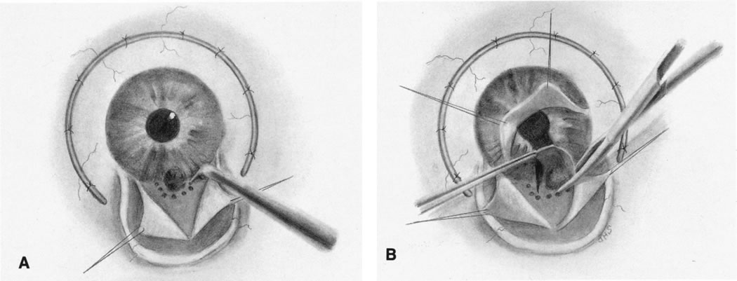
The goal of surgery is to remove the malignant lesion en bloc without contamination and tumor seeding of the remaining tissue.21 The surgical approach is similar to extracapsular cataract extraction. Wide shelving of the incision should be avoided to allow adequate iris root exposure and complete excision. Sutures may be preplaced or placed later. A viscoelastic substance is used to protect the lens and the cornea from surgical trauma (Fig. 1A); however, adequate removal is necessary prior to wound closure. With the cornea retracted by the assistant, Barraquer or Vannas scissors are placed radially through the pupil and a cut is made up to the base on either side of the lesion in normal-appearing iris (Fig. 1B). The section of iris is grasped at the pupil margin, and fine scissors, usually curved Vannas scissors, are used to incise through the iris root (Fig. 1C). A similar approach for circumscribed iris lesions has been described by Shields and Shields,21,31 termed partial lamellar scleroiridectomy.

Fig. 1. A. Corneal-scleral wound is opened, and a viscoelastic substance is placed under the iris to protect the lens. B. The cornea is retracted by an assistant, and iris scissors are used to cut the tumor radially with clear margins. C. Iris section is grasped and excision along the iris root is performed.

To achieve histologic orientation, the specimen should be pinned to a tongue depressor or piece of filter paper prior to placing into fixation solution. Fixed in this manner, proper anatomic margins of the tissue are preserved.

PROGNOSIS

The prognosis for vision is generally good in well-defined iris melanomas, providing surgical excision is without complications.23 Hyphema, corneal decompensation, cataract, and poor intraocular pressure control all can affect outcome. Cataract surgery may be combined with the excision of the iris lesions, but is usually postponed for secondary management. The prognosis for survival in iris melanoma excision is favorable with reported metastasis in less than 4% of patients,32 although more recent reports demonstrate 10% metastatic rates at 20 years.21 Rones and Zimmerman18 reported three deaths from metastases in 125 patients observed for as long as 15 years or more. Diffuse iris melanomas with heavy pigmentation are most likely to metastasize, particularly if nuclei show prominent nucleoli.

Back to Top
CILIARY BODY
Tumors and cysts may involve the ciliary body or be combined with iris lesions. These lesions include glioneuroma, medulloepithelioma, iridociliary cysts, leiomyoma, malignant melanoma, melanocytoma, lacrimal gland choristoma, adenoma of the nonpigmented ciliary epithelium, and nematode granuloma; it is sometimes difficult to differentiate them clinically.33–36 Congenital tumors are more often medulloepitheliomas or, rarely, glioneuromas, which present between ages 2 to 4 years with visual symptoms.37 In recent years, ultrasound biomicroscopy has proven to be a useful tool for diagnosis of iris, ciliary body, and anterior choroidal lesions.38–42 In cases that remain diagnostic dilemmas, iridocyclectomy is useful in permitting surgical excision of suspicious lesions without sacrificing the eye.33 Iridocyclectomy offers the benefit of both treatment and diagnosis as an excisional biopsy for lesions that are small and localized.

INDICATIONS FOR IRIDOCYCLECTOMY

Mass Lesion Growth

The option of observing a ciliary body lesion is determined by its initial status on presentation. Small lesions may be followed for growth if visual functions are intact; however, most melanomas43 and medulloepitheliomas44 result in early enucleation because of advanced stages at presentation with cataract, glaucoma, retinal detachment, and sometimes pain.

Small lesions confined to the iris and ciliary body may be successfully resected, usually when they produce symptoms or documented suspicious growth. Ciliary body melanoma often remains hidden and asymptomatic until attaining a large size, which may obstruct the visual axis or produce glaucoma.

Vail33 noted that iridocyclectomy may be performed with lesions not greater than 4 clock hours, although other authors have stated that lesions from 2.5 to 6 clock hours can be safely excised.20,33,45,46 Intraoperative and postoperative complications, particularly hypotony, are increased proportionally by the size of the tumor. Whether enucleation would result in better survival statistics than iridocyclectomy in larger melanomas is not known. Complications include seeding of the iris by iris melanoma and tumor recurrence.47,48 Thorough systemic and ocular examination should reveal no evidence of tumor extension or metastases prior to excision.

TECHNIQUE OF IRIDOCYCLECTOMY

Limbal Flap Approach

A fornix-based conjunctival flap is made to expose sclera in the area of the tumor. An anterior-hinged limbal-scleral flap is raised four-fifths of the scleral thickness at least 3 mm around the tumor (Fig. 2). Diathermy is applied to the remaining scleral bed surrounding the tumor. The anterior chamber is entered, and the flap is elevated to expose iris and tumor (Fig. 3). The tumor is excised starting at the pupil margin, moving posteriorly, and removing the section in one piece. The sector iridectomy is then closed with fine nonresorbable suture and the scleral flap is closed with interrupted sutures (Fig. 4).

Fig. 2. Fornix-based conjunctival flap is opened. The scleral flap is raised four-fifths of the thickness 3 mm around the tumor.

Fig. 3. Diathermy is placed in scleral bed. The tumor is excised.

Fig. 4. Sector iridectomy and scleral flap are closed with fine interrupted sutures.

T Incision of Stallard

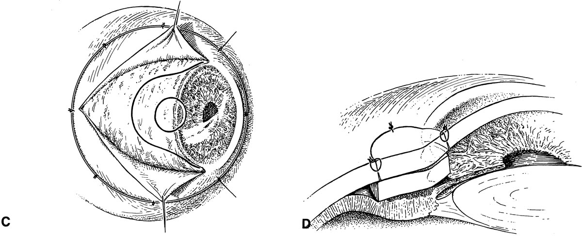
The T incision of Stallard allows larger tumor exposure and excision. A conjunctival flap is opened to expose bare sclera and a Flieringa ring is fixed through episcleral sutures. A T-shaped limbal-scleral incision is then made with a knife and extended with the corneoscleral scissors. Closing sutures may be preplaced. The ciliary body surrounding the lesion is treated with penetrating diathermy (Fig. 5A). With the cornea and scleral flaps reflected, the pupillary margin is grasped and the iris incised radially to the iris root (Fig. 5B). The involved portion is excised with scissors.49,50

Fig. 5. A. A T-shaped incision is made over the tumor to expose the mass outlined by transillumination. Diathermy is placed around the tumor-free margins. B. The pupillary margin is grasped and the iris incised radially to the iris root. The tumor is excised by grasping the pupil margin with Vannas scissors. C. Curved scleral-limbal incision with trephine of Müller. The area over the tumor is trephined through the cornea and sclera. D. The corneoscleral button is depressed, and the scleral graft is sutured in place. The tumor is then excised with the corneoscleral tissue.

Modifications of this technique, such as eliminating the Flieringa ring or including cataract removal, may be made as necessary. Müller45 described a technique using a trephine to replace the overlying corneoscleral tissue over the tumor prior to dissecting the sclera (Fig. 5C). Its advantage is that there is less likelihood of disturbing the tumor mass while excising the tumor (Fig. 5D).

Iridocyclochoroidectomy of Peyman

Iridocyclochoroidectomy of Peyman allows large tumor removal when there is more choroidal involvement posteriorly. After transillumination, localization, and marking on the bare sclera, a Peyman C-shaped cyclectomy basket is sutured in place (Fig. 6A). The scleral flap is fashioned depending on the location of the tumor. A limbal-based scleral flap can be raised with tumors located anteriorly not involving the retina. However, with more posteriorly located tumors that involve the retina, the scleral flap is started at the limbus and dissected posteriorly. A partial-thickness scleral flap is dissected and penetrating diathermy performed around the tumor margins outlined by transillumination (Fig. 6B). The tumor is excised with scissors, using radial iris incisions extending along the diathermy margins (Fig. 6C). The crystalline lens is removed with vitrectomy instruments, cutting all zonular attachments. After suturing the sclera in place with fine nonresorbing sutures (Fig. 6D), a vitrectomy is completed through an opening in the wound, or preferably using a standard pars plana approach. Ocular volume is replaced by intravitreal gas or silicone oil injection. Endolaser retinopexy is used to augment diathermy more posteriorly around the tumor base to ensure chorioretinal adhesion.

Fig. 6. A. Surface localization of the tumor is marked with diathermy. The cyclectomy basket is in place. B. A partial-thickness scleral flap is reflected. Penetrating diathermy is applied around the tumor. C. The tumor is excised. D. The partial-thickness scleral flap is sutured with running sutures.

The major complications of iridocyclectomy and large excisional biopsy include hemorrhage, vitreous loss, cataract and lens dislocation, and retinal detachment. Many authors have expressed surprise, however, at the small amount of hemorrhage that occurs postoperatively, although hemorrhage is the most common complication. It is likely that the penetrating diathermy encircling the excised ciliary body lesion reduces the risk of hemorrhage.

Back to Top
CHOROID
Lesions within the choroid can be difficult to differentiate clinically, although technologic advances in noninvasive imaging have helped to monitor size and growth. The greatest difficulty involves differentiating posterior pole amelanotic lesions within the choroid from amelanotic melanomas, metastatic neoplasms, granulomas (both inflammatory and infectious), and vascular lesions. Reports of enucleation misdiagnosis have decreased from 20% to between 2% and 6% in most large institutions,51,52 0.75% in exceptionally specialized ocular oncology centers,3 and 0.3% in the Collaborative Ocular Melanoma Study of 1527 specimens.53 Most of these decreases have come through the better use of noninvasive techniques and the use of biopsy techniques.

FINE-NEEDLE ASPIRATION BIOPSY

Ophthalmic fine-needle aspiration biopsy has developed along with the evolution of radioactive plaque therapy and more conservative surgical approaches to enucleation. Cytopathologists have achieved definitive diagnoses based on small cellular samples.54,55 The method of choice for fine-needle aspiration biopsy is contingent on the existing anatomic state of the eye, the location of the tumor, size of the tumor, and presence of retinal detachment. The anterior uveal lesion may be approached through the cornea-limbus for iris lesions (Fig. 7), or in aphakic eyes, into the more posterior ciliary body lesions (Fig. 8). In the posterior uveal lesion, either a pars plana transvitreal approach may be made (Fig. 9) or a transscleral approach may be adapted in more anterior lesions or in lesions with overlying retinal detachment (Fig. 10). Most surgeons prefer to use a 25-gauge fine needle with a flexible connector to a 10-mL aspiration syringe to minimize movement and surgical trauma during biopsy (Fig. 11). Some surgeons prefer a spinal tap needle with a trocar and cannula56; however, many surgeons use a standard long 25-gauge needle. It is felt that the excess movement caused by removing the trocar and attaching a syringe and flexible connector may lead to increased complications.

Fig. 7. Iris lesion approached through limbus.

Fig. 8. Aphakic eye may allow limbal fine-needle aspiration biopsy approach to more posterior lesions.

Fig. 9. Transvitreal fine-needle aspiration biopsy of posterior uveal tumor. The 25-gauge needle is attached to flexible tubing.

Fig. 10. Transscleral approach to posterior lesions with overlying retinal detachment. The scleral flap is raised, and transillumination is used to localize the lesion.

Fig. 11. Aspiration device for fine-needle aspiration biopsy. The apparatus is separated from the surgeon's needle by a flexible tube for better control at surgery.

For anterior segment fine-needle aspiration biopsy, the needle is inserted from the opposite side of the mass under direct visualization of the surgical microscope.57 For posterior segment lesions, the transvitreal approach is used, initially making a small partial-thickness 1.5 mm–long sclerotomy with a no. 57 Beaver blade. Only the 25-gauge needle passes through the remaining sclera. Subsequently, the wound is closed with 6-0 or 7-0 Vicryl sutures.

Seeding of tumor cells along the needle tract is thought to be reduced by the transvitreal approach because of the sweeping action of the vitreous.4 Tumor cell seeding along the scleral needle tract has been reported with retinoblastoma and has been identified in needle tracts of aspiration biopsy in the enucleated eye harboring melanoma.58 The transvitreal approach also allows deep penetration of the tumor by the needle through the overlying retina. This, however, increases the risk of rhegmatogenous retinal detachment (see Fig. 9).

Char and Miller59 demonstrated in a retrospective study that fine-needle aspiration biopsy was useful in identifying 9% of lesions that had been presumed to be uveal melanoma on noninvasive testing. They concluded that noninvasive testing was not as accurate as fine-needle aspiration biopsy for smaller lesions. Cohen and colleagues60 demonstrated that fine-needle aspiration biopsy had a diagnostic accuracy of 98% for choroidal lesions 4 mm or greater in height, but only 40% for lesions less than 1.99 mm.

INDICATIONS FOR CHOROIDECTOMY

Careful documentation of tumor growth is important before treating small tumors. Diagnostic biopsy is usually reserved for difficult clinical settings in which the diagnosis is in question. A complete diagnostic evaluation, including serial photography, ultrasonography (A-scan and B-scan), magnetic resonance imaging, and intravenous fluorescein angiography should be undertaken.

The maximum size of tumor that can be resected has not been determined. Large choroidal and ciliary body tumors have been resected; if the tumor is located anteriorly and has greater height and a narrow choroidal base, it is easier to remove. Tumors of up to 17 mm have been resected by Peyman.61,62 Lesions closer to the fovea or the optic nerve may have poor visual and anatomic results if removed by external resection. Postoperative hypotony may be prevented by the use of intravitreal silicone oil and silicone gels.63 Lesions closer than 4 disc diameters to the disc are difficult to remove without significant trauma and may represent cases best approached by an internal resection method.17,64–66 Tumor location within 1 diameter of the optic disc or fovea is significantly associated with local tumor recurrence following choroidectomy.48 The risk of retinal detachment has been reported to range from 9% to 18% following choroidectomy.67,68

TECHNIQUES OF CHOROIDECTOMY

Trapdoor Approach of Stallard

After a conjunctival peritomy, a Flieringa ring is sutured to the bare sclera. A lamellar-scleral flap (50% to 80% thickness) is dissected in a trapdoor fashion over the tumor area. Penetrating diathermy is applied around the lesion with adequate margins. The lesion is excised with scissors (Fig. 12), leaving the retina intact.

Fig. 12. Lamellar-scleral trapdoor with the lesion surrounded by diathermy.

Full-Thickness Eye Wall Resection of Peyman

Preoperatively, the mass lesion is surrounded by rows of heavy photocoagulation, which is performed in two sessions 3 to 4 weeks apart.69–75 The resection is performed 4 weeks after the final session.73

A conjunctival 360-degree peritomy is carried out, and the tumor is localized by transillumination and diathermy. A Peyman basket is sutured to the globe. A partial-thickness scleral flap is dissected around the lesion. Penetrating diathermy is applied around the tumor in the scleral bed. A pars plana sclerotomy is performed for later vitrectomy and sealed with scleral plugs or temporally sutured (Fig. 13A). The tumor is excised with curved Vannas scissors (Fig. l3B). After the scleral flap has been sutured in place (Fig. 13C), a pars plana vitrectomy is performed to remove any vitreous blood and incarcerated vitreous from the excision site (Fig. 13D). Surgery is performed under hypotensive anesthesia to reduce the risk of hemorrhage.72

Fig. 13. A. Pars plana sclerotomy is done. B. The tumor is resected en bloc. C. The scleral flap is sutured in place. D. Pars plana vitrectomy is performed.

Partial Lamellar Sclerouvectomy of Shields

Partial lamellar sclerouvectomy of Shields was modified from Foulds' original technique described in the 1970s.16,44,64 A scleral flap is made over the tumor site.76 The flap is dissected to 80% thickness and opened over the tumor with scissors at one edge. The tumor is lifted with the sclera using fine forceps and bluntly dissected from the retinal tissue with a muscle hook (Fig. 14). An attempt is made to leave the retina and vitreous intact. A surgical margin of 4 mm is outlined around the tumor prior to excision.

Fig. 14. Under the partial-thickness scleral flap, the tumor is lifted and bluntly dissected from the retinal tissue. An attempt is made to leave the retina and vitreous intact.

Internal Retinochoroidectomy of Peyman

Tumors within one to 2 disc diameters of the optic nerve cannot be easily resected by an external approach. These shortcomings have led to the development of internal excisional and biopsy approaches that may prove useful in management of lesions involving the posterior pole.65,66,69

The original technique required placement of barrier photocoagulation surrounding the lesion before retinochoroidectomy65,66; however, the more recent technique of choroidectomy under a retinal flap requires no posterior photocoagulation.2

Three or four pars plana sclerotomies are performed, one reserved for a chandelier light source. Endodiathermy is applied, and a 180-degree arcuate retinotomy is performed to ensure a 1 disc–diameter tumor-free margin (Fig. 15A and B). The retinal flap is dissected and the entire tumor exposed (Fig. 15C). Argon endolaser is applied to the tumor surface and free margins. Tumor and free margins are then dissected and removed. Following tumor removal and complete vitrectomy, the retinal flap is repositioned and an air-fluid exchange is performed. Endolaser is applied to retinotomy edges and the posterior pole (Fig. 15D). Oral aminocaproic acid is begun 24 hours preoperatively and continued for 5 days postoperatively. Hypotensive anesthesia is used during choroidectomy and tumor excision.

Fig. 15. A. Uveal melanoma located in the posterior pole within 2 disc diameters of the optic nerve and within 0.5 disc diameter of the fovea. B. Following endodiathermy, a 180-degree arcuate retinotomy is performed, ensuring a 1 disc–diameter tumor-free margin. C. The retinal flap is dissected, and the entire tumor exposed. Argon endolaser is applied both to tumor surface and free margins. Tumor and free margins are then dissected from surrounding tissue bed and removed. D. Following tumor removal and complete vitrectomy, the retinal flap is repositioned and an air-fluid exchange performed. Endolaser is applied to the retinotomy edges and the posterior pole. (Peyman GA, Nelson NC Jr, Paris CL, et al: Internal choroidectomy of posterior uveal melanomas under a retinal flap. Int Ophthalmol 16:439, 1992. Reprinted by permission of Kluwer Academic Publishers.)

RESULTS OF CHOROIDECTOMY

Shields and colleagues14 reported that the integrity of the globe has remained intact after removal of large ciliary body and choroidal melanomas. Of 95 patients, 26% maintained 20/30 vision after surgery at a mean follow-up of 5 years, and in 74 cases of confirmed melanoma, 91% of patients were alive and well.

In 1984, Peyman and colleagues77 also reported success with eye wall biopsy with long-term follow-up. Two deaths from tumor metastases occurred in the 35 patients harboring melanomas. This represents a 5.7% mortality over a mean follow-up period of more than 5 years. The interval from eye wall resection to determination of metastatic disease was 36 and 42 months in the two patients. Two additional patients died of unrelated causes. The remaining 23 eyes retained good cosmesis.77 To date, it appears that the postoperative mortality for local resection is not significantly different from mortality after enucleation.14–16,64,78 A review of 21 confirmed melanoma patients undergoing internal retinochoroidectomy was reported by Peyman and colleagues.17 The patients underwent surgery between 1984 and 1991. A total of three patients (14%) died of metastasis at 15 months, 57 months, and 84 months. These data agree with other data presented for external eye wall resection of malignant melanoma that show mortality associated with these techniques does not differ from that of patients treated with enucleation.15,16,44,77 Longer follow-up (mean 40.1 months) on patients undergoing internal resection of posterior uveal melanomas demonstrated a combined metastatic and mortality rate of 9.4%, one local recurrence, and a 9% retinal detachment rate.79 Visual acuity of 6/60 or better was maintained by 31.2% of patients.

Back to Top
COMPLICATIONS OF UVEAL EXCISIONAL BIOPSY
The most important complications of uveal excisional biopsy include vitreous hemorrhage, cataract, and retinal detachment.67,68,70,77–79 It is felt that diathermy and vitrectomy may reduce the risk of severe vitreous hemorrhage. Retinal detachment may develop either early or later, requiring further vitrectomy, scleral buckle, laser, and vitreous substitute procedures. Postoperative hypotony may be found in large tumor resections with greater exposed areas of sclera and enhanced uveal-scleral outflow.

Diagnostic biopsy specimens require special attention to ensure that the sample is properly handled. Pinning the sample flat or using cyanoacrylate glue to firm the edges allows proper orientation of the soft sample.1 Biopsy has been useful in diagnosis of early reported cases of Pneumocystis carinii choroiditis,80 metastatic lesions, and amelanotic choroidal lesions. Expanding roles for uveal biopsy may develop as surgical techniques improve and limitations of noninvasive techniques are recognized.

Back to Top
DIAGNOSTIC EYE WALL BIOPSY
Peyman and associates73 reported the use of eye wall biopsy for diagnosis of uveitis in a patient with uveitis and decreased vision in one eye. The other eye, which had no light perception, had been injured with a copper foreign body, and sympathetic ophthalmia was suspected. However, the patient did not wish to undergo enucleation of the blind eye. Diagnostic biopsy (choroidectomy) was performed, and histologic examination of the resected specimen ruled out sympathetic ophthalmia.

Eye wall biopsy (Fig. 16) has been successfully used in nine patients to diagnose retinal and choroidal disease. Long-term follow-up of these patients has shown no untoward effects from such surgery.70 A modified scleral flap version of this technique has been advocated by Constable and coworkers.72

Fig. 16. A. Peyman eye biopsy basket is placed. B. Eye basket is sutured to the globe. C. Trephine marks the site. D. Partial-thickness scleral flap is made. E. Full-thickness scleral trephine is done. F. Surface diathermy is performed. G. Sclerochorioretinal biopsy specimen is taken. H. Vitrectomy is done. I. Sclera is sutured in place. J. Ocular volume is reconstituted by air or fluid injection, and a vitrectomy is performed as in Figure 13D.

Back to Top
VITREOUS BIOPSY
Cytologic examination of vitreous aspirates has been used to make the diagnosis of reticulum cell sarcoma, amyloidosis, or Whipple's disease81 and may be a useful adjunct to uveal biopsy techniques.10,11 Material obtained during vitrectomy has been used to study the changes in retinal pigment epithelium and blood cells. Samples of vitreous can be obtained either by a vitrectomy instrument or a 21-gauge needle inserted through the pars plana. The aspirate can be fixed in 2% glutaraldehyde. The material can later be centrifuged or passed through millipore filters for histologic studies.
Back to Top
REFERENCES

1. Foulds WS, Lee WR, Roxburgh STD, et al: Can chorioretinal biopsy be justified? Trans Ophthalmol Soc UK 104:864, 1985

2. Peyman GA, Nelson NC Jr, Paris CL, et al: Internal choroidectomy of posterior uveal melanomas under a retinal flap. Int Ophthalmol 16:439, 1992

3. Shields JA, Augsburger JJ, Brown GC, et al: The differential diagnosis of posterior uveal melanoma. Ophthalmology 87:518, 1980

4. Jakobiec FA, Coleman DJ, Chattock A, et al: Ultrasonically guided needle biopsy and cytologic diagnosis of solid intraocular tumors. Ophthalmology 86:1662, 1979

5. Char DH, Ljung B, Miller T, et al: Primary intraocular lymphoma (ocular reticulum cell sarcoma): Diagnosis and management. Ophthalmology 95:625, 1988

6. Gregor RJ, Chang CA, Augsburger JJ, et al: Endogenous Nocardia asteroides subretinal abscess diagnosed by transvitreal fine-needle aspiration biopsy. Retina 9:118, 1989

7. Carroll DM, Franklin RM: Vitreous biopsy in uveitis of unknown cause. Retina 1:245, 1981

8. Taylor D, Day S, Tiedemann K, et al: Chorioretinal biopsy in a patient with leukaemia. Br J Ophthalmol 65:489, 1981

9. Augsburger JJ: Fine needle aspiration biopsy of suspected metastatic cancers to the posterior uvea. Trans Am Ophthalmol Soc 86:499, 1988

10. Michels RG, Knox DL, Erozan YS, et al: Intraocular reticulum cell sarcoma: Diagnosis by pars plana vitrectomy. Arch Ophthalmol 93:1331, 1975

11. Kirmani MH, Thomas EL, Rao NA, et al: Intraocular reticulum cell sarcoma: Diagnosis by choroidal biopsy. Br J Ophthalmol 71:748, 1987

12. Peyman GA, Fishman GA, Sanders DR, et al: Histopathology of Goldmann-Favre syndrome obtained by full-thickness eye-wall biopsy. Ann Ophthalmol 9:479, 1977

13. Peyman GA, Fishman GA, Sanders DR, et al: Biopsy of human scleral-chorioretinal tissue. Invest Ophthalmol 14:707, 1975

14. Shields JA, Shields CL, Shah P, et al: Partial lamellar sclerouvectomy for ciliary body and choroidal tumors. Ophthalmology 98:971, 1991

15. Shields JA, Shields CL, Donoso LA: Management of posterior uveal melanoma. Surv Ophthalmol 36: 161, 1991

16. Foulds WS: Experience with local excision of uveal melanomas. Trans Ophthalmol Soc UK 97:412, 1977

17. Lee KJ, Peyman GA, Raichand S: Internal eye wall resection for posterior uveal melanoma. Jpn J Ophthalmol 37:287, 1993

18. Rones B, Zimmerman LE: The prognosis of primary tumors of the iris treated by iridectomy. Arch Ophthalmol 60:193, 1958

19. Kersten RC, Tse DT, Anderson R: Iris melanoma: Nevus or malignancy? Surv Ophthalmol 29:423, 1985

20. Reese AB, Cleasby GW: The treatment of iris melanoma. Am J Ophthalmol 47:118, 1959

21. Shields CL, Shields JA, Materin M, et al: Iris melanoma: risk factors for metastasis in 169 consecutive patients. Ophthalmology 108:172, 2001

22. Demirci H, Shields CL, Shields JA, et al: Diffuse iris melanoma: A report of 25 cases. Ophthalmology 109:1553, 2002

23. Brown D, Boniuk M, Font RL: Diffuse malignant melanoma of iris with metastases. Surv Ophthalmol 34:357, 1990

24. Dick AD, Jagger J, McCartney AC: Refsum's disease: electron microscopy of an iris biopsy. Br J Ophthalmol 74:370, 1990

25. Preac-Mursic V, Pfister HW, Spiegel H, et al: First isolation of Borrelia burgdorferi from an iris biopsy. J Clin Neuroophthalmol 13:155, 1993

26. Moorthy RS, Rao NA, Sidikaro Y, et al: Coccidioidomycosis iridocyclitis. Ophthalmology 101:1923, 1994

27. MacLean H, Clarke MP, Strong NP, et al: Primary ocular relapse in acute lymphoblastic leukemia. Eye 10:719, 1996

28. Karcioglu ZA, Mullaney PB: Diagnosis and management of iris juvenile xanthogranuloma. J Pediatr Ophthalmol Strabismus 34:44, 1997

29. Chan SM, Hutnik CM, Heathcote JG, et al: Iris lymphoma in a pediatric cardiac transplant recipient: Clinicopathologic findings. Ophthalmology 107:1479, 2000

30. Char DH, Crawford JB, Kroll S: Iris melanomas. Diagnostic problems. Ophthalmology 103:251, 1996

31. Shields JA, Shields CL: Surgical approach to lamellar sclerouvectomy for posterior uveal melanomas: The 1986 Schoenberg lecture. Ophthalmic Surg 19:774, 1988

32. Arentsen JJ, Green WR: Melanoma of the iris: Report of 72 cases treated surgically. Ophthalmic Surg 6:23, 1975

33. Vail DT: Iridocyclectomy: a review. Am J Ophthalmol 71:161, 1971

34. LoRusso FJ, Boniuk M, Font RL: Melanocytoma (magnocellular nevus) of the ciliary body: Report of 10 cases and review of the literature. Ophthalmology 107:795, 2000

35. Shields JA, Eagle RCJr , Shields CL: Adenoma of nonpigmented ciliary epithelium with smooth muscle differentiation. Arch Ophthalmol 117:117, 1999

36. Shields JA, Eagle RCJr , Shields CL, et al: Natural course and histopathologic findings of lacrimal gland choristoma of the iris and ciliary body. Am J Ophthalmol 119:219, 1995

37. Broughton WL, Zimmerman LE: A clinicopathologic study of 56 cases of intraocular medulloepitheliomas. Am J Ophthalmol 85:407, 1978

38. Pavlin CJ, McWhae JA, McGowan HD, et al: Ultrasound biomicroscopy of anterior segment tumors. Ophthalmology 99:1220, 1992

39. Augsburger JJ, Affel LL, Benarosh DA: Ultrasound biomicroscopy of cystic lesions of the iris and ciliary body. Trans Am Ophthalmol Soc 94:259, 1996

40. Reminick LR, Finger PT, Ritch R, et al: Ultrasound biomicroscopy in the diagnosis and management of anterior segment tumors. J Am Optom Assoc 69:575, 1998

41. Shields JA, Shields CL, Mercado G, et al: Adenoma of the iris pigment epithelium: A report of 20 cases: the 1998 Pan-American Lecture. Arch Ophthalmol 117:736, 1999

42. Mannino G, Malagola R, Abdolrahimzadeh S, et al: Ultrasound biomicroscopy of the peripheral retina and the ciliary body in degenerative retinoschisis associated with pars plana cysts. Br J Ophthalmol 85:976, 2001

43. Reese AB: Tumors of the Eye, p 286. New York, Paul B. Hoeber, 1951

44. Shields JA, Shields CL: Intraocular Tumors: A Text and Atlas. Philadelphia, WB Saunders, 1992

45. Müller HK, Lund OE, Söllner F et al: Die operative behandlung von tumoren des kammerwinkels und des ciliarkörpers. Doc Ophthalmol 20:500, 1966

46. Barrada A, Peyman GA, Palacio MN: Limitations of iris and ciliary body resection in primates. Retina 4:119, 1984

47. Gupta M, Puri P, Rennie IG: Iris seeding following iridocyclectomy for localised iris melanoma. Eye 15:808, 2001

48. Damato BE, Paul J, Foulds WS: Risk factors for residual and recurrent uveal melanoma after trans-scleral local resection. Br J Ophthalmol 80:102, 1996

49. Stallard HB: Partial cyclectomy: Some further modifications in technique. Br J Ophthalmol 48:1, 1964

50. Flieringa HJ: A method of surgery in the angle of the anterior chamber of the eye. Trans Ophthalmol Soc UK 81:421, 1961

51. Robertson DM, Campbell RJ: Errors in the diagnosis of malignant melanoma of the choroid. Am J Ophthalmol 87:269, 1979

52. Ferry AP: Lesions mistaken for malignant melanoma of the posterior uvea: A clinicopathologic analysis of 100 cases with ophthalmoscopically visible lesions. Arch Ophthalmol 72:463, 1964

53. Histopathologic characteristics of uveal melanomas in eyes enucleated from the Collaborative Ocular Melanoma Study. COMS report no. 6. Am J Ophthalmol 125:745, 1998

54. Augsburger JJ, Shields JA, Folberg RT, et al: Fine needle aspiration biopsy in the diagnosis of intraocular cancer: Cytologic-histologic correlations. Ophthalmology 92:39, 1985

55. Char DH, Kroll SM: Cytomorphometry of uveal melanoma: Fine needle aspiration biopsy versus standard histology. Trans Am Ophthalmol Soc 87:197, 1989

56. Midena E, Segato T, Piermarocchi S, et al: Fine needle aspiration biopsy in ophthalmology. Surv Ophthalmol 29:410, 1985

57. Augsburger JJ, Shields JA: Fine needle aspiration biopsy of solid intraocular tumors: Indications, instrumentation and techniques. Ophthalmic Surg 15:34, 1984

58. Karcioglu ZA, Gordon RA, Karcioglu GL: Tumor seeding in ocular fine needle aspiration biopsy. Ophthalmology 92:1763, 1985

59. Char DH, Miller T: Accuracy of presumed uveal melanoma diagnosis before alternative therapy. Br J Ophthalmol 79:692, 1995

60. Cohen VM, Dinakaran S, Parsons MA, et al: Transvitreal fine needle aspiration biopsy: The influence of intraocular lesion size on diagnostic biopsy result. Eye 15:143, 2001

61. Peyman GA, Ericson ES, Axelrod AJ, et al: Full-thickness eye wall resection in primates: An experimental approach for treatment of choroidal melanoma. Arch Ophthalmol 89:410, 1973

62. Peyman GA, Koziol JE: Limitation of eye wall resection. Can J Ophthalmol 9:328, 1974

63. Peyman GA, Conway MD, Karaĉorlu M, et al: Evaluation of silicone gel as a long-term or permanent vitreous substitute in non-human primates [abstract]. Invest Ophthalmol Vis Sci 33(suppl):899, 1992

64. Foulds WS, Damato BE: Alternatives to enucleation in the management of choroidal melanoma. Aust N Z J Ophthalmol 14:19,1986

65. Peyman GA, Cohen SB: Ab interno resection of uveal melanoma. Int Ophthalmol 9:29, 1986

66. Peyman GA: Internal retinal biopsy: Surgical technique and results. Int Ophthalmol 14:101, 1990

67. Damato B, Groenewald CP, McGalliard JN, et al: Rhegmatogenous retinal detachment after transscleral local resection of choroidal melanoma. Ophthalmology 109:2137, 2002

68. Jonas JB, Groh MJ, Rummelt V, et al: Rhegmatogenous retinal detachment after block excision of epithelial implantation cysts and tumors of the anterior uvea. Ophthalmology 106:1942, 1999

69. Peyman GA, Charles H: Internal eye wall resection in the management of uveal melanoma. Can J Ophthalmol 23:219,1988

70. Peyman GA, Juarez CP, Raichand M: Full-thickness eye-wall biopsy: Long-term results in 9 patients. Br J Ophthalmol 65:722, 1981

71. Peyman GA, Axelrod AJ, Graham RO: Full-thickness eye wall resection: An experimental approach for treatment of choroidal melanoma: Evaluation of cryotherapy, diathermy and photocoagulation. Arch Ophthalmol 91:219, 1974

72. Constable IJ, Chester GH, Horne R, et al: Human chorioretinal biopsy under controlled systemic hypotensive anaesthesia. Br J Ophthalmol 64:559, 1980

73. Peyman GA, Diamond JG, Axelrod AJ: Sclero-chorio-retinal (SCR) resection in humans. Ann Ophthalmol 6:1347, 1974

74. Peyman GA, Nelson PT, Axelrod AJ, et al: Full thickness eye wall resection: Evaluation of preoperative photocoagulation. Invest Ophthalmol 12:262, 1973

75. Peyman GA, May DR, Ericson ES, et al: Full thickness eyewall resection: An experimental approach for treatment of choroidal melanoma. II. Homo- and heterograft. Invest Ophthalmol 11:668, 1972

76. Shields JA, Shields CL: Surgical approach to lamellar sclerouvectomy for posterior uveal melanomas: The 1986 Shoenberg Lecture. Ophthalmic Surg 19:774, 1988

77. Peyman GA, Juarez CP, Diamond JG, et al: Ten years experience with eye wall resection of uveal malignant melanomas. Ophthalmology 91:1720, 1984

78. Zimmerman LE, McLean IW, Foster WD: Does enucleation of the eye containing a malignant melanoma prevent or accelerate the dissemination of tumour cells? Br J Ophthalmol 62:420, 1978

79. Kertes PJ, Johnson JC, Peyman GA: Internal resection of posterior uveal melanomas. Br J Ophthalmol 82:1147, 1998

80. Freeman WR, Gross JG, Labelle J, et al: Pneumocystis carinii choroidopathy: A new clinical entity. Arch Ophthalmol 107:863, 1989

81. Durant WJ, Flood T, Goldberg MF, et al: Vitrectomy and Whipple's disease. Arch Ophthalmol 102:848, 1984

Back to Top