Chapter 46
Toxoplasmosis
KHALID F. TABBARA
Main Menu   Table Of Contents

Search

THE ORGANISM
EPIDEMIOLOGY
ANIMAL MODELS OF TOXOPLASMOSIS
CLINICAL MANIFESTATIONS OF OCULAR TOXOPLASMOSIS
OCULAR FINDINGS
OCULAR COMPLICATIONS
PATHOLOGY
DIFFERENTIAL DIAGNOSIS
LABORATORY DIAGNOSIS OF OCULAR TOXOPLASMOSIS
PRENATAL DIAGNOSIS OF CONGENITAL TOXOPLASMOSIS
SEROLOGIC DIAGNOSIS OF CONGENITAL TOXOPLASMOSIS
ISOLATION OF TOXOPLASMA GONDII
MAGNETIC RESONANCE IMAGING AND COMPUTED TOMOGRAPHY SCANS
POLYMERASE CHAIN REACTION IN THE DIAGNOSIS OF TOXOPLASMOSIS
FLUORESCEIN AND INDOCYANINE GREEN ANGIOGRAPHY
IMAGING TECHNIQUES
IMMUNOLOGY OF TOXOPLASMOSIS
PREVENTION AND TREATMENT
TREATMENT
PREVENTION
REFERENCES

Toxoplasmosis is a systemic disease caused by the organism Toxoplasma gondii. The disease is widespread, affecting both humans and animals. Infection with the organism in immunocompetent individuals leads to a mild or subclinical systemic disease. Most patients have no recognizable symptoms and develop immunity to the organism. In some patients, the primary infection may lead to fever, fatigue, lymphadenopathy, and malaise. After parasitemia, Toxoplasma organisms may reach the posterior segment of the eye through the bloodstream, leading to formation of cysts within the retinal tissue, or they may cause localized, relentless destruction of the retina, which may result in loss of vision. The disease may be congenital or acquired and may show tendency to recur in the eye without evidence of systemic manifestations or increase in the antibody titer. Although the disease may present with the typical clinical picture, it may mimic other entities or present with unusual clinical manifestations, frequently leading to misdiagnosis. Since Toxoplasma is an intracellular parasite, the retina sustains the major damage and primary insult. The vitreous cavity may be invaded by the inflammatory cells carrying with them Toxoplasma organism. Early diagnosis is essential for prompt initiation of therapy, especially when the lesion is close to the macula, optic nerve, or the papillomacular nerve bundle. Clinical findings of toxoplasmosis in immunocompromised patients may show more severe and fulminant manifestations leading to rapid deterioration of vision and serious outcome.
Back to Top
THE ORGANISM
Toxoplasma gondii is an obligate intracellular protozoan belonging to the phylum Apicomplexa, class Sporozoa, order Eucoccidia, and suborder Eimeriina. There are three forms of the parasite: the tachyzoite, the bradyzoite, and the sporozoite. All three forms cause infection in humans.

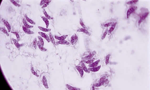
Tachyzoite is the proliferative form of the organism, which used to be known as trophozoite. It has a crescent shape and measures approximately 7 μm in length and 3 μm in width (Fig. 1). The organism is motile with a unique cytoskeletal structure allowing it to twist, wiggle, rotate, and glide. The rostrum of the tachyzoite is known as the conoid, which can extend, retract, tilt, and rotate. These movements allow the tachyzoite to find its target host cell and to penetrate the cell, establishing an intracellular existence. Tachyzoites are easily propagated in peritoneal cavities of mice and in mammalian tissue culture cell lines. The organism has the ability to replicate in all mammalian nucleated cells. In the host cell, the tachyzoite multiplies by endodyogeny; this reproductive process is susceptible to heat, freezing and thawing, desiccation, and gastric enzymes.

Fig. 1. Toxoplasma gondii. Tachyzoites from the peritoneal exudates of infected mice stained with Giemsa (× 100).

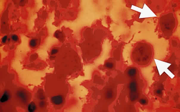
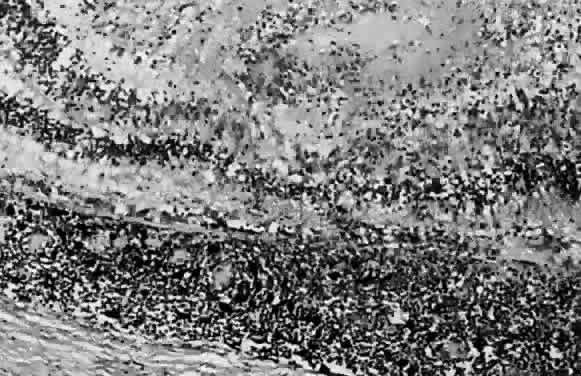
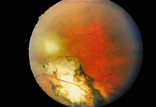
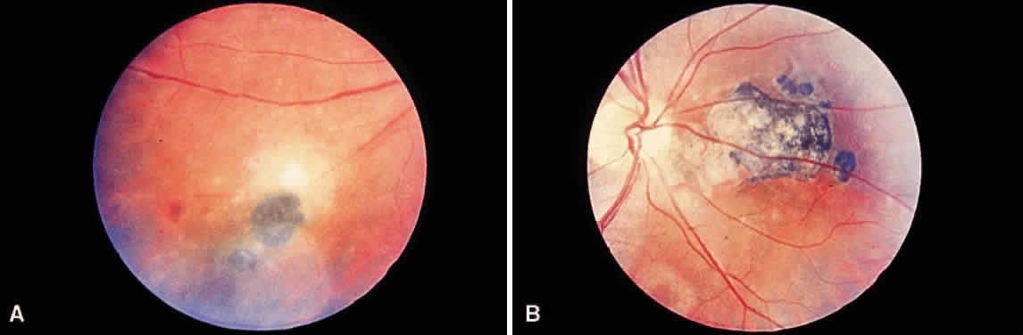
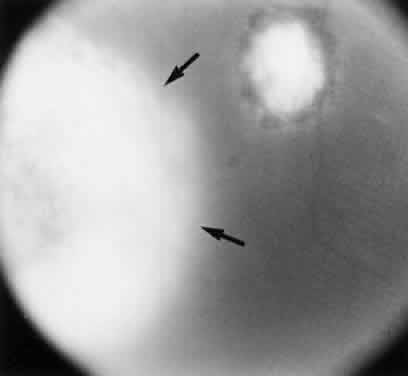
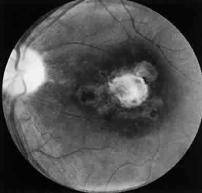
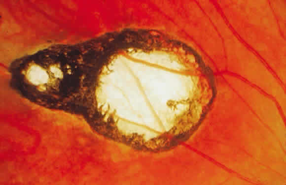
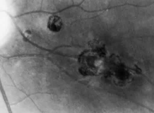
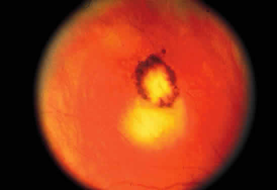
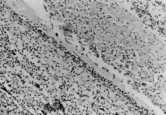
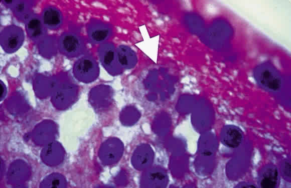
Bradyzoites are slowly metabolizing organisms found in cysts formed within the tissue of the infected host. The size of the Toxoplasma cyst varies, depending on the number of organisms that have multiplied within it. The cyst may reach more than 100 μm in diameter and may contain from 50 to 3000 organisms. The cyst wall is strongly argyrophilic and periodic acid-Schiff positive. It contains constituents that are derived from both the parasite and the host tissue. Constituent from the host tissue compose the outer part of the cyst, whereas those derived from the parasite are in the inner part of the cyst wall. Toxoplasmosis may be found in the inner layers of the retina after episodes of acute retinochoroiditis. The cyst may stay in the retinal tissue for years without showing any signs of invasiveness. Considering that the tissue cyst incorporates elements derived from the host into its outer wall, it is easily tolerated by the host, and no inflammatory reaction is seen around it (Fig. 2). It may remain for years in certain tissues, such as the eye or muscles, without provoking any inflammatory reactions. The bradyzoite inside the cyst derives its nutrition from the slow diffusion of substances through the cyst wall. The number of organisms increases within the cyst in the retina, and once the cyst wall breaks down by mechanical stretching, the bradyzoites escape, convert into tachyzoites, and invade contiguous cells. This process may lead to recurrence of retinitis. Certain immunologic mechanisms of the host may influence the organisms significantly. Immunosuppression coinciding with the rupture of the cyst and release of bradyzoites allows the organisms to become tachyzoites and proliferate in host tissue without restriction. The cyst of the Toxoplasma organism appears to be a defensive stage in its life cycle. The resistance of toxoplasmosis within chronically infected tissues of animals may lead to transmission of the disease by the ingestion of undercooked meat, including mutton, beef, pork, and chicken. Tissue cysts can develop within any organ and are commonly found in infected tissues of brain, eye, heart, skeletal muscles, and lymph nodes. Rupture of tissue cysts causes reactivation of the systemic toxoplasmosis in immune deficiency states, leading to dissemination of Toxoplasma organisms to other organs.

Fig. 2. Toxoplasma bradyzoites inside cysts of tissue sections in chronically infected rabbit's retina (arrows). The animal was killed 6 months after acute Toxoplasma retinochoroiditis after suprachoroidal injection of 1000 organisms (Beverley strain). There was no clinical evidence of inflammation at the time of killing. (H & E, × 400)

Sporozoites are found in the oocyst. Each sporozoite measures 10 to 12 μm in diameter. The sporozoite is produced in the intestines during the enteroepithelial part of the Toxoplasma life cycle. Toxoplasma organisms divide by endodyogeny (asexual reproduction), schizogony (splitting of nuclear material), and sexual reproduction involving the gametocytes. The gametocytes are found throughout the small intestines, especially in the ileum of the definitive host. The male gametocytes produce microgametes, which have the ability to fertilize macrogametes. After fertilization of the macrogamete, the zygote becomes surrounded by an oocyst wall and is shed in the feces of infected cats. Sporogony, which spans approximately 3 to 21 days, occurs within the oocyst outside of the host.1 The sporulated oocysts become infectious. Millions of oocysts may be excreted in the feces of an infected cat. Under conditions of moist and warm soil, the oocyst may remain infectious for more than a year.2 The oocyst appears to be an important method for the transmission of toxoplasmosis. Once ingested, the oocyst wall is digested by the gastric enzymes, trypsin and pepsin, thereby liberating Toxoplasma sporozoites. Shedding of the oocyst begins 1 to 24 days after the ingestion of the parasite by the cat. Sporulation requires 2 to 3 days at 24°C and 14 to 21 days at 11°C and does not occur above 37°C. Dry heat and exposure to temperatures above 66°C render the oocyst noninfectious. Puppies are susceptible to infection with Toxoplasma organisms and tend to shed more oocysts than adult cats.3 The reinfection of cats by Toxoplasma rarely leads to excretion of oocyst.

PENETRATION OF HOST CELL

Since the Toxoplasma organism is an obligate intracellular parasite, it cannot multiply in the extracellular space, and the initial step of entry into the host cell is critical for survival of the organism and the pathogenesis of the disease. The tachyzoite has a well-defined nucleus that is oval or round with a central karyosome. It is able to attain intracellular habitat by active invasion of host cell membrane. This is accomplished by a series of sequential steps involving the release of penetration enhancing factors. The conoid at the rostral end has the ability to thrust outward at time of invasion into the cell. At the time of contact between toxoplasmic plasmalemma and the host cell membrane, the differentiated organelles in the conoid, known as rhoptries, appear to play a role in the parasite penetration of the host cell. As the Toxoplasma organisms penetrate the host cell, a parasitophorous vacuole forms from the host cell membrane. The process of active penetration takes 30 seconds under laboratory conditions. The thrusting of the conoid into the cell membrane probably is accomplished by an actin-like system. The organisms enter the host cell by active invasion, whereas entry into the macrophage can be by phagocytosis or active invasion. In naive macrophages, the organisms have the ability to actively invade the macrophage, which provides a free ride and helps in the dissemination of the infection to various organs, including the eye. In immunized hosts, the organism attains the intracellular status in the macrophage by active phagocytosis. The parasite may enter the retinal pigment epithelial cell by active penetration or phagocytosis. Retinal pigment epithelial cells have the ability to support the multiplication of T. gondii. In most host cells, however, invasion appears to be the predominant means of cellular entry by the organism. Structural differences between invasion and phagocytosis can be recognized by electron-microscopic study.4 Contact between the macrophage and the conoid may augment the penetration of the protozoan into the macrophage. If the macrophage grasps the posterior end of the organism, rhoptry discharge may not occur. The absence of microfilament aggregates under the cell membrane of the macrophage during toxoplasmic penetration is strong evidence that the cellular invasion was a parasitic effected process. Several factors may interfere with the cellular penetration, whether the penetration is by phagocytosis or by active invasion. Coating the organism with immunoglobulin G (IgG) or C'3 stimulates and enhances phagocytosis. In the absence of serum, phagocytosis may proceed, but slowly. Active invasion by the parasites may be inhibited by cytochalasin D, a substance that interferes with actin filament function.5 This cytoplasmic cytoskeletal system in the Toxoplasma organism helps it to thrust forward and move in an undulating motion.

When the parasite attains intracellular habitat, it starts to multiply, leading to host cell destruction and release of live protozoa. Live organisms do not stimulate respiratory burst during phagocytosis, but heat-killed or antibody-coated organisms generate a respiratory burst by the macrophage. The parasite may retain some of the disrupted macrophage plasmalemma adherent to the surface immediately after invasion. A vacuole quickly forms around the parasite that has invaded the host cell. The parasitophore appears to be larger than the organism, with tubules connecting membrane with the plasmalemma of the parasite. Each vacuole contains one parasite, and several organisms may be seen within a single macrophage. The Toxoplasma organism has a remarkable ability to evade the respiratory burst of macrophages by remaining within the vacuole. In activated macrophages, however, the organism has difficulty surviving within the macrophage, and the parasite is killed after fusion between the cell membrane and the internal limiting membrane of the vacuole. The organism is, in general, resistant to hydrogen peroxide, but it is susceptible to oxygen intermediates generated by the xanthine oxidase system.

LIFE CYCLE OF TOXOPLASMA GONDII

The definitive hosts of T. gondii are domestic and wild cats, whereas intermediate hosts encompass a wide variety of animals, including humans. In the definitive host, the parasite has both enteroepithelial and extraintestinal cycles. In the intermediate host, it persists in the extraintestinal cycle only. When oocysts from contaminated soil are ingested by animals, the oocyst wall is digested by the gastric enzymes, and the sporozoites are released. Similarly, when animals—including cats—ingest chronically infected tissues containing bradyzoites, the cyst wall is digested, and bradyzoites are released, leading to infection. Toxoplasma organisms then invade intestinal mucosal cells and initiate the infection (enteroepithelial cycle). In the intestinal mucosa, the organisms undergo asexual reproduction (endodyogeny, endopolygeny, splitting, schizogony) and sexual reproduction (gametogony cycles), culminating in the formation of zygotes, which develop into oocysts. The oocysts are shed in the feces of the definitive host. After oocyst excretion, sporulation occurs; the mature oocysts remain infectious in moist soil for a long time if not subjected to extreme climatic conditions.2 Freezing to -20°C, heating above 66°C, and desiccation are lethal to the cyst. Simultaneous with the enteroepithelial oocyst formation in cats, bradyzoites or sporozoites may invade and disseminate widely to all host tissue through the bloodstream or lymphatics, where they undergo an asexual cycle (extraintestinal cycle), particularly in muscle, heart, brain, lung, lymphoid tissue, retina, and central nervous system (CNS). The organism multiplies rapidly by endodyogeny in infected cells, forming pseudocysts containing tachyzoites. This usually leads to death and disruption of the cell, thereby liberating the tachyzoites, which enter contiguous cells in which they multiply, forming more pseudocysts. The rapidly proliferating tachyzoites are responsible for initial spread of the infection and tissue destruction (acute stage). In response to increased host immunity, tachyzoites transform into the slowly multiplying organisms, bradyzoites, which form true cysts in tissues (chronic stage). These can lie dormant in tissues throughout the life of the host but may cause reactivation of the clinical disease when the host's immunity is suppressed.6

Back to Top
EPIDEMIOLOGY
Toxoplasmosis is a worldwide disease of humans and animals. The disease is common among the healthy adult population. Serologic evidence of Toxoplasma infection ranges from 3% to 70% of the adult population in the United States. Geographic area, age, and socioeconomic factors influence the prevalence of the disease. The prevalence is highest in tropical regions and lowest in cold regions of the world.8 In a seroepidemiologic study on toxoplasmosis, Luyasu and associates9 found, in two population groups in Belgium, a progressive age-related increase with a prevalence rate of Toxoplasma antibodies of up to 60% in pregnant women and up to 77% in blood donors. Among female patients, the prevalence of Toxoplasma antibodies was higher among female blood donors than among pregnant women.9 Choi and colleagues10 report two outbreaks of acute toxoplasmosis involving eight adult patients in Korea. All patients developed acute systemic toxoplasmosis after consumption of uncooked pork. In the first outbreak, three patients developed unilateral chorioretinitis within 3 months of eating a meal consisting of raw spleen and liver of a wild pig. In the second outbreak, 5 of 11 soldiers who ate a meal consisting of liver roll of domestic pig developed lymphadenopathy. All patients had high levels of T. gondii antibodies (1:1024 or greater) and elevated IgM antibodies.10 In Nigeria, Olusi and associates11 determined the prevalence of toxoplasmosis among 606 women of childbearing age. The mean percentage of positive antibodies to T. gondii was 43.7%, ranging from 25% in the 15- to 18-year age group to 71.4% in the 39- to 42-year age group. Their study demonstrates a progressive increase in the prevalence of positive antibodies to T. gondii with age.11

Toxoplasma gondii organisms can be transmitted from mothers who are chronically infected with human immunodeficiency virus (HIV) to the fetus, but this is not common. In a small cohort study of HIV-infected women, Minkoff and associates12 did not observe congenital transmission of T. gondii among those with severe immunodeficiency. Congenital toxoplasmosis, therefore, may occur in patients with acquired immunodeficiency syndrome (AIDS) but is not common. The authors observed only one case of congenital toxoplasmosis among 28 HIV-infected female patients.12

A prospective case-control study designed to identify preventable risk factors for T. gondii infection in pregnancy was conducted in Norway.13 Patients were identified through a serologic screening program consisting of 37,000 pregnant woman through sporadic antenatal testing for Toxoplasma infection. Sixty-three pregnant women with serologic evidence of recent primary T. gondii infection and 128 seronegative control women matched by age, expected date of delivery, stage of pregnancy, and geographic area were enrolled. The following risk factors were found to be significant in patients with maternal Toxoplasma infection: (1) eating of raw and undercooked minced meat product (p = 0.007), (2) eating unwashed raw vegetables and fruits (p = 0.03), (3) eating raw or undercooked mutton (p = 0.005), (4) eating raw or undercooked pork (p = 0.03), (5) cleaning the cat litter box (p = 0.02), and (6) washing the kitchen knives infrequently after preparation of raw meat before handling another food item (p = 0.04). In the univaried analysis, traveling to countries outside of Scandinavia was identified as a significant risk factor in acquiring toxoplasmosis.13 In another study on the seropositivity to Toxoplasma among pregnant woman, Ljungstrom and associates14 found 12% to 26% of their subjects to be positive for Toxoplasma antibodies in various urban and rural localities of Sweden. The estimates of the prevalence of toxoplasmosis and risk of maternal infection were strongly dependent on underlying temporal change in the incidence of the disease. Their studies demonstrate the difficulties in interpretation of horizontal cross-sectional data and the need for longitudinal studies of age prevalence and seroconversion in determining the risk factors of maternal toxoplasmosis. Zadik and coworkers15 found 160 (9.9%) of the 1621 women who had experienced seroconversion to be of childbearing age. The rate of seroconversion in the United Kingdom appears to be less than reported. Meenken and colleagues16 found a positive correlation between ocular toxoplasmosis and an increased in the frequency of HLA BW-62 in patients with severe ocular involvement.

Toxoplasmosis may be acquired in humans by the following modes of transmission:

  Ingestion of undercooked infected meat (chicken, mutton, beef, pork) containing Toxoplasma cysts
  Ingestion of the oocyst from contaminated hands or food (vegetables)6
  Accidental skin penetration and inoculation of tachyzoites
  Allograft organ transplantation: heart,17 kidney,18 liver,19 or bone marrow20
  Congenital transplacental transmission
  Consumption of raw milk21
  Inhalation of oocysts22
  Transcojunctival transmission23
  Water supply contamination24

The tachyzoite occasionally may penetrate mucosal surfaces such as the conjunctiva.23

Toxoplasma-infected animals may develop a subclinical infection and live normally but harbor the encysted form (bradyzoite) of the parasite in their muscular system. Undercooked beef, mutton, chicken and pork have been previously incriminated in the transmission of the disease. Ingestion of sporozoites in oocysts may represent a major mode of transmission of the infection in many countries. The presence of infected puppies around homes may allow the accumulation of numerous oocysts. Teutsch and associates22 report an outbreak of toxoplasmosis in which Toxoplasma oocysts were the source of infection and the presumed mode of transmission was by inhalation.

Food consumed by humans may be contaminated with the oocysts by insects and cockroaches. Furthermore, accidental contamination of hands while disposing of cat feces may represent another mode of transmission of the disease.13 Allograft transplantation also has been shown to transmit Toxoplasma in humans. The consumption of raw milk also may cause transmission of the disease.

An outbreak of toxoplasmosis in Vancouver, British Columbia, involved 110 individuals, including 12 newborns who were infected with T. gondii.24 This is the world's largest recorded outbreak of toxoplasmosis that was found to be caused by a contaminated water supply.

If a woman acquires the infection during pregnancy, the disease may be transmitted to the fetus. The prevalence of congenital toxoplasmosis has been estimated to vary between 1:1000 and 1:10,000. This form of transplacental transmission is of great clinical importance. Toxoplasmic infection in consecutive siblings is rare, but congenital ocular toxoplasmosis has been described in siblings.

Most acquired toxoplasmosis is asymptomatic. The disease may mimic upper respiratory tract infection and infectious mononucleosis. The mild form of the systemic disease may present as a flulike syndrome and is frequently misdiagnosed as a viral infection. Most of the systemic acquired infections are subclinical, as evidenced by serologic testing of asymptomatic patients.

It had been believed that most cases of toxoplasmic retinochoroiditis represented a recrudescence of a congenital disease. Toxoplasmic retinochoroiditis may complicate the course of systemic acquired toxoplasmosis.25–27 I have observed cases of toxoplasmic retinochoroiditis that followed systemic acquired toxoplasmic disease (Fig. 3). Ronday and coworkers28 describe eight cases of unilateral toxoplasmic retinochoroiditis, presumably acquired after toxoplasmosis. Montoya and Remington report 22 adult cases of acute toxoplasmic chorioretinitis that occurred in the setting of acute postnatally acquired toxoplasmosis.29 A patient developed toxoplasmic retinochoroiditis associated with acquired toxoplasmic myocarditis and polymyositis.30 It appears that toxoplasmic retinochoroiditis after acute systemic acquired toxoplasmosis is more common than previously recognized.

Fig. 3. Toxoplasma cysts containing bradyzoites in a human retina (arrows). The eye was enucleated because of absolute glaucoma. The patient had been treated with oral and periocular corticosteroids. (H & E, × 400)

In a community-based survey of toxoplasmosis in Saudi Arabia, Tabbara and associates31 found that 32.7% of the Saudi population had serologic evidence of toxoplasmosis. Retinochoroiditic scars suggestive of ocular toxoplasmosis were found among 2.9% of cases with systemic toxoplasmosis.31

Acquired toxoplasmosis has been reported among family members.32

The prevalence of acquired toxoplasmosis during pregnancy is 0.2% to 1%.33 Congenital infection develops in 30% to 50% of infants born to mothers with acquired toxoplasmosis during pregnancy.34,35

Immune mothers protect their infants from successive fetal transmission. Mothers who deliver one child with congenital toxoplasmosis will have little or no future fetal transmission of the disease.

If the mother develops Toxoplasma infection, the risk to the fetus is 4%: 3% of the infants will manifest clinical infection of toxoplasmosis, and 1% will have subclinical infection.36

Back to Top
ANIMAL MODELS OF TOXOPLASMOSIS
Animal models of infectious disease of the retina and choroid should have retinal lesions that morphologically and histopathologically resemble lesions seen in humans. Furthermore, the natural course of the disease in the selected animal model should be similar to the disease seen in humans. Several animal models of Toxoplasma retinochoroiditis have been described. Injection of Toxoplasma organisms (Beverley strain) into the suprachoroidal space of pigmented rabbits produced focal retinochoroiditis with no encephalitis. The animal model was used to study the pathogenesis and effects of therapeutic modalities on the course of toxoplasmic retinochoroiditis. Murine Toxoplasma retinitis may be induced by intraperitoneal injection of T. gondii. Mice develop encephalitis and retinitis.

An animal model of ocular toxoplasmosis was produced in a nonhuman primate, the cynomolgus monkey, by Culbertson and coworkers.37 The retinochoroiditis was induced by direct inoculation of Toxoplasma organisms into the retina through the pars plana under direct microscopic visualization. The focal necrotizing retinitis lesions had morphologic and histopathologic resemblance to the lesion seen in humans, and the course of retinochoroiditis closely resembled the course of the disease in humans. The cynomolgus monkey was found to be resistant to the RH strain of T. gondii and developed no evidence of systemic disease and no encephalitis. Healing of the retinochoroiditis was associated with scar formation with atrophic scar and variable pigmentation. Histologic studies reveal mononuclear inflammatory reaction with evidence of areas of necrosis seen in the retina (Fig. 4). The monkey appears to be a good animal model for the study of ocular toxoplasmosis. Pavesio and associates report an animal model of acquired retinochoroiditis in hamsters.38

Fig. 4. Section of the retina of a cynomolgus monkey infected with T. gondii shows extensive retinal necrosis with disruption of the normal retinal architecture, mononuclear cell infiltration, and severe choroiditis. (H & E, × 100)

Back to Top
CLINICAL MANIFESTATIONS OF OCULAR TOXOPLASMOSIS
The most frequent form of infection with T. gondii is subclinical, and the disease is discovered by serologic testing for antibodies to Toxoplasma organisms. Clinically, the disease presents in children or adults who are unimmunized and contract the infection or by the reactivation of a latent infection. The signs and symptoms of toxoplasmosis may mimic a variety of other disorders. The following are clinical entities of toxoplasmosis: congenital toxoplasmosis, acquired systemic toxoplasmosis, toxoplasmosis in the immunocompromised host, acquired or reactivation of a latent infection, and ocular toxoplasmosis after either congenital or acquired systemic disease.

CONGENITAL TOXOPLASMOSIS

Congenital toxoplasmosis results from transplacental transmission of T. gondii. The incidence of congenital infection varies with the trimester during which the mother becomes infected. The lowest incidence occurs in the first trimester (15% to 20%), and the highest incidence is in the third trimester (59%).38 Acquired toxoplasmosis during pregnancy may lead to spontaneous abortion, prematurity, or stillbirth. Most infected newborns, however, are delivered asymptomatic at birth. When primary Toxoplasma infection is acquired during pregnancy, the organism may be transferred to the fetus in up to 61% of the cases. The clinical spectrum of congenital toxoplasmosis varies in severity, depending on the trimester of pregnancy during which the maternal Toxoplasma infection was acquired. Ninety percent of infected infants have no clinical signs of the disease, but the infection is confirmed by the detection of antibodies in the sera of the newborn.39 Infection acquired by the mother in the second trimester produced infection in 21% of newborns; of those who were infected, 74% had no clinical evidence of the disease.40

Mets and coworkers41 determined the natural history of treated and untreated cases of congenital toxoplasmosis. In a longitudinal study, 76 newborns were treated with pyrimethamine and sulfadiazine for 1 year, and 18 patients who were not treated during their first year of life were entered in the study as historical controls. The most frequently encountered ocular finding was retinochoroiditic scars, which occurred in 58% of treated cases and 82% of the controls. Macular scars were seen in 54% of the treated patients and 76% of the historical controls. Visual impairments were observed in 29% of the treated patients. Recurrences were observed in 13% of the treated patients and 44% of the untreated patients. This study suggests that early diagnosis and prompt treatment of cases with congenital toxoplasmosis may prevent complications of congenital toxoplasmosis and induce rapid rehabilitation. Ocular complications of congenital toxoplasmosis include cataract, amblyopia, phthisis bulbi, optic atrophy, retinal detachment, macular scars, and macular membranes.

It had been believed that the women who are at risk of delivering an infected newborn are those who acquired the infection just before or during gestation. Recent reports, however, suggest that a chronically infected woman with toxoplasmosis may transmit the disease to her offspring. Vogel and associates42 report a case of congenital toxoplasmosis transmitted from an immunologically competent mother infected before her conception.42 Polymerase chain reaction (PCR) for B1 gene of T. gondii was present in her lymphoid biopsy specimens.

Rarely, congenital toxoplasmosis may be transmitted from an immunocompetent mother infected before conception. The cerebrospinal fluid (CSF) may show mononuclear pleocytosis and elevated protein content. Patients may develop microcephaly, hydrocephalus, intracranial calcifications, and bone lesions.43

Toxoplasmosis acquired in the first trimester results in congenital infection in 14% of infants, but the disease usually is severe and may result in stillbirth.44

Clinical manifestations of congenital toxoplasmosis consist of retinochoroiditis, fever, hepatosplenomegaly, lymphadenopathy, jaundice, skin rash, petechiae, myocarditis, encephalitis, and retinochoroiditis. Patients may later develop hydrocephaly or microcephaly, cerebral calcifications, convulsions, and psychomotor retardation. The most common clinical manifestation of congenital toxoplasmosis is retinochoroiditis. The microcephaly, which is a severe form of disease, may occur after injury to the brain by the infectious process. The calcifications tend to be scattered throughout the brain substance in toxoplasmosis (Fig. 5), whereas in cytomegalic inclusion disease the calcifications tend to be paraventricular. This finding, however, varies and is not absolute.

Fig. 5. Cerebral calcification in congenital toxoplasmosis. (Christenson L, Beeman H, Allen A: Cytomegalic inclusion disease. Arch Ophthalmol 57:90, 1957)

The infection in the mother usually is asymptomatic, but infection in the fetus is expressed as a disease with varying severity, from a subclinical disease to a fulminant course with fatal outcome. The infection can terminate in fetal or neonatal death, but most infants survive, and those who acquire the disease show sequelae of the CNS infection. The clinical manifestations of congenital toxoplasmosis are related to several factors, including the age of the fetus at the time of infection, the virulence of the Toxoplasma organism, and the status of maternal and fetal host defense mechanisms. Premature infants often experience severe CNS and ocular disease in the first 3 months of life.44 Full-term infants who acquire the infection in utero develop milder disease and show generalized sign of infection, such as hepatosplenomegaly and lymphadenopathy, in the first few months of life. Infants with subclinical infection at birth may develop signs and symptoms of toxoplasmosis if the patients are observed into adolescence.45 In a clinical evaluation during childhood (up to a mean age of 8 years), 11 of 13 infected children who were normal at birth experienced sequelae, and in each of the children, the initial clinical manifestation was a retinochoroiditis, which appeared at a mean age of 3.7 years. Three children developed unilateral blindness, whereas the remainder had retinochoroiditis without visual loss. Five of the 11 children developed neurologic abnormalities; 1 child had delayed psychomotor development, microcephaly, and seizures; and 2 children had minor cerebellar signs.45

The differential diagnosis of congenital toxoplasmosis includes cytomegalic inclusion disease, rubella, syphilis, and herpes virus infection.

ACQUIRED TOXOPLASMOSIS

Adult acute acquired toxoplasmosis presents as an acute febrile illness associates with cervical lymphadenopathy. Hilar and submental lymph node enlargement also may occur, as well as enlargement of other lymph node groups. Only 10% to 20% of Toxoplasma infections in the adult are symptomatic.46 Patients develop malaise, myalgia, weakness, fatigue, headache, sore throat, and maculopapular rash with sparing of the palms and soles. Hepatosplenomegaly, lymphocytosis with the presence of atypical forms of lymphocytes, and hilar lymphadenopathy may occur. Acquired toxoplasmosis may present as fever of unknown origin with or without abdominal pain. The course of the disease in the immunocompetent host is self-limited and often is benign. The signs and symptoms may persist for extended periods, and the generalized fatigue and malaise may occur for several months. The CNS involvement by Toxoplasma may occur in immunologically normal patients and in patients with AIDS.47,48 Encephalitis, pneumonitis, or myocarditis can result in fatal outcome. The clinical manifestations of toxoplasmosis may mimic many diseases, including Hodgkin's disease and infectious mononucleosis.49 Less than 1% of the infectious mononucleosis is caused by infection with T. gondii.49 Laboratory diagnosis is essential in confirming the clinical diagnosis of toxoplasmosis.

Retinochoroiditis may occur in patients with acquired toxoplasmosis and can be the sole clinical manifestation of the disease.6,26,27,32,50–53 Weiss and coworkers report a patient with Toxoplasma retinochoroiditis who presented with the ocular lesion as the initial manifestation of AIDS.47 The retinochoroiditis may be a presenting manifestation of adult acquired toxoplasmosis or may occur several months to 2 years after the onset of acquired toxoplasmosis.

Outbreaks of systemic toxoplasmosis among families have been reported.32,54 Masur and colleagues found that six of the seven members of a household investigated for toxoplasmosis demonstrated high antibody titers consistent with a recent infection, and five of these members were symptomatic.32 The most common manifestations were fever and lymphadenopathy, which developed from 7 to 18 days after a exposure to a common source—ingestion of infected meat. One patient developed vision-threatening retinochoroiditis 129 days after infection with toxoplasmosis.

Similar, well-documented cases of retinochoroiditis caused by Toxoplasma after acquired systemic toxoplasmosis have been reported.27,26,51–54 In an outbreak of systemic toxoplasmosis that occurred in October 1977 in Atlanta, Georgia, 37 patients became ill or had serologic evidence of acute infection.22 Epidemiologic studies suggest that infected cats in the riding stable were the source of the infection. Aerosolization of oocysts and hand-to-mouth contact were the presumptive means of transmission. All patients were examined and followed for a year without evidence of Toxoplasma retinochoroiditis. In a 4-year follow-up study, only one patient had shown evidence of ocular disease consisting of an acute retinochoroiditis.27 This suggests that many sporadic cases of acute retinochoroiditis result from or may follow an episode of acquired toxoplasmosis (Fig. 6). Notice that not all cases of retinochoroiditis occur simultaneously with an episode of acquired systemic illness but may manifest any time after the onset of the disease, from a few weeks to years after the acute episode of acquired systemic toxoplasmosis.27 The case described in Figure 6 is one such example.

Fig. 6. Toxoplasma retinochoroiditis after acute systemic toxoplasmosis. A 36-year-old man presented with history of seeing floaters in the right eye of 1 week's duration. The onset of his ocular symptoms occurred 56 days after a flulike illness with postauricular lymphadenopathy. Acute and convalescent serum specimens were obtained and showed an increase in Toxoplasma antibody titers from negative to 1:1024. A. Single focus of retinochoroiditis below the macular area of the right eye (vision: 20/200 [metric equivalent 6/60]). The left eye was normal with 20/20 (6/6) vision. B. Fluorescein angiography (early late phase) indicates leakage from retinal capillaries. C. Two months after a 3-week course of clindamycin, sulfadiazine, and systemic corticosteroids, the lesion completely resolved. The patient's vision decreased because of epiretinal gliotic membrane formation, causing macular pucker and subretinal neovascularization.

TOXOPLASMOSIS AMONG PATIENTS WITH AIDS

One of the most common parasitic infections in patients with AIDS is toxoplasmosis. The disease may be a recrudescence of a latent infection or a newly acquired toxoplasmic infection. In cases of recrudescence of the disease, it may result from rupture of the cyst and conversion of the bradyzoites into the tachyzoites within the tissue. This may occur in the retina or elsewhere in the body. Toxoplasmic encephalitis is a fatal disorder if not treated early or promptly. Clinical diagnosis and therapy of toxoplasmosis in patients with AIDS pose a challenge to the managing clinician. T. gondii organisms may infect the nervous system and form cysts in the tissue of normal individuals after systemic toxoplasmosis. Release of bradyzoites from the cysts leads to proliferation and foci of the infection. In the CNS, toxoplasmic encephalitis may present as a mass lesion in the brain, toxoplasmoma. The differential diagnosis of such a focal lesion in patients with HIV infections include toxoplasmosis in 60% of the cases, primary CNS lymphoma in 25%, and multifocal leukoencephalopathy in 15%. Other infections are less frequently encountered. Vyas and Ebright55 report a case of acute myelopathy caused by toxoplasmosis of the spinal cord in a patient with AIDS. The patient recovered completely with treatment for toxoplasmosis. The patient had perineuritis with sensory loss and urinary bladder dysfunction and localized pain. Most patients with spinal cord toxoplasmosis show abnormalities of the spinal cord and brain on magnetic resonance imaging (MRI). Treatment for toxoplasmosis results in clinical and the radiographic improvement.

TOXOPLASMOSIS IN THE IMMUNOSUPPRESSED HOST

Toxoplasmosis is becoming an important cause of mortality and morbidity in patients undergoing immunosuppressive therapy for malignancies, organ transplantation, or autoimmune disorders. Patients with impaired immunity such as those with lymphoma, leukemia, malignancies, and AIDS also are at increased risk of acquiring severe infection by T. gondii. Patients with hairy cell leukemia also are highly susceptible to toxoplasmosis. It is likely that clinically apparent toxoplasmosis in these patients most often is a consequence of reactivation of latent infection.56–58 The clinical manifestations of toxoplasmosis in the immunodeficient host may mimic other opportunistic pathogens. Patients may present with fever, encephalitis, myocarditis, and pneumonitis, which are the most common and serious of the clinical manifestations. More than 50% of these patients have CNS involvement. Occasionally, the encephalitis may mimic a mass lesion in the brain.

Toxoplasmic encephalitis is the most common infection of the brain in patients with AIDS. Toxoplasmic retinochoroiditis occurs in 1% to 3% of AIDS patients.

Back to Top
OCULAR FINDINGS
Toxoplasma gondii is the most common cause of infection of the retina. Ocular findings include involvement of the retina, choroid, retinal vessels, macula, optic nerve, vitreous, and anterior uvea. The ocular findings in toxoplasmosis are summarized in Table 1.


TABLE 1. Clinical Manifestations of Ocular Toxoplasmosis

  Typical Manifestations
  Focus of retinitis surrounded by fuzzy retinal edema
  Pigmented atrophic retinochoroiditic scar adjacent to the lesion or elsewhere in the fundus
  Vitreous cells and exudates
  Focal retinal vasculitis
  Hyperemia of the optic nerve head
  Cells and flare in the anterior chamber (rarely, mutton-fat keratic precipitates)
  In patients with recurrent ocular toxoplasmosis: anterior segment findings, including posterior synechiae, secondary cataract, and secondary glaucoma
  Atypical Manifestations
  Juxtapapillar retinitis
  Retrobulbar neuritis
  Rhegmatogenous retinal detachment
  Pars planitis
  Punctate outer retinitis
  Serous macular detachment
  Retinal branch artery or vein occlusion
  Retinal or subretinal neovascularization
  Choroidoretinal vascular anastomosis
  Panuveitis


RETINA-CHOROID

Ocular toxoplasmosis most commonly presents as a focus of necrotizing retinitis (Fig. 7) involving the inner layers of the retina and associated with a whitish fluffy lesion surrounded by retinal edema.6,59 Cells are seen in the vitreous overlying the lesion. The focus of retinitis may be large (Fig. 8) or small (Fig. 9). The retina is the primary site for the multiplying parasites, whereas the choroid and sclera may be the sites of contiguous inflammation. When the choroid is involved by the inflammatory reaction, the lesion is referred to as retinochoroiditis. The organisms are rarely seen in the choroid. The retinal lesion may be small and single or large and multiple and may reach several disc diameters in size. Large granulomas in the posterior segment of the eye frequently are seen in patients with ocular toxoplasmosis (Fig. 10). This is associated with extensive and marked vitreous reaction that precludes visualization of the retina, and the granuloma appears as a yellowish mass surrounded by a red reflex. Punctate outer retinal involvement may be seen in some patients with toxoplasmosis.60 These appear as multifocal, gray-white lesions in the deep retina and at the level of the retinal pigment epithelium accompanied by no overlying vitreous reactions. The lesions may resolve to form fine, granular white dots. This is the earliest finding seen in the rabbit models of ocular toxoplasmosis after the injection of Toxoplasma organisms in the suprachoroidal space. The lesions in the animal model may start as deep outer retinal punctate lesions that later involve the inner layers of the retina and cause the typical focus of retinochoroiditis. Involvement of the macular area is common in patients with congenital toxoplasmosis, and exudative retinal detachment may occur in severe cases. Clumps of inflammatory cells may be seen in the vitreous or over the detached posterior vitreous face. The focus of retinitis may be a manifestation of congenital toxoplasmosis or may be associated with or follow an episode of acquired systemic toxoplasmosis. Patients presenting with recurrent toxoplasmic retinochoroiditis usually are in the second or third decade, but the disorder may occur at any age. Healing of the retinitis is associated with a decrease in retinal edema and flattening of the lesion with evidence of scar formation surrounded by variable amounts of pigment (Fig. 11). The lesion may appear as a punchedout scar with underlying sclera resulting from extensive retinal and choroidal necrosis surrounded by pigment proliferation (Fig. 12), it may become a conglomerate or proliferated retinal pigment cells (Fig. 13), or it may be small and appear as a pigment clump in the retina. The retinochoroiditic scar may harbor the Toxoplasma cysts. Healing also is associated with decrease in the vitreous cells and improvement in visual acuity. Immunologic suppression is associated with recurrence of retinochoroiditis (Fig. 14). Recurrent toxoplasmic retinitis frequently appears as “satellites” or occurs adjacent to a previous scar. Old, inactive lesions often appear to be a conglomeration of previous multiple inflammatory foci (see Fig. 13).

Fig. 7. A focus of Toxoplasma retinitis (arrow) close to healed retinochoroiditic scars in a 32-year-old man.

Fig. 8. Severe Toxoplasma retinochoroiditis with large granuloma and overlying vitreous cells. A healed retinochoroiditic scar is seen in close proximity to the granuloma.

Fig. 9. A. A small focus of Toxoplasma retinochoroiditis adjacent to a healed retinochoroiditic scar near the equator in the right eye of a 23-year-old man. The Toxoplasma antibody titer IgG was 1:64 (indirect fluorescence antibody test). Such small lesions with minimal vitreous reaction may be observed and followed without treatment. B. Left eye of same patient showing a healed pigmented retinochoroiditic scar. Recurrences of Toxoplasma retinochoroiditis are not associated with an increase in antibody titer (B, Courtesy of Dr. John Cavender)

Fig. 10. Large Toxoplasma retinochoroiditic granuloma with vitreous exudates (arrows). The lesion appears as a pale, elevated mass in a background of red reflex. A small punched-out retinochoroiditic scar and fundus details are hazily seen.

Fig. 11. Macular retinochoroiditic scar in a 6-year-old child with healed congenital ocular toxoplasmosis.

Fig. 12. Typical punched-out Toxoplasma retinochoroiditic scar surrounded by pigmentation. The central whitish area is the sclera and results from extensive necrosis of the retina and choroid. Toxoplasma cysts may remain viable in the retina throughout life.

Fig. 13. Multiple retinochoroiditic scars with pigment proliferation seen in the retina of a 28-year-old woman who had three previous episodes of Toxoplasma retinochoroiditis.

Fig. 14. Toxoplasma retinochoroiditis in a 28-year-old kidney transplant recipient who had been observed to have a healed, inactive retinochoroiditic scar on a routine ophthalmologic examination before his kidney transplantation. The patient developed retinochoroiditis after he was started on azathioprine (Immuran), 250 mg orally daily, and prednisone, 80 mg orally daily, after renal transplantation. Serologic tests revealed the Toxoplasma IgG antibody titer to be 1:64 (indirect fluorescence antibody test). The titer remained the same 6 months later. This case reaffirms the fact that immunologic suppression may lead to reactivation of Toxoplasma retinitis. Recurrences of Toxoplasma retinochoroiditis are not associated with an increase in antibody titers.

Retinal vasculitis is common in cases of active retinochoroiditis. Theodossiadis and associates observed vascular involvement in all 64 cases of toxoplasmic retinochoroiditis, and 59 (92%) had vasculitis in the same quadrant of retinal vasculitis. Vasculitis may lead to vascular occlusion and retinal hemorrhages.61

Lesions in the macular area probably are established as a result of entrapment of free-swimming organisms or parasite containing macrophages in the terminal capillaries of the periofoveal retina. A similar entrapment in the peripapillary capillary network may account for the juxtapapillary lesion but also is characteristic of the disease. Cystoid macular edema occurs when lesions are close to the macular area.62 In elderly patients, an atypical, severe form of necrotizing retinochoroiditis may occur. The disease may manifest as multifocal or diffuse necrotizing retinitis.63

OPTIC NERVE

The CNS is frequently involved in toxoplasmosis. The optic nerve may present with optic neuritis or papillitis associated with edema. Direct extension of cerebral infection through the sheath of the optic nerve also may lead to optic neuritis or papillitis. In one case, Toxoplasma organisms were isolated from the CSF of a patient who had both a meningoencephalitis and a juxtapapillary retinochoroiditis.64 Manschot and Daamen65 found numerous T. gondii organisms in the optic nerves of an infant who died of congenital toxoplasmosis. Eichenwald found four infants who had optic neuritis in a series of 140 cases of congenital toxoplasmosis.66 In six patients with ocular toxoplasmosis, the active site of inflammation was found to be the optic nerve head.67 The patients presented with severe papillitis, vitreous inflammation, and sector or nerve fiber bundle field defects. In these patients, the typical foci of retinitis suggesting toxoplasmosis were absent, and the initial diagnosis was incorrect. Therefore, patients with Toxoplasma papillitis may present without evidence of a focus of retinitis. Willerson and coworkers68 describe an adult with ocular toxoplasmosis who they believed had an active focus of inflammation within the optic nerve head. The patient also had an area of active retinitis in the macula. The diagnosis may be hard to make when patients present with severe papillitis and no evidence of active retinal lesion.

VITREOUS

Toxoplasma gondii is an obligate intracellular parasite and, therefore, the organism does not invade the acellular vitreous cavity. Posterior vitreous detachment commonly is seen in patients with posterior segment inflammation from toxoplasmosis, and patients may develop precipitates of inflammatory cells on the posterior vitreous face. Inflammatory cells are seen in patients with active retinitis and retinochoroiditis. Vitreous strands may be seen, and rare solid cylindrical lines have been described by Schlaegel.69 Large lesions of Toxoplasma retinochoroiditis may be associated with vitreous cells. In patients with papillitis, vitreous cells are seen in clumps over the optic nerve head.

ANTERIOR UVEA

Anterior uveitis (granulomatous or nongranulomatous) may be associated with Toxoplasma retinochoroiditis. In immunocompetent patients, the anterior uveal inflammation is a hypersensitivity reaction to the Toxoplasma antigens. The parasite has not been recovered from aqueous fluid of immunocompetent hosts. Rehder and associates report a case of acute unilateral iridocyclitis in a patient with AIDS.70 At autopsy, Toxoplasma organisms were found in the iris tissue.70

Back to Top
OCULAR COMPLICATIONS
Severe inflammatory changes within the globe secondary to toxoplasmosis may lead to several complications. Fuchs' heterochromic iridocyclitis has been described in 13 patients who had focal necrotizing retinochoroiditis characteristic of ocular toxoplasmosis.71 The reason for the association of Fuchs' heterochromic iridocyclitis and toxoplasmosis is not clear. Eleven of the 13 patients had keratic precipitates, and none of the patients had ciliary injection or posterior synechiae. Six of the 13 patients had iris atrophy, 2 had glaucoma, and 4 had lesions in the fellow eye.71 Severe anterior uveitis may lead to peripheral anterior synechiae and secondary glaucoma in patients with chronic ocular toxoplasmosis.

Subretinal neovascularization may complicate the course of Toxoplasma retinochoroiditis. Gilbert72 describes two unusual cases of ocular toxoplasmosis in which the patients developed retinal and optic nerve neovascularization in association with ocular toxoplasmosis. The neovascularization regressed with resolution of the inflammation. The reason for neovascularization of the optic nerve and the retina is not well understood. Retinal ischemia associated with severe retinal vasculitis may predispose to neovascularization of the retina. On the other hand, inflammatory reactions alone may cause neovascularization of the retina. Retinal vasculitis, whether it is arteritis or phlebitis, may lead to retinal hemorrhages.72 Retinal tears may be seen that can lead to rhegmatogenous retinal detachment. Optic papillitis and optic neuritis may eventuate in optic atrophy. Juxtapapillary retinochoroiditis may lead to nerve fiber bundle field defects.

The vitreous may show thick, vitreous stands and vitreous membranes that may require vitrectomy. Vascular anastomoses may be seen in patients with ocular toxoplasmosis. Vascular anastomoses between the retinal and choroidal circulation can occur through the damaged Bruch's membrane and fundus scars resulting from ocular toxoplasmosis. Cerebral blindness and oculomotor nerve palsies have been described in patients with ocular toxoplasmosis,73,74 as have retinal branch artery occlusion,75 subretinal neovascularization,76,77 and choroidal vascularization.78,79

Long-term ocular and neurologic involvement may occur in severe cases of congenital toxoplasmosis.16

Atypical forms of ocular toxoplasmosis are observed in immunosuppressed individuals80 and elderly patients.63

TOXOPLASMOSIS IN IMMUNOCOMPROMISED PATIENTS

In patients with AIDS, ocular toxoplasmic retinochoroiditis may represent a reactivation of a latent infection or a newly acquired disease. The clinical manifestations of ocular toxoplasmosis in patients with AIDS may be severe. Patients may have single or multiple lesions of retinitis. The disease may be unilateral or bilateral and may present in a fulminant manner. Retinal hemorrhages may occur, and patients may develop papillitis. Toxoplasmosis accounts for 1% to 3% of ocular infections in patients with AIDS.81 A variety of clinical manifestations can be confused with toxoplasmosis, including cytomegalovirus retinitis, herpes simplex, and herpes zoster retinitis. The ocular disease caused by toxoplasmosis may be the first manifestation of intracranial and disseminated T. gondii infection in AIDS patients. Therefore, prompt recognition and early treatment with appropriate antiparasitic drugs will prevent widespread tissue destruction and may decrease the chances of dissemination. Serologic diagnosis of toxoplasmosis in AIDS may not be helpful, and the clinician may have to rely on clinical findings and results of PCR testing. A nested multiplex PCR assay has been designed for the simultaneous detection of Epstein-Barr virus and T. gondii DNA from the ocular fluid of AIDS patients.82 T. gondii DNA was detected in eight of eight patients with toxoplasmic encephalitis and in zero of six patients without toxoplasmosis in patients with AIDS. PCR appears to be a helpful diagnostic test to confirm the presence of toxoplasmosis in patients with AIDS. Optic nerve involvement and papillitis in ocular toxoplasmosis may be the initial manifestations of AIDS.

Back to Top
PATHOLOGY
Active retinochoroiditis is characterized by an inflammatory process consisting of a zonal granuloma with intense central necrosis surrounded by successive layers of mononuclear cell infiltration (Fig. 15). The cells consist predominantly of lymphocytes and macrophages with occasional epithelioid cells. Plasma cells may be seen to accumulate in the periphery of the lesion and become more numerous during the healing stage. They are known to secrete antibodies leading to the destruction of the free parasites in the extracellular space and may induce cyst formation by parasites. The formation of cyst is regarded as a defensive action by the parasite in response to the release of antibodies in the ocular tissues. The encysted form of the organism does not provoke inflammatory reaction and may be tolerated in the tissues for many years. Involvement of the retinal pigment epithelium leads to proliferation of the retinal pigment epithelial cells, and healing of the lesion is associated with scar formation. The retinal pigment epithelial cells may act as phagocytes. Toxoplasma cysts may be seen in areas adjacent to the retinochoroiditic scars many years after an episode of retinochoroiditis. The cysts remain dormant in the retina, and the recovery of viable organisms can be accomplished if homogenates of infected retinal tissues are injected into the peritoneal cavities of mice.

Fig. 15. Tissue section of a human retina infected with Toxoplasma. Notice extensive necrosis and destruction of the normal layers of the retina and mononuclear cell infiltration (H & E, × 100).

Back to Top
DIFFERENTIAL DIAGNOSIS
The differential diagnosis of ocular toxoplasmosis includes several disorders that may lead to inflammation of the posterior segment of the eye. The retina and choroid are targets of insults by a variety of immunologic, infectious, and malignant disorders.83,84 The structural damage sustained by ocular tissue depends on the cause of the disease and the afflicted layer.

The diagnosis of posterior uveitis can be established on the basis of an accurate medical history, including geographic location of race, sex, personal habits, laterality, and mode of onset of the disease. The morphologic features of the lesion and the association with systemic disease help in arriving at an accurate diagnosis of posterior uveitis. The recent increase in the number of patients with AIDS has contributed to a corresponding increase in the incidence of agents that cause inflammations in the posterior segment of the eye. Several systemic and ocular diseases that lead to posterior uveitis may mimic toxoplasmosis. Tables 2 and 3 outline the infectious and noninfectious disorders that may afflict the posterior segment of the eye and lead to posterior uveitis. Certain clinical manifestations of posterior uveitis may point out the nature of the diagnostic entity (Tables 3 and 4; see Table 2). To narrow the differential diagnosis to a short list, it may be necessary to retrieve the facts from the medical and social history and to dissect the clinical observation systematically, then focus on the entities that may lead to the ocular disease. The laboratory test results are used to refine the clinical diagnosis and rule out other disorders that cause inflammation of the posterior segment. The signs and symptoms of the presenting disorder may vary from one entity to another. The lesions in the posterior segment can be focal, multifocal, geographic, or diffuse. Those that caused clouding of the overlying vitreous should be differentiated from those that never induce vitreous cells. In addition, the lesions that are regularly associated with retinal vasculitis or with serous detachment of the retina must be separately designated. The type and distribution of the vitreous opacities also must be specified. Inflammatory lesions of the posterior segment are insidious in onset, but some may be abrupt and accompanied by visual loss with rapid progression, such as the case in acute retinal necrosis. As a general rule, such diseases are accompanied by anterior uveitis, which in turn may be associated with secondary glaucoma. In a patient with posterior uveitis, the age, gender, geographic location, and race of patient, as well as the mode of onset and laterality of the condition, should be considered. Active ocular toxoplasmosis usually is a unilateral disease in immunocompetent hosts. Gender is an important factor in the differential diagnosis of posterior uveitis. Certain disorders occur more commonly in male patients, and some occur in childhood, whereas others occur in adulthood (Table 5). Behçet's disease is more common in men than women, whereas Vogt-Koyanagi-Harada syndrome occurs with equal frequency among male and female patients. Both Behçet's disease and Vogt-Koyanagi-Harada syndrome are common in Japan, the Middle East, and Brazil and are uncommon in northern European countries and the United States. Birdshot retinochoroidopathy is more common in Europe and North America, and rare in the Middle East and Japan. Similarly, human T-cell lymphotropic virus type 1 infections are common in southern Japan and some Caribbean countries but rare in the Middle East and Europe. Sarcoidosis is common among blacks in the United States but is uncommon among blacks in South Africa. Genetic and environmental factors appear to play a role in the pathogenesis of posterior uveitis. Cysticercosis occurs in Central America but does not occur in the Middle East because Jews and Moslems do not eat pork. Brucellosis still occurs sporadically in developing countries and rarely in developed countries. A history of penetrating wound is important to elicit the diagnosis of sympathetic ophthalmia or an occult intraocular foreign body.


TABLE 2. Differential Diagnosis of Posterior Uveitis: Anterior Segment Signs


Anterior Segment SignsCauses of Posterior Uveitis
Anterior granulomatous uveitisBrucellosis
 Sarcoidosis
 Lyme disease
 Tuberculosis
 Syphilis
 Toxoplasmosis
 Vogt-Koyanagi-Harada (VKH) syndrome
 Fungal infections
Anterior nongranulomatous uveitis Behçet's disease
 Acute multifocal placoid pigment epitheliopathy
 Masquerade syndrome
Conjunctival noduleSarcoidosis
 Tuberculosis
 Syphilis
 Behçet's disease
 Acquired immunodeficiency syndrome
ScleritisVaricella-zoster virus
 Herpes simplex
 Tuberculosis
 Brucellosis
 Lyme disease
Hypopyon or pseudo-hypopyonBehçet's disease
 Syphilis
 Masquerade syndrome
 Lyme endogenous bacterial endophthalmitis
 VKH syndrome
 Toxoplasmosis
 Tuberculosis
 Sarcoidosis
 Herpetic necrotizing retinitis (acute retinal necrosis)


TABLE 3. Differential Diagnosis of Posterior Uveitis: Posterior Segment Signs


Posterior SegmentCauses of Posterior Uveitis
Choroiditis DiffuseSympathetic ophthalmia, leukemia, VKH syndrome
GeographicTuberculosis, serpiginous choroiditis Mycobacterium avium complex, herpetic necrotizing retinitis
Nummular multifocal white-dot syndromeHepatitis C, histoplasmosis, VKH syndrome, Epstein-Barr virus, viral infection, coxsackievirus, multiple evanescent white-dot syndrome, birdshot retinochoroidopathy, CNS lymphoma, lymphoma, sympathetic ophthalmia, sarcoidosis, Behçet's disease, histoplasmosis, acute multifocal placoid pigment epitheliopathy
RetinitisToxoplasmosis, CMV retinitis, rubella infection, candidiasis, nocardiasis
Retinal vasculitisHerpetic necrotizing retinitis, toxoplasmosis, CMV retinitis, Lyme disease, Behçet's disease, sarcoidosis, syphilis, tuberculosis, systemic lupus erythematosus, polyarteritis nodosa, Wegener's disease, Waldenström macroglobulinemia
Retinochoroiditic scarsHistoplasmosis, toxoplasmosis, toxocariasis, tuberculosis, syphilis, VKH syndrome
VitritisToxoplasmosis, Behçet's disease, syphilis, tuberculosis, toxocariasis, others
Optic nerve involvement (hyperemia, papillitis)VKH syndrome, tuberculosis, sarcoidosis, syphilis, toxoplasmosis
Subretinal neovascularizationToxoplasmosis, VKH syndrome, histoplasmosis
Exudative retinal detachmentVKH syndrome, sympathetic ophthalmia, Lyme disease
Retinal pigment migrationVKH syndrome, syphilis, rubella
Posterior granulomaSarcoidosis, toxoplasmosis, toxocariasis, tuberculosis

VKH, Vogt-Koyanagi-Harada syndrome; CNS, central nervous system; CMV, cytomegalovirus.


TABLE 4. Differentiation Between Herpetic Necrotizing Retinitis, Cytomegalovirus Retinitis, and Toxoplasmic Retinochoroiditis


FeatureHerpetic Necrotizing RetinitisCytomegalovirus RetinitisOcular Toxoplasmosis
CourseRapidSlowInsiduous
Visual lossSevere visualVariableVariable
LesionsMultifocal1–3 lesions/eye1–3 lesions/eye
Retinal detachmentDetachment in mostDetachment in 20%Rare
No. of CD4+ cells per micrometer0–1000–50Normal (except in AIDS)
Retinal lesionDeep retinal clearing around blood vesselsDry granular border and perivascular heathingNecrotizing retinitis healed, scars may be seen
VasculitisNo vascular occlusionVascular occlusionVascular occlusion
TreatmentFoscarnet with ganciclovirFoscarnet (or ganciclovir) 

AIDS, acquired immunodeficiency syndrome.


TABLE 5. Differential Diagnosis of Posterior Uveitis According to Age


Age Group (y)Clinical Entities
<4Toxoplasmosis
 Masquerade syndrome (retinoblastoma, leukemia)
5–15Toxocariasis
 Toxoplasmosis
 Subacute sclerosing panencephalitis
 Masquerade syndrome
 Sympathetic ophthalmia
 Cytomegalovirus retinitis
 Lyme disease
16–40Sarcoidosis
  Behçet's disease
 Vogt-Koyanagi-Harada syndrome
 Sympathetic ophthalmia
 Lyme disease
 Candidiasis
 Aspergillosis
>40Herpetic necrotizing retinopathy (acute retinal necrosis and progressive outer retinal necrosis)
 Central nervous system lymphoma
 Tuberculosis
 Cryptococcus
 Endogenous fungal infections
 AIDS patients (any age)
 Cytomegalovirus retinitis
 Herpetic necrotizing retinopathy (acute retinal necrosis and progressive outer retinal necrosis)
 Tuberculosis
 Toxoplasmosis
 Lymphoma
 Mycobacteriu avium complex infection
 Coccidiodomycosis
 Pneumocystis carinii infection
 Candidiasis
 Cryptococcosis

AIDS, acquired immunodeficiency syndrome.


Ocular toxoplasmosis has a worldwide distribution and affects nationalities and ethnic groups, affecting men and women with equal frequency.

Rarely, patients with ocular toxoplasmosis present with an exudative form of toxoplasmic retinochoroiditis, which may be misdiagnosed as Coats' disease.85

Back to Top
LABORATORY DIAGNOSIS OF OCULAR TOXOPLASMOSIS
Laboratory diagnosis of ocular toxoplasmosis relies on serologic evidence of exposure to Toxoplasma. Serum antibodies to T. gondii may be detected by various methods that determine antibody levels to Toxoplasma. These tests include the Sabin-Feldman dye test, enzyme-linked immunosorbent assay (ELISA), indirect fluorescence antibody (IFA) test, indirect hemagglutination test, and the agglutination test. The complement fixation and precipitating tests are of limited use in the laboratory diagnosis of toxoplasmosis. The most widely used serologic tests for the diagnosis of Toxoplasma include the ELISA and IFA test. IgG, IgM, IgA and IgE anti-Toxoplasma antibodies have been determined.86–96
Back to Top
PRENATAL DIAGNOSIS OF CONGENITAL TOXOPLASMOSIS
The prenatal diagnosis of congenital toxoplasmosis may be established by obtaining fetal blood sampling at 24 weeks of gestation.36 It is possible to detect nonspecific laboratory signs of fetal infection in the fetal blood specimens with specific antibodies of fetal origin and parasitemia by inoculation of the fetal blood sample into mice. The amniotic fluid samples also are inoculated into mice, and Toxoplasma organisms may be present when the fetus is infected. Ultrasonic examination may detect enlargement of the cerebral ventricles in the fetus thought to have toxoplasmosis. Such cases of toxoplasmic encephalitis in the fetus may result in foci of necrosis, allowing a reliable diagnosis.
Back to Top
SEROLOGIC DIAGNOSIS OF CONGENITAL TOXOPLASMOSIS
Congenital infection in the fetus may be confirmed by a rise in gamma glutamyltranspeptidase and lactate the dehydrogenase. In addition, the infected fetus develops a decrease in platelets and a rise in IgM antibodies to Toxoplasma organisms. Eosinophils are found to be increased in the fetus with congenital toxoplasmosis when compared with normal controls. Routine neonatal screening for toxoplasmosis has been recommended.97 Routine screening can identify subclinical congenital infections, and early treatment may reduce the severe, long-term sequelae of the disease. Congenital toxoplasmosis can be made by the determination of anti-P30 IgA antibodies.98 A platelia-toxo IgA kit may be used. Decoster and associates found that detection of anti-P30 antibodies by a platelia-toxo IgA kit is a sensitive and specific method for the early diagnosis of congenital Toxoplasma infection.98 Determination of IgM antibodies has been performed by indirect fluorescence antibody test or ELISA.

Prenatal screening of toxoplasmosis has been performed using an immuno-1 Toxoplasma IgG assay (Bayer Diagnostics, Tarrytown, NY, U.S.A.). The results were compared with another assay, the IMX Toxoplasma IgG assay (Abbott Laboratories, Chicago, IL, U.S.A.). A total of 298 specimens were analyzed. Immuno-1 is a random-access analyzer with minimum hands-on time requirements and may have the advantage in the overall laboratory efficiency. The results of the immuno-1 assay are comparable with the IMX assay.87

The presence IgE antibodies to Toxoplasma was determined by using the immunosorbent-agglutination assay: IgE antibodies appear to be detectable during the acute Toxoplasma infection or during its recurrence. Detection of IgE antibodies in immmunosuppressed patients with reactivation of latent T. gondii infection correlates with the disease activity. Although detection of IgE antibodies seems to correlate with early acute or reactivation of toxoplasmosis, negative IgE results do not exclude the possibility of an acute form of toxoplasmosis.86

A commercial polyclonal antibody to T. gondii may cross-react with Neospora caninum in a paraffin-embedded tissue section.99 An indirect immunofluorescence assay with total antihuman immunoglobulin conjugate (IgG, IgA, IgM) can be used for the joint detection of IgA and IgM antibodies, provided that the serum IgG has been previously absorbed with antihuman IgG. The determination of IgG, IgA, and IgM antibodies by using a single technique (indirect immunofluorescence assay) provides a useful initial tool for the diagnosis of toxoplasmosis.88

Fine-needle aspirates of lymph nodes in cases of acquired systemic toxoplasmosis are associated with ocular toxoplasmosis. Patients may present with Toxoplasma lymphadenitis. Fine-needle aspiration cytology smears and paraffin sections may help in the diagnosis of toxoplasmosis. Typical histopathologic features of the lymph node aspirate can be seen with toxoplasmosis. Liesenfeld and associates found false-positive results in IgM Toxoplasma antibody tests using the platelia-toxo IgM test.89 Liesenfeld and associates studied the Abbott toxo IMX system for detecting IgG and IgM Toxoplasma antibodies.90 The data highlight the importance of confirmatory tests for the diagnosis of recently acquired infection with T. gondii. When compared with the dye test and IgM ELISA, the toxo IgG and IgM IMX assays revealed high overall agreement in the retrospective and prospective studies.92 Specific IgG antibodies to T. gondii suggest previous exposure to the parasite. The presence of IgM antibodies is associated with the acute infection or systemic reactivation. In general, an increase in the IgG and IgM antibodies does not occur in recurrences of toxoplasmic retinochoroiditis. IgA-specific antibodies have been found to be useful in the diagnosis of congenital toxoplasmosis (acute and chronic) and reactivation of toxoplasmosis. Specific IgA anti-Toxoplasma antibodies generally disappear more rapidly than IgM-specific antibodies. Seroconversion of patients thought to have toxoplasmosis is evident by a significant rise in antibody titer in paired sera obtained at the time of the infection and 4 to 6 weeks after the infection. An increase in the titer may indicate or confirm the diagnosis of systemic toxoplasmosis. In patients with ocular toxoplasmosis, seroconversion may be difficult to demonstrate because infection in the retina may occur weeks or months after the initial episode of systemic toxoplasmosis. Furthermore, recurrences of toxoplasmic retinochoroiditis are not associated with increase in the antibody titer.

In patients with AIDS, IgG and IgM antibodies alone often are insufficient to assess the risk of active disease. In children with congenital toxoplasmosis, specific IgA antibodies were detected more frequently than IgM antibodies.96 In contrast in a population of AIDS subjects with clinical toxoplasmosis, tests for IgA had poor sensitivity and high specificity. The two tests for specific IgA antibodies include agglutination tests and immunoenzymatic complex recognizing the protein P-30 of T. gondii.2,94,96 In patients with AIDS, IgA- and IgE-specific anti-Toxoplasma antibodies were not helpful in the diagnosis of toxoplasmosis.94 Specific IgE antibodies to Toxoplasma are useful in the diagnosis of toxoplasmosis in conjunction with other classic tests such as IgG and IgM. IgE antibodies to Toxoplasma were found in 33% of cases with AIDS.93

Back to Top
ISOLATION OF TOXOPLASMA GONDII
The definitive diagnosis of toxoplasmosis includes the isolation of T. gondii in ocular tissues. Ocular specimens may be inoculated into the peritoneal cavities of mice, and subsequent demonstration of T. gondii in the mice peritoneal exudates and the brain is indicative of the disease. The presence of toxoplasmosis in tissue specimens of patients with toxoplasmic retinochoroiditis may suggest the diagnosis but does not prove the acute infection unless the organism has been isolated.
Back to Top
MAGNETIC RESONANCE IMAGING AND COMPUTED TOMOGRAPHY SCANS
Magnetic resonance imaging has been shown to be more sensitive than computed tomography (CT) scan in 50% of patients with toxoplasmic encephalitis. Several features have been proposed to help distinguish Toxoplasma lesions from other causes of encephalitis such as lymphoma, but the differentiation may difficult using imaging techniques alone. MRI and CT scan should be performed when a patient is believed to have toxoplasmic encephalitis in association with toxoplasmic retinochoroiditis. All patients with AIDS who present with toxoplasmic retinochoroiditis should undergo MRI and CT scan.100

Detection of Toxoplasma antigen in ocular fluids has been used in immunocompetent patients. The test is available in only a few laboratories and so is not widely used.101 The detection of circulating T. gondii antigen can aid in the diagnosis of systemic toxoplasmosis in humans, especially in immunocompromised patients whose immune response is impaired.102 Circulating antigens of T. gondii have been detected by ELISA in patients with toxoplasmosis.102

Back to Top
POLYMERASE CHAIN REACTION IN THE DIAGNOSIS OF TOXOPLASMOSIS
Polymerase chain reaction may be helpful in the diagnosis of toxoplasmosis. PCR is applicable to fresh or restored ocular tissues or fluids. It can be difficult to distinguish active infection from latent infection. Bradyzoites of T. gondii may be found in cyst and tissue and may lead to false-positive diagnosis. PCR is a highly sensitive tool for detecting Toxoplasma genes. Applying PCR to ocular fluids, however, avoids the problem of differentiating between inactive lesions and active disease, since, in inactive cases, Toxoplasma organisms are rarely found in ocular fluids such as the vitreous. Vitreous specimens and retinochoroidal biopsy specimens may be subjected to PCR for the diagnosis of toxoplasmosis.103,104 PCR on aqueous humor of patients with ocular toxoplasmosis using T. gondii B1 gene with a length of 194bp showed a low detection rate.105 PCR, therefore, is not a sensitive test for the diagnosis of ocular toxoplasmosis on aqueous humor specimens in immunocompetent individuals. In patients with AIDS, however, PCR analysis appears to be superior to local antibody production in determining the cause of retinitis when combined with other diagnostic tests.106 Results of PCR were positive in 31% of immunocompetent patients with active toxoplasmic retinochoroiditis. The test was performed on aqueous humor. On the other hand, quantitative PCR on vitreous specimens is useful in the diagnosis of ocular toxoplasmosis.107 Eggers and associates108 found PCR on CSF specimens to be of limited value in the diagnosis of toxoplasmic encephalitis in patients with AIDS. Genotypes of T. gondii strains associated with human toxoplasmosis were determined by nested PCR assay. Specimens were obtained from host tissue, and three types were detected. Human disease was mostly associated with type II strains of T. gondii. Nested PCR analysis at the SAG 2 locus provides rapid assignment of T. gondii to a specific genotype.109

Garweg and coworkers105 found restricted applicability of PCR for the diagnosis of ocular toxoplasmosis.

Back to Top
FLUORESCEIN AND INDOCYANINE GREEN ANGIOGRAPHY
Fluorescein angiographic characteristic of the lesion include the presence a dark hypofluorescent center of the lesion surrounded by an area of hyperfluorescence. Angiographic evidence of arterial occlusion may be observed. In patients with retinal vasculitis, dilation of the retinal vein and staining of the vascular wall are seen. The area of scar may show hypofluorescence in the early phase of the fluorescein angiography and may show late leakage around the edges of the scar. The optic nerve head may show leakage of dye with hyperflourescence in the late stages.110 Patients with retinal vasculitis also may show changes on fluorescein angiography with staining of the blood vessel wall. Indocyanine green angiography has been used in cases of recurrent toxoplasmic retinochoroiditis. Indocyanine green angiography was found to be useful in assessing the extent of the involvement of recurrent toxoplasmic retinochoroiditis and the evolution of lesions.111 In 10 of 12 eyes with recurrent toxoplasmic retinochoroiditis, indocyanine green angiography showed multiple satellite dark spots not seen by fluorescein angiography or clinical examination of the fundus.111
Back to Top
IMAGING TECHNIQUES
Ultrasound, CT scan, and MRI have been used to detect signs of intrauterine toxoplasmic infection in newborns.112 The radiologic signs were scarce, and ultrasound findings combined with maternal serologic study were found to be significantly related to the clinical outcome. X-ray of the skull may help to detect intracranial calcification in cases of congenital toxoplasmosis.
Back to Top
IMMUNOLOGY OF TOXOPLASMOSIS
Cell-mediated immunity plays a major role in the control and eradication of intracellular parasites. T-helper cells (Th1 and Th2) may play a role in the pathogenesis of toxoplasmosis. The Th1 lymphocytes are predominantly responsible for the cell-mediated responses with secretions of cytokines, including interferon gamma (IFN-γ), interleukin (IL)-12, and IL-2, whereas the Th2 lymphocytes mediate the humoral responses. The two groups of cytokines may be responsible for the manifestations of the various clinical forms of human toxoplasmosis. Ocular toxoplasmosis in immunocompetent patients could be attributed to a T-helper (Th1) cellular response, whereas congenital toxoplasmosis and toxoplasmic encephalitis in immunocompromised patients, as well as active chronic toxoplasmosis with persistent lymphadenopathy, may be characterized by a predominant Th2 response. Confirmation of this kind of immunologic imbalance requires laboratory testing.113 Gomez Marin and associates examined the role of IFN-γ in protection against T. gondii in the monocytoid cell line THP1.113 The investigators showed that the addition of IFN-γ to cultured infected THP1 cells reduced the number of parasitized cells without altering intracellular multiplication during the first 24 hours. The penetration of Toxoplasma into host cells requires secretory phospholipase A2. The use of specific inhibitors to phospholipase A2 can reduce the number of infected cells at 6 hours.114 IFN-γ appears to inhibit phospholipase A2 production by the parasite and may inhibit invasion of host cells.114 T. gondii is able to evade the naive host immune response by induction of soluble immunosuppressive factors that allow the parasite to become established during the acute phase of the infection.115 T. gondii appears to produce soluble factors leading to immunosuppression. Toxoplasma-infected spleen macrophages can release soluble factors that mediate a transient immunosuppression. Part of the soluble factor-mediated suppression is attributed to a IFN-γ-dependent pathway.

Meenken and associates16 studied the HLA typing in congenital toxoplasmosis and found that patients with ocular toxoplasmosis had an increased frequency of the HLA-BW 62 antigen, which was correlated with severe forms of ocular involvement. The long-term ocular and neurologic involvement in severe congenital toxoplasmosis also has been studied.116 A cross-sectional retrospective study of 17 patients with severe congenital toxoplasmosis was performed. All patients had evidence of toxoplasmic retinochoroiditis. The most common abnormal ocular feature was optic nerve atrophy, which affected 83% of the cases. A visual acuity of 0.1 was observed in 85% of the cases. In addition, some patients had strabismus and microphthalmus. Cataract and iris abnormalities also were observed in half of the cases. Some patients also developed obstructive hydrocephalus and endocrinologic abnormalities.

The pattern of expression of bradyzoite-specific proteins were studied in mouse brain during infection with T. gondii.117 Parasites found in the brain 6 days after ingestion of cysts expressed trachyzoite-specific proteins. On the other hand, bradyzoite-specific proteins were expressed 9 days after the infection. Reactivation of toxoplasmosis was studied in mouse brain using corticosteroids for immunosuppression.117 Multiplication of the parasites in the foci was observed, suggesting that the immunosuppression triggered the release of parasites from preexisting cysts, but the factors including bradyzoite development remained intact. Yang and associates studied the differential regulation of HLA-DR expression and antigen expression in T. gondii-infected melanoma cells by IL-6 and IFN-γ. They found that the there was differential regulation of HLA-DR expression and antigen presentation in T. gondii-infected melanoma cells by IL-6 and IFN-γ. Since the retinal pigment epithelium produces IL-6, retinal pigment epithelium cells may play a role in the antigen presentation of T. gondii to the immune system.118 Denkens and associates119 studied the role of CD4+ , NK1.1+ , T lymphocytes and major histocompatibility complex class II-independent helper cells in the generation of CD8+ effector function against intracellular infection. The investigators used MHC class 2 knock-out mice. Mice were vaccinated with a mutant strain of T. gondii after challenge with lethal parasites RH strain of T. gondii. The knock-out mice displayed absence of CD4+ effector cells but were able to generate CD8+ lymphocytes effector cells (suppressor-killer cells). The CD8+ cells were able to mediate partial protection through IFN-γ secretion. These results demonstrate that in the absence of conventional MHC class 2-restricted CD4+ T lymphocytes, CD8 priming persists and mediates partial protective immunity against T. gondii. Furthermore, the data argue that CD4+ , NK1.1+ cells, previously implicated in the initiation of Th2 responses through their production of IL-4, also play a role as alternative IL-2-secreting helper cells in Th1-mediated host resistance to infection.

Toxoplasma gondii has to be checked and kept at bay by the immune system. It is a highly infectious intracellular parasite. It can rapidly overwhelm the host and cause rapid destruction of tissues. The organism has the ability to cause encephalitis in patients with AIDS. T. gondii induces a potent IFN-γ-dependent, cell-mediated immunity early in infection that controls the replication of protozoan and facilitates the transformation into the dormant cyst stage.119 The protective IFN-γ is derived from three sources: the natural killer cells, and CD4+ and CD8+ T lymphocytes. The parasite is responsible for the early induction of T cells by triggering the production of IL-12, tumor necrosis factor alpha, and IL-1 in macrophages. This response also can promote HIV replication in the same cells. The potential lethal response later is regulated by triggering IL-10 and by inducing allergy in the superantigen-stimulated T-cell population.120

Howe and Sibley found three clonal lineages of T. gondii that correlate the parasite genotype with human disease.121 The genetic structure of T. gondii was determined by multilocus restriction, fragment-length polymorphism analysis at six loci in 106 independent isolates from human and animals.121 Three widespread clonal lineages were detected. Although strains from all three lineages were isolated from humans, most cases of human toxoplasmosis were associated with strains of type II genotype. This specific clonal lineage correlation with human toxoplasmosis has important implications for the development of future vaccines.

Olle and coworkers122 found that when Swiss-Webster mice chronically infected with Toxoplasma were treated with polyclonal rabbit antibody directed against the murine IFN-γ, it caused reactivation of the retinochoroiditis in 5 to 30 days after treatment. The lesions included single foci of retinochoroiditis, multifocal lesions, or diffuse areas of necrosis. Histologic examination showed no organisms in the retinal vessels, but the lesions were restricted to the retina and ciliary body. The retinal damage resulted from the multiplication of the organism or occurred secondary to a hypersensitivity phenomenon. Treatment with anti-IFN-γ was sufficient to reactivate chronic infection. IFN-γ appears to play a major role in triggering the cyst formation and the conversion of the tachyzoites to bradyzoites.119,122

Back to Top
PREVENTION AND TREATMENT
No vaccine is available for toxoplasmosis in humans. T. gondii infection is an important cause of abortion and mortality in sheep and goats throughout the world. A live vaccine using a nonpersistent strain of T. gondii is available in New Zealand, the United Kingdom, and Europe. The vaccine prevents abortion caused by T. gondii infection in sheep. A live vaccine using a mutant strain of T. gondii (T-263) is being developed in the United States to reduce oocyst shedding by cats. Freezing to -12°C, cooking to an internal temperature of 70°C, or using gamma radiation (5 kGy) kill tissue cysts in meat.123 T. gondii oocysts can be killed by radiation. Alpha radiation is an effective means of killing T. gondii oocysts at levels of .25 kGy or more. Mice inoculated with irradiated oocysts at .20 kGy and .40 kGy were partially protected when challenged orally with lethal dosages of nonirradiated oocysts.

In patients with AIDS, prophylactic treatment to prevent toxoplasmosis and Pneumocystis carinii infection has been advocated. Trimethoprim-sulfamethoxazole may be given three times weekly and has been found to be an effective and well-tolerated regimen for the prophylaxis of T. gondii and P. carinii infections in patients given a dapsonepyrimethamine combination.

Back to Top
TREATMENT
Several agents have been shown to be effective against the tachyzoite of T. gondii, but there is no chemotherapeutic agent that is effective against the encysted form (bradyzoite) of the parasite (Table 6).


TABLE 6. Guidelines for the Treatment of Ocular Toxoplasmosis

  Ocular Toxoplasmosis (Adult)
  Pyrimethamine, 75 mg orally for 2 consecutive days followed by 25 mg orally daily for 4 wk
  Sulfadiazine or trisulfapyrimidine, 2 g orally as a loading dose followed by 1 g orally four times daily for 4 wk
  Folinic acid, 3 mg intramuscularly or orally twice a wk
  Force fluids and give sodium bicarbonate 1 tsp three times daily
  Corticosteroids to be used only when vision is threatened: prednisone, 1 mg to 1.5 mg/kg/d, gradually tapered over a period of 4 wk
  Alternate Regimen
  Clindamycin, 300 mg orally four times daily for 4 wk
  Sulfadiazine or trisulfapyrimidine, 2 g orally as a loading dose followed by 1 g four times daily for 1 mo
  Other drugs: azithromycin, minocycline, spiramycin, atovaquone, rifabutin
  Congenital Toxoplasmosis
  Pyrimethamine, 1 mg/kg/d orally once every 3 d, and sulfa-diazine, 50 mg to 100 mg/kg/d orally in two divided doses for 3 wk
  Corticosteroids for vision-threatening lesions: 1 mg/kg/d orally in two divided doses; the dose should be tapered progressively and later discontinued
  Folinic acid, 3 mg twice weekly during treatment with pyrimethamine


Pyrimethamine (Daraprim), a diaminopyrimidine, and sulfonamides have been shown to be active against the tachyzoite and are synergistic in combination (see Table 6). Pyrimethamine and sulfonamide operate on different steps in the synthesis of nucleic acid, causing sequential blockage. Pyrimethamine is readily absorbable from the gastrointestinal tract because of its lipid solubility and is capable of entering most cells in the body. The concentration of pyrimethamine in CSF has been found to be approximately 25% of the plasma concentration. Pyrimethamine may be given at 75 mg orally as a loading dose, followed by 25 mg orally daily. This is combined with 2 g of sulfadiazine orally as a loading dose, followed by 1 g orally every 6 hours. The treatment regimen should be continued for 4 weeks.6 For congenital toxoplasmosis, pyrimethamine may be given at 1 mg/kg/day orally to a maximal dose of 25 mg/day. The dose should be reduced to 0.5 mg/kg/day after 4 days of treatment. Treatment with pyrimethamine can be given to the newborn once every 3 days. This is because the half-life of pyrimethamine is 2 to 3 days. In view of the adverse effects of pyrimethamine on the hematopoietic system, folinic acid should be given at a dosage of 3 mg orally twice a week, and the white blood cells and platelet counts should be closely monitored.

Sulfadiazine may be given (in conjunction with pyrimethamine) to the newborn at a dosage of 50 to 100 mg/kg/day orally in four divided doses over 4 weeks. Patients should be given fluids liberally, and the urine should be kept alkaline by having the patient take 1 teaspoonful of sodium bicarbonate daily. Trisulfapyrimidine may be given instead of sulfadiazine. This is a mixture of equal parts of sulfadiazine, sulfamerazine, and sulfamethazine. Other sulfonamides, including sulfisoxazole, are less active and should not be used.

Clindamycin has been shown to be effective treating ocular toxoplasmosis in humans and animals.124–126 Clindamycin may be a therapeutic alternative to pyrimethamine. In view of the high ocular tissue absorption of clindamycin, it can be given at the dosage of 300 mg orally four times daily for 4 weeks. The drug can be combined with sulfadiazine or trisulfapyrimidine, with an initial loading dose of 2 mg orally followed by 1 g every 6 hours for 1 month. Clindamycin does not cross the blood-brain barrier and, therefore, should not be used to treat congenital toxoplasmosis or given to patients with toxoplasmic encephalitis. Clindamycin may produce pseudomembranous colitis, which results from the production of a toxin by clindamycin-resistant Clostridium difficile organisms.

Other Antibiotics

Among the other chemotherapeutic agents, spiramycin has been found to be effective in treating ocular toxoplasmosis, and recurrences are common.40 Minocycline was found to be effective in treating murine toxoplasmosis,127 and it also was found to ameliorate the course of Toxoplasma retinochoroiditis and to decrease the incidence of encephalitis in the rabbit model of ocular toxoplasmosis.128

Atovaquone has been investigated as an alternative oral agent in treating mild to moderate toxoplasmosis in patients with AIDS. A dosage of 750 mg given three times daily was investigated. In a group of patients with toxoplasmosis unresponsive to conventional agents, atovaquone at 750 mg given orally four times daily produced complete or partial response after 6 weeks of treatment. Atovaquone appears to be a useful option for treating patients with toxoplasmosis.129

In Europe, spiramycin has been used extensively for treating toxoplasmosis. It also has been advocated for treating congenital toxoplasmosis. Spiramycin may lead to prolongation of the QT interval.

Spiramycin may cause arrhythmias in neonates with congenital toxoplasmosis.130 During therapy, seven of eight newborns developed a rare abnormality consisting of thickening of the left ventricular posterior wall, similar to that observed in patients with congenital long QT syndrome. This abnormality disappeared after drug withdrawal. Antibiotic therapy with spiramycin in the neonatal period may cause prolongation of the QT interval and an increase in QT dispersion. This may affect ventricular repolarization and may lead to cardiac arrest. Specific anti-Toxoplasma therapy is given to pregnant woman who have an infected fetus. Spiramycin, 3 g orally/day, has been recommended until recovery.

The most effective therapeutic regimen of AIDS-related toxoplasmosis is the combination therapy of pyrimethamine and sulfadiazine. In patients who cannot tolerate sulfadiazine therapy because of adverse effects or allergy, pyrimethamine can be combined with clindamycin therapy.131 Intermittent treatment with trimethoprim-sulfamethoxazole has been shown to be an effective and well-tolerated regimen for the primary prophylaxis of infection with both T. gondii and P. carinii in patient with AIDS. Twice weekly dapsone-pyrimethamine appears to be a safe and effective alternative.132

DRUG THERAPY IN PREGNANCY

For women who acquire the infection during pregnancy, spiramycin, a macrolide antibiotic that is available in Europe but not in the United States, decreases the incidence of congenital toxoplasmosis when administered early in the course of the disease.40 Notice that pyrimethamine is teratogenic and cannot be used in the first two trimesters of pregnancy. Thus, sulfadiazine and trisulfapyrimidine are the only agents available in the United States for treating women with acute toxoplasmosis who are in the first two trimesters of pregnancy. Because of the possibility of transmitting the infection to the fetus and the probability of severe damage from the infection in early fetal life, it is recommended that therapeutic abortion be considered for patients with confirmed diagnosis of acquired systemic toxoplasmosis during pregnancy.

CORTICOSTEROIDS

Because of hypersensitivity reactions to Toxoplasma, antigens play a significant role in the pathogenesis of ocular toxoplasmosis. Corticosteroids usually are added to the therapeutic regimen when vision-threatening lesions are observed. Vision-threatening lesions are lesions that involve the macula or the maculopapillary bundle or optic nerve. An antimicrobial regimen always should be used in conjunction with corticosteroid therapy to prevent proliferation of the tachyzoite with subsequent retinal necrosis. Corticosteroids may be used at a dosage of 1 to 2 mg/kg/day for 4 to 5 days. The dose should be tapered and discontinued over 4 weeks. Periocular injections of corticosteroids are not recommended because of the possibility of profound local immunologic suppression of the ocular structures with subsequent proliferation and invasion of the retinal tissue by the tachyzoite.

VITRECTOMY

Patients with ocular toxoplasmosis who develop vitreous exudates and vitreous membranes causing a decrease in vision and retinal traction may be helped by vitrectomy. The procedure is aimed at clearing the visual axis and the cellular debris, as well as removing any traction from the surface of the retina. However, the vitrectomy should be be performed when the inflammatory changes have subsided.

PHOTOCOAGULATION AND CRYOTHERAPY

Both photocoagulation and cryotherapy have been used in the treatment of ocular toxoplasmosis.6 Both photocoagulation and cryotherapy may cause destruction of the Toxoplasma cyst and the tachyzoites in the retina, but the procedures are ablative. On the other hand, photocoagulation of active lesions may be associated with serious complications. The active lesion usually is whitish and fluffy and associated with vitreous reaction, making photocoagulation difficult to achieve. Furthermore, retinal hemorrhages, vitreous hemorrhages, or retinal detachment may complicate the photocoagulation.133 Since Toxoplasma cysts may occur in areas of the retina that appear to be otherwise normal by ophthalmoscopic examination,134 cryotherapy and photocoagulation cannot eradicate the Toxoplasma cyst from the retina.

Back to Top
PREVENTION
In view of our understanding of the epidemiology, life cycle, and mode of transmission of T. gondii, preventive measures to minimize human contact with the infectious form of the parasite are important in controlling both the acquired and the congenital form of the disease. Cooking meat to temperatures in excess of 66°C destroys the tissue cyst and prevents transmission. Freezing meat to -20°C for 24 hours or more kills the cyst form of the parasite. Avoiding areas contaminated with cat feces, such as litter pans in homes, and sandboxes and soil around houses where cats are present, is an important preventive measure. Sandboxes should be covered when not in use to avoid contact with cat excreta containing the oocyst. Hands should be washed with soap and water after handling raw or undercooked meat or cats. Pregnant women who are identified to be negative serologically for toxoplasmosis should be advised not to come into contact with cats and to get rid of cats during their pregnancy. Laboratory workers should be advised to wear gloves when handling infected needles and when working with viable Toxoplasma organisms. Blood transfusion constitutes another mode of transmission of T. gondii. Persons who are Toxoplasma antibody-positive should be excluded as donors of organs or blood transfusions to Toxoplasma antibody-negative recipients. Screening tests for toxoplasmosis are recommended to identify the unimmunized woman of childbearing age and warn her against these modes of transmission of toxoplasmosis.

Laser photocoagulation around the foci of Toxoplasma retinochoroiditis was studied.134 Thirty-five patients with Toxoplasma retinochoroiditis receiving medical treatment and then treated with laser photocoagulation around the foci were evaluated retrospectively for the rest of recurrence of retinochoroiditis. The recurrence rate was 13% at 1 year, 15% at 2 years, 16% at 3 years, and 21% at 5 years. Since Toxoplasma cysts may exist in the retina that looks normal by clinical examination, it is difficult to localize the Toxoplasma cyst. Photocoagulation, therefore, cannot prevent complete recurrences of toxoplasmic retinochoroiditis.

Back to Top
REFERENCES

1. Shoukrey N, Tabbara KF: Eye related parasitic diseases. In Tabbara KF, Hyndiuk RA (eds): Infections of the Eye. Boston: Little, Brown, 1996

2. Ruiz A, Frenkel JK, Cerdas L: Isolation of Toxoplasma from soil. J Parasitol 59:204, 1972

3. Dubey JP, Johnstone I: Fatal neonatal toxoplasmosis in cats. J Am Hosp Assoc 18:461, 1982

4. Nichols BA, O'Connor GR: Penetration of mouse peritoneal macrophages by the protozoon Toxoplasma gondii: New evidence for active invasion and phagocytosis. Lab Invest 44:324, 1981

5. Ryning FW, Remington JS: Effect of cytochalasin D on Toxoplasma gondii cell entry. Infect Immun 20:739, 1978

6. Tabbara KF: Ocular toxoplasmosis. In Tabbara KF, Hyndiuk RA (eds): Infections of the Eye, 2nd ed, p 635. Boston, Little, Brown, 1996

8. Feldman HA, Miller LT: Serological study of ocular toxoplasmosis. Am J Dis Child 86:487, 1953

9. Luyasu V, Robert A, Lissenko D et al: A seroepidemiological study on toxoplasmosis. Acta Nilica Belg 52:3,1997

10. Choi WY, Nam HW, Kwak NH et al: Foodborne outbreaks of human toxoplasmosis. J Infect Dis 175:1280, 1997

11. Olusi T, Gross U, Ajayi J: High incidence of toxoplasmosis during pregnancy in Nigeria. Scand J Infect Dis 28:645, 1996

12. Minkoff H, Remington JS, Holman S et al: Vertical transmission of Toxoplasma by human immunodeficiency virus-infected women. Am J Obstet Gynecol 176:444, 1997

13. Kapperud G, Jenum PA, Stray-Pedersen B et al: Risk factors for Toxoplasma gondii infection in pregnancy: Results of a prospective case-control study in Norway. Am J Epidemiol 144:405, 1996

14. Ljungstrom I, Gille E, Nokes J et al: Seroepidemiology of Toxoplasma gondii among pregnant women in different parts of Sweden. Eur J Epidemiol 11:149, 1995

15. Zadik PM, Kudesia G, Siddons AD: Low incidence of primary infection with Toxoplasma among women in Sheffield: A seroconversion study. Br J Obstet Gynecol 102:608, 1995

16. Meenken C, Rothova A, de Waal LP et al: HLA typing in congenital toxoplasmosis. Br J Ophthalmol 79:494, 1995

17. Gallino A, Maggiorini M, Kiowski W et al: Toxoplasmosis in heart transplant recipients. Eur J Clin Microbiol Infect Dis 15:389, 1996

18. Renoult E, Georges E, Biava MF et al: Toxoplasmosis in kidney transplant recipients: Report of six cases and review. Clin Infect Dis 24:625, 1997

19. Mayes JT, O'Connor BJ, Avery R et al: Transmission of Toxoplasma gondii infection by liver transplantation. Clin Infect Dis 21:511, 1995

20. Chandrasekar PH, Momin F: Disseminated toxoplasmosis in marrow recipients: A report of three cases and a review of the literature. Bone Marrow Transplant 19:685, 1997

21. Sacks JJ, Roberto RR, Brooks NF: Toxoplasmosis infection associated with raw goats milk. JAMA 248:1728, 1982

22. Teutsch SM, Juranek DD, Sulzer A et al: Epidemic toxoplasmosis associated with infected cats. N Engl J Med 300:695, 1979

23. O'Connor GR: The mucosae as possible sites of entrance for Toxoplasma gondii. In O'Connor GR (ed): Immunologic Diseases of the Mucous Membranes, p 71. New York, Masson, 1980

24. Mullens A: “I think we have a problem in Victoria”: MDs respond quickly to toxoplasmosis outbreak in BC (published erratum appears in Can Med Assoc J 155:165, 1996). Can Med Assoc J 154:1721, 1996

25. Abel PA, Vermin SHE, Hofeldt AJ: Presumed toxoplasmic retinochoroiditis in four siblings. Am J Ophthalmol 94: 656, 1982

26. Saari M, Vuorre I, Neiminen H et al: Acquired toxoplasmic chorioretinitis. Arch Ophthalmol 94:1485, 1976

27. Akstein RB, Wilson LA, Teutsch SM: Acquired toxoplasmosis. Ophthalmology 89:1299, 1982

28. Ronday MJ, Luyendijk L, Baarsma GS et al: Presumed acquired ocular toxoplasmosis. Arch Ophthalmol 113: 1524, 1995

29. Montoya JG, Remington JS: Toxoplasmic choroiditis in the setting of acute acquired toxoplasmosis. Clin Infect Dis 15:389, 1996

30. Montoya JG, Jordan R, Lingammneni S et al: Toxoplasmic myocarditis and polymyositis in patients with acute acquired toxoplasmosis diagnosed during life. Clin Infect Dis 24:676, 1997

31. Tabbara KF, Al-Omar OM, Tawfiq AQ, Al Shammary F: Toxoplasmosis in Saudi Arabia. Saudi Med J 20:46, 1999

32. Masur H, Jones TC, Lempert JA et al: Outbreak of toxoplasmosis in a family and documentation of acquired retinochoroiditis. Am J Med 64:396, 1978

33. Wong SY, Remington JS: Toxoplasmosis in pregnancy. Clin Infect Dis 18:853, 1994

34. Desmonts G, Couvreur J: Toxoplasmosis in pregnancy and its transmission to the fetus. Bull NY Acad Med 50:146, 1974

35. Koppe JG, Klooterman GJ, de Roever-Bonnet H et al: Toxoplasmosis and pregnancy with a long-term follow-up of the children. Eur J Obstet Gynecol Reprod Biol 4:101, 1974

36. Desmonts G, Daffos F, Forestier F et al: Prenatal diagnosis of congenital toxoplasmosis. Lancet i:500, 1985

37. Culbertson WW, Tabbara KF, O'Connor GR: Experimental ocular toxoplasmosis in primates. Arch Ophthalmol 100:321, 1982

38. Pavesio CE, Chiappino ML, Gormley P et al: Acquired retinochoroiditis in hamsters inoculated with ME 49 strain Toxoplasma. Invest Ophthalmol Vis Sci 36:2166, 1995

39. Desmonts G, Forestier F, Thulliez P et al: Prenatal diagnosis of congenital toxoplasmosis. Lancet i:500, 1985

40. McCabe RE, Remington JS: Toxoplasmosis. Trop Geogr Med 281, 1984

41. Mets MB, Holfels E, Boyer KM et al: Eye manifestations of congenital toxoplasmosis. Am J Ophthalmol 123:1, 1997

42. Vogel N, Kiristis M, Michael E et al: Congenital toxoplasmosis transmitted from an immunologically competent mother infected before conception. Clin Infect Dis 23: 1055, 1996

43. Barron SD, Pass RF: Infectious causes of hydrops fetalis. Semin Perinatol 19:493, 1995

44. Remington JS, Desmonts G: Toxoplasmosis. In Remington JS, Klein JO (eds): Infectious Diseases of the Fetus and Newborn Infant 2nd ed, p 143. Philadelphia, WB Saunders, 1983

45. Wilson CB, Remington JS, Stagno et al: Development of adverse sequelae in children born with subclinical congenital Toxoplasma infection. Pediatrics 66:767, 1980

46. Remington JS: Toxoplasmosis in the adult. Bull NY Acad Med 50:211, 1974

47. Weiss A, Margo CE, Ledford DK et al: Toxoplasmic retinochoroiditis as an initial manifestations of AIDS. Am J Ophthalmol 104:572, 1986

48. Parke DW II, Font RL: Diffuse toxoplasmic retinochoroiditis as an initial manifestations of AIDS. Arch Ophthalmol 104:571, 1986

49. Evans AS: Infectious mononucleosis and related syndromes. Am J Med Sci 276:325, 1978

50. Perkins ES: Ocular toxoplasmosis. Br J Ophthalmol 57:1, 1973

51. Michelson JB, Shields JA, Federman JL et al: Retinitis secondary to acquired systemic toxoplasmosis with isolation of the parasite. Am J Ophthalmol 86:548, 1978

52. Gump DW, Holden RA: Acquired chorioretinitis due to toxoplasmosis. Ann Intern Med 90:58, 1979

53. Michelson JB, Shields JA, Federman JL et al: Retinitis secondary to acquired systemic toxoplasmosis with isolation of the parasite. Am J Ophthalmol 86:548, 1978

54. Luft BJ, Remington JS: Acute Toxoplasma infection among family members of patients with acute lymphadenopathic toxoplasmosis. Arch Intern Med 144:53, 1984

55. Vyas R, Ebright JR: Toxoplasmosis of the spinal cord in a patient with AIDS: Case report and review. Clin Infect Dis 25:1061, 1996

56. Peacock JE, Folds J, Orringer E et al: Toxoplasma gondii and the compromised host: Antibody response in the absence of clinical manifestations of disease. Arch Intern Med 143:1235, 1983

57. Shepp DH, Hackmann RC, Conley FK et al: Toxoplasma gondii reactivation identified by detection of parasitemia in tissue culture. Ann Intern Med 103:218, 1985

58. Remington JS, Anderson SE: Diagnosis and treatment of pneumocytosis and toxoplasmosis in the immunosuppressed host. Transplant Proc 5:1263, 1973

59. O'Connor GR: Manifestations and management of ocular toxoplasmosis. Bull NY Acad Med 50:192,1964

60. Doft BH, Gass JDM: Punctate outer retinal toxoplasmosis. Arch Ophthalmol 103:1322, 1985

61. Theodossiadis P, Kokolakis S, Ladas I et al: Retinal vascular involvement in acute toxoplasmic retinochoroiditis. Int Ophthalmol 19:19, 1995

62. Schlaegel TF Jr, Webber JC: The macula in ocular toxoplasmosis. Arch Ophthalmol 102:697, 1984

63. Johnson MW, Greven GM, Jaffe GJ et al: Atypical severe toxoplasmic retinochoroiditis in elderly patients. Ophthalmology 104:48, 1997

64. Uchida Y, Kakehashi Y, Kameyama K: Juxtapapillary retinochoroiditis with a psychiatric disorder possibly caused by Toxoplasma: A case note. Am J Ophthalmol 86:791, 1978

65. Manschot WA, Daamen CBF: Connatal ocular toxoplasmosis. Arch Ophthalmol 74:48, 1965

66. Eichenwald H: A study of congenital toxoplasmosis. In Siim JC (ed): Human Toxoplasmosis. Copenhagen, Munksgaard, 1960

67. Folk JC, Lobes LA: Presumed toxoplasmic papillitis. Ophthalmology 91:64, 1984

68. Willerson D Jr, Aaberg TM, Reeser F et al: Unusual ocular presentation of acute toxoplasmosis. Br J Ophthalmol 61: 693,1977

69. Schlaegel TF Jr: Wet snow. Arch Ophthalmol 89:169, 1973

70. Rehder Jr, Burnier MB, Pavesio CE et al: Acute unilateral toxoplasmic iridocyclitis in an AIDS patient. Am J Ophthalmol 106:740, 1988

71. De Abreu MT, Belfort R Jr, Hirata PS: Fuchs' heterochromic cyclitis and ocular toxoplasmosis. Am J Ophthalmol 93:739, 1982

72. Gilbert HD: Unusual presentation of acute ocular toxoplasmosis. Graefes Arch Clin Exp Ophthalmol 215:53, 1980

73. Wilson WB, Sharpe JA, Deck JH: Cerebral blindness and oculometer nerve palsies in toxoplasmosis. Arch Ophthalmol 98:512, 1980

74. Perry DD, Marritt JC, Greenwood RS et al: Congenital toxoplasmosis associated with acquired oculomotor nerve (CN III) palsy. J Pediatr Ophthalmol Strabismus 19:265, 1982

75. Braunstein RA, Gass JDM: Branch artery obstruction caused by acute toxoplasmosis. Arch Ophthalmol 98:512, 1980

76. Kayazawa F: Subretinal neovascularization associated with presumed toxoplasmic retinochoroidal scar. Ann Ophthalmol 14:819, 1982

77. Gaynon MN, Baldry EE, Strahlman ER et al: Toxoplasmic neovascularization. Am J Ophthalmol 98:585, 1984

78. Fine SL, Owens SL, Haller JA et al: Choroidal neovascularization as a late complication of ocular toxoplasmosis. Am J Ophthalmol 91:318, 1981

79. Doft BH: Choroidoretinal vascular anastomosis. Arch Ophthalmol 101:1053, 1983

80. Wei ME, Campbell SH, Taylor C: Precipitous visual loss secondary to optic nerve toxoplasmosis as an unusual presentation of AIDS: Aust N Z J Ophthalmol 24:75, 1996

81. Rodgers CA, Harris JR: Ocular toxoplasmosis in HIV infection (editorial). Int J STD AIDS 7:307, 1996

82. Roberts TC, Storch GA: Multiplex PCR for diagnosis of AIDS-related central nervous system lymphoma and toxoplasmosis. J Clin Microbiol 35:268, 1997

83. Nicolle C, Manceaux L: Sur une infection à corps de Leishman (ou organismes voisins du gondi). Compt Rent Acad Sci Paris 147:763, 1907

84. Tabbara KF: Ocular toxoplasmosis. Int Ophthalmol 14: 349, 1990

85. Frezzoti R, Berengo A, Guerra R, Cavallini F: Toxoplasmic Coats' retinitis: A parasitologically proved case. Am J Ophthalmol 59:1099, 1965

86. Gross U, Keksel O, Darde ML: Value of detecting immunoglubulin E antibodies for the serological diagnosis of Toxoplasma gondii infection. Clin Diagn Lab Immunol 4:247, 1997

87. Rao LV, James OA, Mann LM et al: Evaluation of immuno-1 Toxoplasma IgG assay in the prenatal screening of toxoplasmosis. Diagn Microbiol Infect Dis 27:13, 1997

88. Arcavi M, Orfus G, Griemberg G: Diagnosis of toxoplasmosis by joint detection of immunoglobulin A and immunoglobulin M. J Clin Microbiol 35:1450, 1997

89. Liesenfeld O, Press C, Montoya JG et al: False-positive results in immunoglobulin M (IgM) Toxoplasma antibody tests and importance of confirmatory testing: The platelia toxo IgM test. J Clin Microbiol 35:174, 1997

90. Liesenfeld O, Press C, Flanders R et al: Study of Abbott Toxo Imx system for detection of immunoglobulin G and immunoglobulin M Toxoplasma antibodies: Value of confirmatory testing for diagnosis of acute toxoplasmosis. J Clin Microbiol 34:2526, 1996

91. Kumolosasi E, Bonhomme A, Beorchia A et al: Kinetics study of the localization and quantitation of target antigens of immunoglobulin antibodies: Value of confirmatory testing for diagnosis of acute toxoplasmosis. Parasitol Res 82:402, 1996

92. Decoster A, Lecolier B: Bicentric evaluation of access toxo immunoglobulin M (IgM) and IgG assays and Imx toxo IgM and IgG assays and comparison with platelia toxo IgM and IgG assays. J Clin Microbiol 34:1606, 1996

93. Asburn D, Joss AW, Pennington TH, Ho-Yen DO: Specificity and usefulness of an Ig E immunosorbent agglutination assay for toxoplasmosis. J Clin Pathol 48:64, 1995

94. Pinon JM, Foudrinier F, Mougeot G et al: Evaluation of risk and diagnosis value of quantitative assays for anti-Toxoplasma gondii immunoglobulin A (IgA), IgE, and IgM and analytic study of specific IgG in immunodeficient patients. J Clin Microbiol 33:878, 1995

95. Decoster A, Gontier P, Dehecq et al: Detection of anti-Toxoplasma immunoglobulin A antibodies by platelia toxo IgA directed against P30 and by Imx toxo IgA for diagnosis of acquired and congenital toxoplasmosis. J Clin Microbiol 33:2206, 1995

96. Foudrinier F, Marx-Chemla C, Aubert D et al: Value of specific immunoglobulin A detection by two immunocapture assays in the diagnosis of toxoplasmosis. Eur J Clin Microbiol Infect Dis 14:585, 1995

97. Guerina NG, Hsu HW, Meissner HC et al: Neonatal serologic screening and early treatment for congenital Toxo-plasma gondii infection. N Engl J Med 330:1858, 1994

98. Decoster A, Slizewics B, Simon J et al: Platelia toxo IgA: A new kit for early diagnosis of congenital toxoplasmosis by detection of anti-P30 immunoglobulin A antibodies. J Clin Microbiol 29:2292, 1991

99. Sundermann CA, Estridge BH, Branton MS et al: Immunohistochemical diagnosis of Toxoplasma gondii: Potential for cross-reactivity with Neospora caninum. J Parasitol 83: 440, 1997

100. Raffi F, Aboulker JP, Michelet C et al: A prospective study of criteria for the diagnosis of toxoplasmic encephalitis in 186 AIDS patients: The BIOTOXO Study Group. AIDS 11:1777, 1997

101. Rollins DF, Tabbara KF, O'Connor GR et al: Detection of Toxoplasma antigen and antibody in ocular fluids in experimental ocular toxoplasmosis. Arch Ophthalmol 101: 455, 1983

102. Hafid J, Tran Manh Sung R, Raberin H et al: Detection of circulating antigens of Toxoplasma gondii in human infection. Am J Trop Med Hyg 52:336, 1995

103. Muller N, Zimmermann V, Hentrich B, Gottstein B: Diagnosis of Neospora caninum and Toxoplasma gondii infection by PCR and DNA hybridization immunoassay. J Clin Microbiol 34:2850, 1996

104. Angel SO, Matraijt M, Margarit J et al: Screening of active toxoplasmosis in patients by DNA hybridization with the ABGTg7 probe in blood samples. J Clin Microbiol 35: 591, 1997

105. Garweg J, Boehnke M, Koerner F: Restricted applicability of the polymerase chain reaction for the diagnosis of ocular toxoplasmosis. German J Ophthalmol 5:104, 1996

106. deBoer JH, Verhagen C, Bruinenberg M et al: Serologic and polymerase chain reaction analysis of intraocular fluids in the diagnosis of infectious uveitis. Am J Ophthalmol 21:650, 1996

107. Norsoe K, Tokushima T, Yano A: Quantitative polymerase chain reaction in diagnosing ocular toxoplasmosis. Am J Ophthalmol 121:441, 1996

108. Eggers C, Gross U, Klinker H et al: Limited value of cerebrospinal fluid for direct detection of Toxoplasma gondii in toxoplasmic encephalitis associated with AIDS. J Neurol 242:644, 1995

109. Howe DK, Honore S, Derouin F, Sibley LD: Determination of genotypes of Toxoplasma gondii strains isolated from patients with toxoplasmosis. J Clin Microbiol 35:1411, 1997

110. Garweg J, BoehnkeM, Koerner F: Restricted applicability of the polymerase chain reaction for the diagnosis of ocular toxoplasmosis. German J Ophthalmol 5:104, 1996

111. Auer C, Bernasconi O, Herbot CP: Toxoplasmic retinochoroiditis: New insights provided by indocyanine green angiography. Am J Ophthalmol 123:131, 1997

112. Virkola K, Lappalainen M, Valanne L, Koskiniemi M: Radiological signs in newborns exposed to primary Toxoplasma infection in utero. Pedtr Radiol 27:133, 1997

113. Gomez Marin JE, Pinon JM, Bonhomme A, Guenounou M: Does human toxoplasmosis involve an imbalance in T1/T2 cytokines? Med Hypotheses 48:161, 1997

114. Gomez-Marin JE, Bonhomme A, Geunounou M, Pinon JM: Role of interferon-gamma against invasion by Toxoplasma gondii in a human monocytic cell line (THP1): Involvement of the parasite's secretory phopholipase A2. Cell Immunol 162:218, 1996

115. Channon JY, Kasper LH: Toxoplasma gondii-induced immune suppression by human peripheral blood monocytes: Role of gamma interferon. Infect Immun 64:1181, 1996

116. Meenken C, Assies J, van Nieuwenhuizen O et al: Long-term ocular and neurological involvement in severe congenital toxoplasmosis. Br J Ophthalmol 79:581, 1995

117. Odaert H, Soete M, Fortier B et al: Stage conversion of Toxoplasma gondii in mouse brain during infection and immunodepression. Parasitol Res 82:28, 1996

118. Yang TH, Aosai F, Norsoe K et al: Differential regulation of HLA-DR expression and antigent presentation in Toxoplasma gondii-infected melanoma cell by interleukin 6 interferon gamma. Microbiol Immunol 40:443, 1996

119. Denkens EY, Scharton-Kersten T, Barbieri S et al: A role for DC+ NK1.1+ T lymphocytes as major histocompatibilty complex class II independent helper cells in the general of CD8+ effector function against intracellular infection. J Exp Med 184:131, 1996

120. Sher A, Denkens EY, Gazzinelli RT: Induction and regulation of host cell-mediated immunity by Toxoplasma gondii: Review. Ciba Found Symp 195:95 [discussion 104], 1995

121. Howe DK, Sibley LD: Toxoplasma gondii comprises three clonal lineages: Correlation of parasite genotype with human disease. J Infect Dis 172:1561, 1995

122. Olle P, Bessieres MH, Malecaze F, Sequela JP: The evolution of ocular toxoplasmosis in anti-interferon gamma treated mice. Curr Eye Res 15:701, 1996

123. Dubey JP: Strategies to reduce transmission of Toxoplasma gondii to animals and humans. Vet Parasitol 64:65, 1996

124. Tabbara KF, O'Connor GR: Treatment of ocular toxoplasmosis with clindamycin and sulfadiazine. Ophthalmology 87:129, 1980

125. Lakhanpal V, Schocket SS, Nirankari VS: Clindamycin in the treatment of toxoplasmic retinochoroiditis. Am J Ophthalmol 95:605, 1983

126. Tate GW, Martin RG: Clindamycin in the treatment of ocular toxoplasmosis. Can J Ophthalmol 12:188, 1977

127. Tabbara KF, Sakuragi S, O'Connor GR: Minocycline in the chemotherapy of murine toxoplasmosis. Parasitology 84:297, 1982

128. Rollins DF, Tabbara KF, Ghosheh R et al: Minocycline in experimental ocular toxoplasmosis in the rabbit. Am J Ophthalmol 93:361, 1982

129. Spencer CM, Goa KL: Atovaquone: A review of its pharmacological properties and therapeutic efficacy in opportunistic infections. Drugs 50:176, 1995

130. Stramba-Badiale M, Nador F, Porta N et al: QT interval prolongation and risk of life-threatening arrhytmias during toxoplasmosis prophylaxis with spiramycin in neonates. Am Heart J 133:108, 1997

131. Behbahani R, Moshfeghi M, Baxter JD: Therapeutic approaches for AIDS-related toxoplasmosis. Ann Pharmacother 29:760, 1995

132. Podzamczer D, Salazar A, Jimenez J et al: Intermittent trimethoprim-sulfamethoxazole compared with dapsone-pyrimethamine for the simultaneous primary prophylaxis of Pneumocystis pneumonia and toxoplasmosis in patients infected with HIV. Ann Intern Med 122:755, 1995

133. Ghartey KN, Brockhurst RJ: Photocoagulation of active toxoplasmic retinochoroiditis. Am J Ophthalmol 89:858, 1980

134. Desmettre T, Labelette P, Fortier B et al: Laser photocoagulation around the foci of Toxoplasma retinochoroiditis: A descriptive statistical analysis of 35 patients with long term follow-up. Ophthalmologica 202:90, 1996

Back to Top