Chapter 58
Tuberculosis and Atypical Mycobacteria
JEAN DESCHÊNES, N. KEVIN WADE and RICHARD LALONDE
Main Menu   Table Of Contents

Search

EPIDEMIOLOGY
PATHOGENESIS AND HISTOPATHOLOGY
DIAGNOSIS
OCULAR MANIFESTATIONS
PREVENTION
TREATMENT
OPHTHALMIC SIDE EFFECTS OF ANTITUBERCULOUS THERAPY
IMMUNOLOGY OF TUBERCULOSIS
ATYPICAL MYCOBACTERIA (MOTT)
REFERENCES

Tuberculosis (TB) is a chronic infection caused by three species of mycobacteria, Mycobacterium tuberculosis, Mycobacterium bovis, and Mycobacterium africanum.1 M. tuberculosis is the major cause of TB in humans. M. africanum sometimes causes pulmonary TB in humans in Africa. M. bovis is uncommon; it is transmitted from cattle by ingestion of nonpasteurized milk. Illness from M. tuberculosis occurs either from direct bacterial invasion of any organ in the body, including the eye, or from abnormal immune reactions initially provoked by mycobacterial products.

After acquisition of M. tuberculosis, most persons remain asymptomatic, but the infection persists permanently in a latent or dormant state. Intact immunity is required to maintain latency. Tuberculous disease (active TB) occurs when microorganisms are reactivated and begin replicating, usually through a failure of the immune system. The worldwide pandemic of human immunodeficiency virus (HIV) infection has caused a parallel pandemic of immunosuppression and a resulting resurgence of active TB worldwide. Latent infection with M. tuberculosis and active infection with HIV both are highly prevalent in many parts of the developing world. Thus, dual infection is very common, and HIV infection dramatically increases the rate of reactivation of TB.

Back to Top
EPIDEMIOLOGY
For nearly the past century, the morbidity and mortality rates from TB had been declining progressively in the United States. However, for the first time in a century, the incidence of TB had, between 1986 and 1992, increased dramatically.2 Since 1993, because of major public health control efforts, TB rates in the United States again have stabilized and returned to their former downward trend.3 The transient alarming increase in TB rates was explained largely by the impact of the increasing number of persons infected with HIV. In the United States, 10 to 15 million persons have an asymptomatic latent tuberculous infection,4 and, annually, 25,000 to 30,000 new cases of active disease and 2800 deaths are reported. Globally, one third of the world's population has a latent tuberculous infection, 20 million have active tuberculous disease, and 8 million additional persons become infected with M. tuberculosis every year. TB and HIV both are highly prevalent in most developing countries. Because of the TB-HIV interaction, TB caused more deaths in the world in 1995 than in any other year in human history. Active TB currently kills more adults—men and women—than all other infectious diseases combined, and it creates more orphaned children than any other infection.5,6

Since 1950, TB has been treated successfully with antituberculous drugs. However, the control of this disease has become compromised by the emergence and transmission of strains of M. tuberculosis resistant to multiple drugs (MDR-TB). Up to 50 million people have been infected with MDR-TB.5

M. tuberculosis most commonly is spread by infectious aerosols from persons with active TB of the respiratory system. Conditions that bring individuals in close contact, such as crowding, favor the spread of TB. The risk of transmission to family contacts of an active case of pulmonary TB is 30% to 50%. Many outbreaks of TB have occurred in hospitals when the diagnosis of active pulmonary disease was delayed and respiratory isolation was not used. HIV fosters the reactivation of latent TB, increases the likelihood of active disease of the airways, and thus, indirectly increases the probability that TB will spread in the community.

Back to Top
PATHOGENESIS AND HISTOPATHOLOGY

PRIMARY INFECTION

M. tuberculosis is acquired from infected aerosols. The organism lodges in the lungs, where it establishes a microscopic primary focus of infection, from which it spreads to the local lymphatic nodes in the mediastinum. At this time, the microorganism probably disseminates extensively through the blood stream to all organ systems, but it does so in such small numbers that organ damage and clinical symptoms do not occur. Occasionally (less than 5% of cases), the primary focus of infection is arrested not successfully, and primary symptomatic active TB develops. The immune response to the primary focus usually restricts effectively any further bacterial replication, and it forces the organism into a state of dormancy. Latent foci are established wherever the organism has lodged during the period of lymphatic and hematogenous dissemination. Reactivation may occur at any of those sites, more commonly in the first 2 years after acquisition of the infection. It most commonly occurs in the upper pulmonary lobes. Occasionally, multiple foci of TB are active simultaneously, a condition known as miliary TB, which is associated with severe illness and a high mortality rate. About 15% of persons with M. tuberculosis have active TB during their lifetime.

Primary TB is divided into two groups, primary infection and primary disease. Primary infection usually occurs in the lungs, but mycobacteria also can enter the body via the skin, genitourinary tract, and alimentary tract.7 The disease is not transmitted easily. Even when conditions favor transmission (e.g., long exposure to household contacts), only 22% of contacts become infected.8 M. tuberculosis elicits humoral and cellular immune responses. Antibodies are not protective. Cellular immunity is complex. When effectors of cellular immunity are fully operational, bacterial replication is blocked and the extent of disease limited. Cellular immunity elicits a rather typical inflammatory process characterized by granulomas (also called tubercles), where macrophages are transformed to giant epithelioid cells. Necrosis at the center of granulomas has a cheesy appearance on tissue examination, and it is thus called caseous necrosis. Granulomas ultimately fibrose and become calcified; then they can become visible as flecks of calcification on the chest radiograph.

The host develops a cell-mediated immune response to the bacilli within 4 to 6 weeks of initial infection. This type IV hypersensitivity reaction consists of increased macrophage activity around bacilli with phagocytosis of tubercle bacilli and transformation of macrophages into larger epithelioid histiocyte cells. These cells surround the foci of bacilli and form granulomatous tubercles. The center of the tubercles undergoes caseation necrosis. If the cell-mediated immune response is effective, multiplication of the bacilli decreases and dissemination ceases. The primary site of infection then heals by fibrosis and calcification. In the lungs, these calcified lesions may show on chest radiographs and serve as a marker of previous tuberculous infection.

PRIMARY DISEASE

Primary infection must be differentiated from primary disease. Primary infection results in granuloma formation and a dormant stage in which the body's immune system keeps the tubercle bacilli sequestered. Persons with primary infection are asymptomatic and noninfectious. Sometimes, the initial cell-mediated immune response is not sufficient, and primary infection leads to primary disease. In primary disease, areas of caseation undergo liquefaction due to hydrolytic enzymes from macrophages. Bacilli undergo multiplication and spread through the lungs and into the bloodstream. Such individuals are highly contagious. Massive bloodstream dissemination (miliary TB) can occur.

SECONDARY DISEASE (REACTIVATION TUBERCULOSIS)

In 5% to 15% of infected patients, the dormant foci from the primary infection break down (liquefaction necrosis and cavitation), causing dispersion of tubercle bacilli. This secondary disease, or reactivation TB, occurs because of a decrease in the cellular immune response. Reactivation TB involves both pulmonary and extrapulmonary disease. Extrapulmonary TB includes scrofula (painless lymphadenitis, usually cervical), peritonitis, pericarditis, genitourinary involvement, spondylitis (Pott's disease), articular and osseous TB, chronic skin nodules (lupus vulgaris), central nervous system (CNS) involvement (meningitis, intracranial tuberculoma), and ocular infection.

TUBERCULOSIS AND ACQUIRED IMMUNE DEFICIENCY SYNDROME

Although the most common mycobacterial infection in acquired immune deficiency syndrome (AIDS) is caused by Mycobacterium avium complex, in groups at risk for AIDS in which TB is prevalent, M. tuberculosis disease is more common. Pulmonary and extrapulmonary TB are increased in HIV-1 infection and in HIV-2 infection to a lesser degree.9 In the past decade, the prevention and eradication of HIV has come to the forefront in the battle against TB. HIV has profound effects on TB. HIV-induced immunosuppression increases the risk of reactivation of latent foci of TB and accelerates the subsequent progression of tuberculous disease. Furthermore, rapidly progressive disease may increase the community spread of TB, including MDR-TB. TB in HIV-positive individuals is less frequently cavitary, more disseminated, and often extrapulmonary. The purified protein derivative (PPD) response is less, and chest radiograph changes are less characteristic.

Alternatively, TB influences the course of HIV disease. Active TB infection elevates HIV plasma virus load (RNA). Treatment of TB also has been shown to raise virus load.10 Activation of lymphocytes by TB infection increases tumor necrosis factor (TNF)-α and coactivates latent HIV infection, leading to an accelerated loss of CD4 lymphocytes. Although multiple opportunistic infections decrease survival in AIDS, TB has a particularly profound effect because of its often early onset while CD4 counts are high, difficulty in diagnosis, and persistent adverse effects on the immunoregulation of HIV.

General guidelines in the management of HIV infection recommend that a PPD be done at HIV diagnosis while immune responsiveness still is present. A 5-mm or greater induration should be considered positive in this population.4 At a CD4 lymphocyte count of less than 200 cells/μL, prophylactic therapy against Mycobacterium avium intracellulare (MAI) is recommended.11 Protease inhibitor therapy has multiple drug interactions with antituberculous therapy, and both may require medication review.12 Should mycobacterial infection develop in the eye, it may be atypical, with minimal inflammation and absent vitritis.13

Back to Top
DIAGNOSIS
The diagnosis of TB has changed little since the days of Koch and Roentgen. Acid-fast microscopy of sputum, urine, morning gastric aspirates, or biopsy specimens followed by culture confirmation remains the cornerstone of the diagnosis of TB. New rapid culture techniques, polymerase chain reaction (PCR), and sensitivity testing all have roles to play in the diagnosis of TB. Chest radiographs may support the clinical diagnosis.

A useful classification system for studying ocular TB divides cases into those that fulfill certainty, probability, or possibility of TB diagnosis.14 This classification is similar to other classifications for diseases in which the key tissues are hard to sample, such as infectious endocarditis and multiple sclerosis. Certainty implies recovery of mycobacteria from ocular specimens and resolution of disease with antituberculous drugs. Probability implies strong clinical suspicion and resolution with antituberculous drugs. Possibility is used for cases in which assessment of resolution is impossible because of inadequate follow up. Classically, chest radiographs can show calcified tuberculoma, multinodular infiltrates with cavitation in the upper segments of one or both lungs, or, in miliary TB, a “miliary pattern.” However, TB may produce any form of pulmonary radiographic abnormality, especially in immunosuppressed persons.15 Lordotic chest radiograph views allow better assessment of the lung apices. Tuberculous activity should be assessed with serial films.16 Computed tomography (CT) and magnetic resonance imaging (MRI) of the head and neck may show low attenuation or low signal intensity with rim enhancement. Direct ocular involvement may reveal a unilateral choroidal mass. This can be differentiated from a retinal detachment or subretinal exudate with contrast enhanced MRI or B-scan ultrasound.17,18 When the clinical and radiologic findings are suggestive of TB, the diagnosis of ocular TB is supported further by a moderate to strongly positive PPD skin test.19 A patient with uveitis and a positive PPD test result has a 1% likelihood of having TB.17 This low probability means that the PPD is not useful in the routine evaluation of patients with uveitis. Its indiscriminate use may lead to improper diagnosis, increased costs, and, occasionally, inappropriate therapy. In a retrospective study of patients with tuberculous uveitis, 67% of patients had positive results.20 PPD should be used in the screening evaluation of patients only if there is information that would lead to a high pretest likelihood of TB. A history of weight loss and night sweats, history of chronic or recurrent pulmonary infection or past exposure to TB, and a suggestive chest radiograph increase the likelihood of the diagnosis.

PPD is the form of tuberculin used for diagnostic purposes. It is obtained by precipitation of protein from autoclaved culture filtrates of a virulent strain of M. tuberculosis (H37Rv). The most common form of testing is the Mantoux test. In the Mantoux test, 0.1 ml PPD containing 5 international units tuberculin is injected intradermally to raise a bleb. On analyzing the result at 48 to 72 hours, the important feature is the presence of induration measured by palpation21 and not erythema. Induration of 5 mm in diameter or greater is classified as positive in close contacts of patients with active TB, persons with HIV infection, and persons with fibrotic chest radiographs consistent with healed TB. An induration of 10 mm or greater is considered positive in those who do not meet the aforementioned criteria but have any of the following risk factors: injecting drug users, medical conditions increasing the risk of latent TB reactivation, residents and employees of high-risk congregate settings, recently arrived (less than 5 years) foreign-born persons from countries with a high prevalence of TB, some medically underserved populations, high-risk local racial or ethnic minorities, children younger than 4 years of age, and all nonadults exposed to those in high-risk categories. An induration of 15 mm or greater is defined as positive in persons who do not meet any of the aforementioned criteria.4 Doubtful reactions should be retested at another site of injection. A positive response does not necessarily indicate active disease but may indicate a history of past exposure. False-negative responses occur in up to 50% of critically ill patients with TB.21 If a person has past negative tuberculin test results and now tests positive, the recent acquisition of disease is suggested and the person is termed a “converter.” When TB ocular disease is suspected and results of an intermediate strength tuberculin test (5 international units/0.1 ml) are negative, then a “second strength” test (100 or 250 international units) should be done before a negative test is considered conclusive.22 A two-stage tuberculin skin test (5 international units then a repeat 5 international units given 1 to 3 weeks later) is of little value in patients with HIV infection.23 Multipuncture skin testing panels have a lower sensitivity for delayed-type hypersensitivity (DTH) to TB but have the theoretical advantage of host anergy testing to a spectrum of common antigens. Unfortunately, responses may be antigen specific, and reaction to common environmental antigens and not tuberculin may falsely imply that a patient has not had exposure to mycobacterial antigens. The complex immune response to PPD is demonstrated further by the fact that PPD rarely may trigger severe bilateral uveitis.24

A dilemma occurs when a patient has a suspicion of ophthalmic TB, a positive tuberculin test, but no evidence of systemic TB. Such cases occur often in the literature.25,26 In such cases, it is difficult to be certain whether the uveitis truly is tuberculous in origin, and a therapeutic trial of isoniazid (INH) 300 mg/day has been suggested. An improvement within 1 to 3 weeks was considered to add weight to the diagnosis of ocular TB and to indicate that a full course of antituberculous therapy should be continued.27 A trial of INH in suspected ocular TB seldom is used by current uveitis specialists. Awareness of the rare optic neuropathy caused by INH, the other potential systemic effects, the theoretical risk of contributing to MDR-TB, the lower pretest likelihood of TB in the modern age, and the possibility of uveitis improvement despite INH make this therapeutic trial less than satisfactory. The reliability of this test also is clouded because such patients often are being treated concurrently with cycloplegic agents or corticosteroids and may be responding to these medications and not the INH. In addition, the natural history of tuberculous uveitis often is one of a waxing-and-waning course, and improvement may be related to the natural course of the disease.28 Furthermore, in the cases described by Schlaegel, many patients had uveitic syndromes recognized since as not tuberculous. Finally, 5% of TB isolates are resistant to INH. Any strong suspicion of ocular TB where TB isolates cannot be obtained should be offered an appropriate course of multidrug antituberculous chemotherapy under the supervision of a clinician conversant with these medications.

The diagnosis of TB is confirmed by the culture of the mycobacteria. Mycobacteria are strictly aerobic bacilli, nonspore-forming and nonmotile. The high lipid content of the cell wall enables binding with certain dyes (e.g., fuchsin); the resulting complexes resist decolorization with acid alcohols, and thus, these organisms are called acid fast. Commonly used acid-fast stains are the Ziehl-Neelsen, Kinyoun stains, and fluorochrome techniques. Fluorochrome staining is the preferred method of staining because it is more sensitive and can stain nonviable organisms, whereas the Ziehl-Neelsen and Kinyoun stains may not stain nonviable organisms.

These organisms are slow growing, with a generation time of about 20 hours. The most commonly used culture medium is the egg-based Lowenstein-Jensen medium. M. tuberculosis grows best at 35°C to 37°C in 5% to 10% carbon dioxide (CO2). Niacin production characterizes M. tuberculosis. Cultures may become positive at 18 to 24 days with egg-based media or slightly earlier with agar media. Cultures should be examined weekly for 6 to 8 weeks before being discarded as negative. More rapid methods for detection of mycobacteria currently are available. The Bactec system rapidly detects mycobacteria by monitoring the amount of radioactive CO2 produced when the organism metabolizes specific radiolabeled substrates. With such techniques, detection time can be reduced by 9 days compared with solid media culture techniques.3 PCR has been used successfully to detect TB in patients with no systemic manifestations of disease.29 Despite this, PCR has been disappointing because of errors from contamination, amplification inhibitors, and lower yield with specimens that stain negatively for mycobacteria. Research into the use of techniques such as gas-liquid chromatography, thin-layer chromatography, enzyme-linked immunosorbent assay (ELISA) testing, PCR, transcription-mediated amplification, nucleic acid hybridization, and DNA probes offer hope of more rapid diagnosis.3,30–32 Restriction fragment length polymorphisms may be used to trace strains of M. tuberculosis during outbreaks.33

Back to Top
OCULAR MANIFESTATIONS
In patients with chronic ocular inflammation and a compatible history, TB should be considered, especially if the inflammation is unresponsive to conventional antiinflammatory therapy. The most common form of ocular involvement is uveitis34; however, any of the ocular tissues can be involved. Often there is detectable systemic disease,35 but eye disease also can occur without clinically evident extraocular TB. The incidence of tuberculous ocular involvement in systemic TB is 1% to 2%.34,36 However, the true incidence may be higher because the facilities to repeatedly screen patients for ocular manifestations seldom are in place where TB is most prevalent. Ocular studies with adequate microbiological support in the postchemoprophylaxis, AIDS-influenced, modern era of TB are lacking.

The mechanisms of ocular and adnexal involvement include active infection and immunogenic disease.

Active infection can occur as a primary ocular infection caused by introduction into the eye by contaminated hands or fomites, or exposure to dust or sputum particles laden with bacilli.37 More commonly, active ocular or adnexal infections occur secondarily by hematogenous spread of tubercle bacilli from a distant site of active primary or secondary stage systemic disease or by contiguous spread (e.g., nasal sinuses, meninges). The result is granuloma (tubercle) formation that can involve any part of the eye, orbit, or visual pathways.

EYELIDS

Primary infection of the eyelid is characterized by ulceration and lymphadenopathy. However, lid involvement usually occurs as lupus vulgaris, a slowly progressive, chronic process without regional lymphadenopathy, which begins as a small tubercle under the epithelium. The nodule becomes more superficial in location, varying in size from pinpoint to pea-sized, with surrounding erythema.38 The nodules increase in numbers, forming a lupus-patch. Large areas can become involved and result in severe cicatrization, causing ectropion and corneal exposure.

ORBIT

Orbital involvement can include cellulitis, periostitis, osteomyelitis abscess formation, and chronic dacryoadenitis. Tuberculous orbital involvement may displace the globe, cause irregular destruction of the orbital walls, and induce hyperostosis.39 These lesions usually are of metastatic origin. Fistula formation can occur.40 Dacryocystitis can be primary or secondary to adjacent osseous TB.

CONJUNCTIVA

Tuberculosis of the conjunctiva is very rare. Infection can be primary, in which the source is exogenous (e.g., airborne particles, trauma). Secondary infection can be endogenous via the bloodstream or by direct extension from an adjacent focus. Bulbar conjunctival nodules in a patient with a history of TB should make one suspicious of conjunctival TB.41 Conjunctivitis may start slowly as a granulomatous lesion or present as an acute purulent or pseudomembranous conjunctivitis. Primary conjunctivitis can be accompanied by visible lymph node involvement, that is, Parinaud's oculoglandular syndrome. Gross lymph node involvement is not typical of secondary infections42 but may occur.43

CORNEA

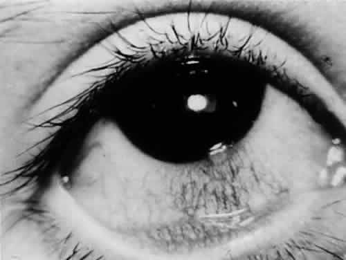
Corneal involvement usually is allergic in origin (phlyctenulosis, interstitial keratitis) or is secondary to spread from adjacent structures (sclerokeratitis). Primary infectious keratitis has been described but is rare. Phlyctenular keratoconjunctivitis is the most common form of external ocular TB (Fig. 1). Phlyctenulosis may be caused by tubercle protein hypersensitivity.44

Fig. 1. Limbic phlyctenulosis in a patient with systemic TB. An elevated infiltrate is seen at the limbus with a fornix-based leash of blood vessels. Tuberculous phlyctenulosis responds well to topical corticosteroids.

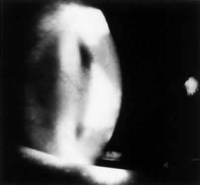
Interstitial keratitis typically affects one eye (Fig. 2). The tuberculoproteins of the cell wall of M. tuberculosis are responsible for causing allergic reactions in the cornea, producing interstitial keratitis.45 The clinical course is prolonged and may involve frequent attacks of inflammation. Infiltration in TB usually is peripheral and sectoral and spares the central cornea (Fig. 3). Often there is associated scleritis. TB usually affects the superficial and middle layers of corneal stroma; vascularization follows, with the vessels usually located in the anterior stroma. Residual localized opacification ensues because of necrosis in areas of infiltration. These features are in contrast to luetic interstitial keratitis, which involves the stroma more posteriorly, affects the central and peripheral cornea, and tends to leave less opacification because corneal infiltration usually is not as severe.46

Fig. 2. Tuberculous interstitial keratitis usually is unilateral. The stromal reaction is intense, and deep corneal neovascularization is noted.

Fig. 3. Tuberculous interstitial keratitis has a prolonged course and leaves residual dense opacification. The central cornea usually is not affected.

Primary corneal keratitis caused by M. tuberculosis varies in clinical appearance, and thus, diagnosis is difficult to make on clinical grounds.47 Secondary keratitis can result from direct spread from tuberculous scleritis or anterior segment infection.48

SCLERA AND EPISCLERA

Episcleritis is the result of hypersensitivity reaction to tuberculoprotein. Duke-Elder49 described epibulbar disease to be either ulcerative, nodular, hypertrophic papillary, or polypoid. Deep nodular scleritis results from direct invasion of the sclera by the tubercle bacillus. Tuberculous scleritis usually elicits pain as a prominent feature, as is seen typically in scleritis associated with autoimmune diseases. Clinically, the scleritis consists of one or more indurated nodules that appear fixed to the sclera and are associated with marked injection. Eventually, the nodule turns yellow as it undergoes caseation, and eventually ulceration follows. Involvement of the adjacent ciliary body and iris can result in an associated granulomatous anterior uveitis. Scleral perforation may occur.50,51

The scleritis is refractory to treatment with topical corticosteroids, suggesting that the scleritis is the result of direct invasion of the tubercle bacillus and is not a hypersensitivity reaction.52

UVEITIS

Tuberculosis once was considered a common cause of uveitis, but it currently is uncommon. Its incidence accounts for about 0.2% of all uveitis cases. Anterior uveitis usually is granulomatous and is characterized by mutton-fat keratic precipitates, Koeppe and Busacca nodules on the iris. Hypopyon can occur. Less commonly, the anterior uveitis can be nongranulomatous, with an acute, recurrent, or chronic course. Topical corticosteroids may convert an anterior granulomatous uveitis to a nongranulomatous form.53 Conglomerate tubercles of the iris resulting from coalescing of miliary tubercles of the iris rarely are seen but have been reported.54 The possibility of TB should be entertained for any steroid nonresponsive uveitis.

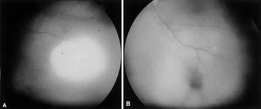
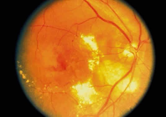
The choroid has a particular propensity for involvement by TB. Choroidal granulomas can be solitary or—as in miliary TB—multiple, causing a disseminated choroiditis. Choroidal granulomas are yellowish-white nodules with indistinct borders, ranging in diameter size from 0.5 mm to 2 cm. They may be seen in patients without active systemic disease (Figs. 4 and 5). Although A-scan findings are variable, B-scan typically reveals a solid elevated choroidal mass with an absent scleral echo due to absorption by inflammatory cells.55 Fluorescein angiography generally shows early hyperfluorescence with leakage at the margins and late central leakage. After treatment, there may be loss of early hyperfluorescence and late staining. Choroidal neovascularization can occur.55–57 Endobiopsy has been performed safely for diagnosis.58

Fig. 4. Tuberculous choroiditis does not have a pathognomonic appearance. The presenting symptoms usually are blurred vision and floaters. A. This patient had a 2-month history of decreased vision and presented with a large choroidal infiltrate. Systemic TB was proved by culture of Mycobacterium tuberculosis. B. The lesion responded dramatically to systemic antituberculous medications.

Fig. 5. Latent choroidal tubercle in a patient with multidrug resistant TB.

Exudative retinitis and retinal vasculitis can follow. Involvement of the posterior uvea sometimes can resolve spontaneously. The resulting chorioretinal scars could be confused with old toxoplasmosis or histoplasmosis scars and may lead to loss of central vision.59 Serum angiotensin-converting enzyme often is measured to investigate possible sarcoidosis but may be positive in 38.9% of miliary TB cases.60 Care should be taken because these diseases may be confused clinically.

Tuberculous choroiditis is more common in patients with AIDS than in the general population.61,62 Vitritis may be absent.13 Other causes of choroidal nodules in patients with AIDS are Pneumocystis carinii, fungi, atypical mycobacteria, syphilis, and lymphoma. Uveitis also may be caused by the use of rifabutin for MAI.63

Almenoff et al64 demonstrated mycobacterial cell wall-deficient forms in 19 of 20 patients with sarcoid and none of 20 controls. This and other similar work suggests that mycobacteria may play a broader role in etiologies of uveitis than previously suspected.

RETINA

Tuberculosis of the retina may occur from endogenous spread from a distant source. The inflammation commences in the vessel layer of the retina and may take two forms. In the miliary type, there is formation of small tubercles that remain localized and eventually heal (superficial exudative retinitis). In the second form, there may be massive retinitis with extensive grey-white lesions and heavy vitreous reaction. Severe endophthalmitis can ensue. Care should be taken to distinguish these lesions from retinoblastoma in children.54,65,66 Retinal periphlebitis may result from direct infection of the retina by tubercle bacilli.44 This has been reported to cause a central retinal vein occlusion.67 Disc swelling, macular star, and retinal folds may occur in a neuroretinitis pattern.25 TB often affects the retina by spread from adjacent involved choroid. The choroid usually is involved in miliary TB.68 Eale's disease may result from hypersensitivity to tuberculin protein and other antigens.44

OPTIC NERVE

Optic nerve involvement may accompany uveitis or tuberculous meningitis, either as a direct infiltration of the nerve, as an inflammatory disc edema, or bilaterally because of increased intracranial pressure (papilledema). Optic atrophy after TB meningitis may be more common in patients with cerebrospinal fluid protein content greater than 75 mg/dL.69 The addition of dexamethasone to the treatment regimen of tuberculous meningitis may help prevent optic atrophy. Optic neuropathy also may occur as a result of antituberculous treatment with ethambutol and INH.

CENTRAL NEUROOPHTHALMIC PATHWAYS

Tuberculous meningitis typically causes a basilar meningitis and may involve any of the cranial nerves. It should enter into the differential diagnosis whenever there is a pattern of multiple cranial nerve involvement. Alternatively, disease within the CNS may lead to focal cranial mononeuropathies or other neuroophthalmic syndromes, such as internuclear ophthalmoplegia,71 bilateral internuclear ophthalmoplegia,72 chiasmal or junctional visual loss,73,74 gaze palsy,75 and third cranial nerve palsy.69

A tuberculoma may behave as a space-occupying lesion and has been reported involving the cavernous sinus.76 CNS TB must make one vigilant for local ocular disease.77 Conversely, ocular disease must make one aware of the possibility of CNS involvement. The ophthalmologist may be of great assistance in diagnosing systemic or miliary TB in the choroid in critically ill patients when laboratory support is unavailable or delayed.59

Back to Top
PREVENTION
TB can be prevented by improving sanitary living conditions, avoiding overcrowding, and having good personal hygiene. Bacille Calmette-Guérin vaccine (BCG) can be used in persons with negative tuberculin test results who are exposed to significant risk of TB infection.78 Because BCG vaccine is a live culture preparation of M. bovis, it should not be given to immunosuppressed individuals because of the possibility of causing systemic infection. BCG is not used commonly in North America because of the low incidence of TB and because it results in a positive tuberculin test result, thus minimizing the diagnostic value of this test. A very strongly positive PPD many years after BCG vaccination must make one suspicious of possible TB exposure. The main purpose of prophylactic therapy is to prevent latent infection from progressing to clinical disease. The usual regimen is oral INH for 6 to 12 months (300 mg/day for adults and 10 mg/kg/day [maximum 300 mg/day] for children). Twelve months of therapy are recommended for persons with HIV infection or chest radiograph findings compatible with past TB. Indications for chemoprophylaxis include79: persons with HIV infection with positive PPD test results; close contacts of persons with newly diagnosed infectious TB; recent tuberculin test converters; persons whose chest radiographs suggest old healed TB and positive PPD; and HIV-seronegative intravenous drug abusers with induration 10 mm or more on PPD testing. For those persons exposed to INH-resistant organisms, 12 months of rifampin is recommended.80
Back to Top
TREATMENT
The major thrust of treatment of any case of ocular TB is the treatment of the systemic infection. The therapy should be directed by an internist. Because of the high rate of mutation, a single drug should not be used; at least two drugs (e.g., INH and rifampicin) should be used to prevent development of resistant strains. Also, the addition of a single drug to a failing regimen can lead to resistance. Premature cessation of treatment results in inadequate treatment and emergence of resistant strains. To improve compliance, drug regimens must be convenient, simple, and long acting.81 The patient treated with antituberculous drug treatment is no longer infectious even though sputum may yield acid-fast bacilli on culture for 2 months or more.

For uncomplicated pulmonary and extrapulmonary TB, the recommended treatment is INH plus rifampicin daily for 6 months, with pyrazinamide for the first 2 months. Pyridoxine is added to prevent peripheral neuropathy. In disseminated TB, TB meningitis, and TB in AIDS, INH plus rifampicin plus ethambutol (or pyrazinamide) should be given for at least 9 months. The World Health Organization is recommending “directly observed treatment, short course” (DOTS) to cure 95% of all TB for as little as $11/patient. In patients with AIDS, standard antituberculous therapy is effective, but the ideal duration of therapy is unknown; it probably is longer than for AIDS patients who do not have AIDS.82

For those exposed to drug-resistant organisms or those recently emigrated from countries where drug-resistant organisms are prevalent (Southeast Asia, Mexico, the Philippines) the regimen of INH plus rifampicin plus ethambutol plus pyrazinamide is recommended for the first 2 months of therapy; the therapy then should be adjusted according to culture results. With 3 months of INH, rifampicin, and pyrazinamide, 80% of patients are cured.

There is a place for corticosteroids in certain systemic tuberculous conditions (e.g., meningitis, pericarditis, severe systemic toxicity). Systemic corticosteroids also may be needed to preserve vision in which severe intraocular inflammation is caused by ocular TB. Although there is disagreement in the literature, as a rule one must remember that the possibility exists that the immunosuppressive effect of corticosteroids might exacerbate systemic TB. Thus, these patients require concurrent antituberculous therapy and careful follow-up. The dose of corticosteroid should be as low as possible. One should consider the use of sub-Tenon's corticosteroids in these patients to avoid the use of systemic corticosteroids. However, it is possible that this could exacerbate the ocular infectious process. In addition, the long duration of action of repository corticosteroids could extend the duration of any deterioration. Topical corticosteroids can be used safely for anterior uveitis, interstitial keratitis, and phlyctenulosis.

Clearly, if global TB is to be controlled, it requires greater resources, international coordination, and an increased political commitment.83

Back to Top
OPHTHALMIC SIDE EFFECTS OF ANTITUBERCULOUS THERAPY
INH (Nydrazid) optic neuropathy14,82 may have an additive effect on the better recognized ethambutol toxic optic neuropathy.84 INH also is known to cause optic neuropathy alone.85 INH has also been reported to cause subepithelial corneal infiltrates, with recurrence on reexposure.86 Extra caution should be exercised in groups at risk for INH toxicity, such as those with preexisting liver disease, increased age, other hepatotoxic medications, and excessive use of alcohol. Fifty percent of whites and blacks are slow acetylators and are more prone to toxic effects from the drug. Drug interactions occur with phenytoin and disulfiram. False-positive diabetic urine glucose test results may occur. Concurrent administration of pyridoxine prevents peripheral neuropathy and may prevent optic neuropathy.85

Ethambutol (Myambutol, Etibi) may cause decreased acuity in 6% of patients. Optic neuropathy is the side effect most concerning to the ophthalmologist. Neuropathy can occur in an axial or periaxial pattern.87 The incidence of this reaction is dose related and is observed in 15% of patients receiving 50 mg/kg/day, in 5% of those receiving 25 mg/kg/day, and in less than 1% of those taking 15 mg/kg/day or less.85 Occasionally, visual loss may be irreversible and may follow standard dosages.4,79,81 Macular edema and pigmentation also have been reported.88 The correct method for screening patients remains controversial among ophthalmologists. Some texts recommend monthly examinations, which may be excessive.89 A baseline examination is recommended, and appropriate screening for optic neuropathy includes a history of visual disturbance, an examination for Snellen acuity in each eye, and color vision testing for red/green deficits. Other screening tests that have proven useful are the Farnsworth D-15, automated threshold perimetry, optic nerve head photography,90 and Arden contrast sensitivity plates.91 also have been advocated to screen for ethambutol optic neuropathy.92,93 Renal impairment may increase the risk of toxic optic neuropathy from ethambutol.94 Peripheral neuropathy may occur before the onset of optic neuropathy and should signal the need for caution with ethambutol.95 Recovery from ethambutol toxic optic neuropathy generally occurs in 3 to 4 months. Although central acuity may recover completely, other measures of visual function may remain abnormal. Recovery has been shown to occur most completely to least completely in the following order96:

  • Visual acuity
  • Visual evoked potentials
  • Color sense
  • Critical flicker fusion
  • Visual field
  • Contrast sensitivity
  • Pupil cycle time

There is strong ophthalmic evidence that ethambutol should be replaced by less toxic available therapies for TB.97,98 At the very least, patients should be warned to stop the drug immediately if any visual symptoms occur.99

Rifampin (Rifadin, Rimactane) may cause a reddish-orange discoloration of tears. This is harmless. Soft contact lenses may become discolored.

Rifabutin is used to prevent M. avium complex (MAC) disease in susceptible individuals (CD4 counts of less than 200 cells/mm3). It has been associated with uveitis, which may be difficult to differentiate from other causes of uveitis in patients with AIDS. Soft contact lenses may become permanently stained because this medication discolors tears. Uveitis is unusual at the recommended oral dosage of 300 mg/day, but becomes common as the total daily dose approaches 1 g.63 Baseline patient body weight of less than 55 kg significantly increases the risk of uveitis, although therapy with fluconazole, a drug known to raise serum rifabutin concentrations, does not increase the risk of uveitis. Inflammation may be severe, and 7 of 28 patients in one study had hypopyon. The uveitis may be unilateral or bilateral, anterior, intermediate, or posterior.100

Streptomycin, pyrazinamide, and para-aminosalicyclic acid (PAS) have no known ophthalmic complications. The following medications have prescribing information detailing ophthalmic symptoms which are not detailed in the literature. Ethionamide may cause blurred vision and diplopia. Cycloserine and rifampin may cause visual disturbances through unknown mechanisms.85

Back to Top
IMMUNOLOGY OF TUBERCULOSIS
In 1921, the first attenuated strain of M. bovis was given to humans as a vaccine to prevent TB. Since then, our understanding of the immune system's response to TB has advanced considerably and is increasingly important in dealing with HIV coinfection.

The immune response to TB is both systemic and local. The immune attack on a TB granuloma and PPD skin testing are examples of local reactions, whereas vaccine development is a systemic preventative response. Immunogenic disease involves a type IV hypersensitivity reaction to tuberculoproteins. Hypersensitivity is responsible for interstitial keratitis, phlyctenular disease, episcleritis, granulomatous uveitis, and retinal vasculitis.

The Th1 CD4 subset of lymphocytes is predominantly active in eradicating TB bacilli, and the Th2 CD4 subset may allow TB infection to persist. Both coexist during infection with TB and help to determine the host response and explain skin test observations. The transient depression of a DTH response in active TB likely is caused by the production of inhibitory cytokines by peripheral blood mononuclear cells. Similarly, the balance between activating and inhibiting cytokines may influence the systemic course of TB infection. CD4 lymphocytes maintain quiescence of latent TB foci by active immunologic surveillance. Cytokines such as interleukin (IL)-2, IL-12, interferon (IFN)-γ, and TNF-α common to the Th1 CD4 activation have the potential for enhancing immune responsiveness to TB. Alternatively, IL-10 and transforming growth factor (TGF)-β tend to limit the host's eradication of TB. The demonstration that DTH and protective immunity to TB are distinct phenomena determined by different antigens has relevance for the development of vaccines.

Enhancing an individual's preexisting immunity to TB has been done historically with BCG although clearly, novel approaches are needed. BCG is known to have its best efficacy in preventing systemic dissemination and extrapulmonary TB in infants and children. It also has a modest ability to prevent primary infection with TB. Repeated administration of BCG may boost immunity to TB but is fraught with the limitations of a live attenuated bacteria—particularly in the immunosuppressed patient. A booster of purified subunit TB antigens such as ESAT-6 or 85BAg can heighten the immune response to TB, but maximal clinical benefit is likely to be achieved with a multivalent vaccine.101 Vaccination with auxotrophic mutants,102 low-dose immunotherapy with IL-2,103 heat-killed Mycobacterium vaccae, IFN-γ,104 thalidomide (a specific TNF-α inhibitor),105 and pentoxifylline (a nonspecific cytokine inhibitor)106 all have had some success in controlling TB infection. The recognition of HLA-DR2 susceptibility to TB107 and specific gene mutations such as the interferon-γ receptor108 and the defective production of TNF-α,109 which predispose to progressive disease, are important in regulating the host response to TB.

Eale's disease represents an example of a local ocular immune response believed to be secondary to immunity to a capsular protein on the tubercle bacillus. Most patients are PPD positive. The disease has been well characterized by Das and others,110 but its exact immunopathogenesis remains unknown.

Back to Top
ATYPICAL MYCOBACTERIA (MOTT)
Mycobacteria other than M. tuberculosis, M. bovis, M. africanum, Mycobacterium microti, and Mycobacterium leprae are grouped as atypical mycobacteria. This group most accurately is named mycobacteria other than tubercle bacilli (MOTT). Because these organisms seldom cause disease in hosts with normal immune responses, they also are called opportunistic mycobacteria. Unlike M. tuberculosis, which is an obligate human pathogen, the atypical bacteria are distributed widely in the environment.

There are more than 50 species altogether. Because some of these species share similar biochemical, serologic, and pathogenic characteristics, they often are grouped together and identified as a complex. Atypical mycobacteria most commonly isolated in human disease are, in decreasing frequency, M. avium complex, Mycobacterium fortuitum, Mycobacterium kansasii, Mycobacterium chelonae, and Mycobacterium scrofulaceum.

These facultative pathogens cause opportunistic disease in humans. Predisposing factors to infection include trauma, injections, surgery, and immunosuppression. The most common clinical diseases associated with the atypical mycobacteria are chronic pulmonary disease, lymphadenitis, and soft-tissue infections.111 Endophthalmitis has been reported to occur.64

Disseminated disease can occur in immunocompromised persons and, in particular, those infected with HIV. With the onset of the AIDS epidemic, disseminated atypical mycobacterial disease is more common; the most important agents are M. avium complex, M. kansasii, and M. fortuitum. M. fortuitum and M. chelonae (M. fortuitum complex) are the major causes of atypical mycobacterial infection of the eye.112 Both are classed as rapidly growing mycobacteria.111 Other atypical mycobacteria as causative agents of ocular disease are rare but have been reported.113,114 Not all recovery of nontuberculous mycobacteria represents disease—some simply may represent colonization. Atypical mycobacteria must be considered in any case of keratitis, scleritis, uveitis, or endophthalmitis in which onset is insidious or delayed, if standard cultures are negative, postsurgically, after trauma, and in immunocompromised persons.

Back to Top
REFERENCES

1. Wayne LG: Microbiology of tubercle bacilli. Am Rev Respir Dis 125:31, 1982

2. Warren CW, Enarson DN, Chaisson RE et al: Symposium: Tuberculosis. Ann RCPSC 22:455, 1989

3. Shafer RW, Edlin BR: Tuberculosis in patients infected with human immunodeficiency virus: Perspective on the past decade. Clin Infect Dis 22:683, 1996

4. Centers for Disease Control: Screening for tuberculosis and tuberculosis infection in high-risk populations. MMWR Morb Mortal Wkly Rep 44:18, 1995

5. World Health Organization. Executive Summary: WHO Report on the Tuberculosis Epidemic 1996 (GENERIC). Geneva: WHO, 1996

6. World Health Organization. WHO Attacks Gloal Neglect of Tuberculosis Crisis (GENERIC). Geneva, WHO, 1993

7. Peiffer RJ, Lewen RM, Yin H: Tuberculosis. In Gold DH, Weingeist TA (eds): The Eye in Systemic Disease, p. 197. Philadelphia: JB Lippincott, 1990

8. Griffith DE, Wallace RJ: Environmental mycobacterial diseases. In Kelly WN, DeVita VT et al (eds): Textbook of Internal Medicine, p. 1578. Philadelphia: JB Lippincott, 1989

9. Gnaore E, Sassan-Morokro M, Kassim S et al: A comparison of clinical features in tuberculosis associated with infection with human immunodeficiency viruses 1 and 2. Trans R S Trop Med Hyg 87:57, 1993

10. Michael N, Whalen C, Johnson J et al: Comparison of HIV1 viral load between HIV-infected patients with and without tuberculosis. Anonymous 3rd Conference on Retroviruses and Opportunistic Infections: Program and Abstracts (Washington, DC), Alexandria: Infectious Diseases Society of America, 1996

11. el-Sadr W, Oleske JM, Agins BD: Managing early HIV infection. In Clinical Practice Guideline—Quick Reference Guide for Clinicians 1 Washington, DC: Agency for Health Care Policy and Research, 1994

12. Torres G: Tuberculosis therapy and the new protease inhibitors: Treatment issues. GMHC Treatment Educ Advocacy 10:11, 1996

13. Perez BE, Montero RM, Mendez RJ: Tuberculous choroiditis and acquired immunodeficiency syndrome. Ann Ophthalmol 26:50, 1994

14. Bouza E, Merino P, Munoz P et al: Ocular tuberculosis: A prospective study in a general hospital. Medicine 76:53, 1997

15. Anonymous: American Thoracic Society. Diagnostic standards and classification of tuberculosis. Am Rev Respir Dis 142:725, 1990 [published erratum appears in Am Rev Respir Dis 1990 Dec;142(6 Pt 1):1470]

16. Daniel TM: Tuberculosis. In Isselbacher KJ, Braunwald E, Wilson JD et al (eds): Harrison's Principles of Internal Medicine, p. 710. New York: McGraw-Hill, 1994

17. Rosenbaum JT, Wernick R: The utility of routine screening of patients with uveitis for systemic lupus erythematosus or tuberculosis: A Bayesian analysis. Arch Ophthalmol 108:1291, 1990

18. Moon WK, Han MH, Chang KH et al: CT and MR imaging of head and neck tuberculosis. Radiographics 17:391, 1997

19. Panova IE, Tarasova LN, Teplova SN et al: Immunological aspects of tuberculosis uveitis pathogenesis [Russian]. Problemy Tuberkuleza 51, 1997

20. Abrams J, Schlaegel TFJ: The tuberculin skin test in the diagnosis of tuberculous uveitis. Am J Ophthalmol 96:295, 1983

21. Huebner RE, Schein MF, Bass JB Jr: The tuberculin skin test. Clin Infect Dis 17:968, 1993

22. Smith RE, Nozik RA: In Anonymous: Uveitis: A Clinical Approach to Diagnosis and Management, p. 215. Baltimore: Williams and Wilkins, 1989

23. Webster CT, Gordin FM, Matts JP et al: Two-stage tuberculin skin testing in individuals with human immunodeficiency virus infection. Am J Respir Crit Care Med 151:805, 1995

24. Burgoyne CF, Verstraeten TC, Friberg TR: Tuberculin skin-test-induced uveitis in the absence of tuberculosis. Graefes Arch Clin Exp Ophthalmol 229:232, 1991

25. Berinstein DM, Gentile RC, McCormick SA et al: Primary choroidal tuberculoma. Arch Ophthalmol 115:430, 1997

26. Meaney TPJ, Ogunsola AB: Tuberculosis presenting as an orbital mass lesion in childhood. Eye 9:649, 1995

27. Abrams J, Schlaegel TFJ: The role of the isoniazid therapeutic test in tuberculous uveitis. Am J Ophthalmol 94:511, 1982

28. Nussenblatt RB, Palestine AG: In Anonymous: Uveitis: Fundamentals and Clinical Practice, p. 396. Chicago: Year Book, 1989

29. Haring G, Nolle B, Kiehne K et al: Chronic granulomatous chorioretinitis in tuberculosis [German]. Klinische Monatsblatter fur Augenheilkunde 209:376, 1996

30. Forbes BA: Critical assessment of gene amplification approaches on the diagnosis of tuberculosis. Immunol Invest 26:105, 1997

31. Heifets L: Mycobacteriology laboratory. Clin Chest Med 18:35, 1997

32. Ling ML: Molecular biology in medicine: laboratory diagnosis of tuberculosis. Ann Acad Med Singapore 25:94, 1996

33. Daley CL, Small PM, Schecter GF et al: An outbreak of tuberculosis with accelerated progression among persons infected with the human immunodeficiency virus. N Engl J Med 326:231, 1992

34. Donahue HC: Ophthalmologic experience in a tuberculosis sanatorium. Am J Ophthalmol 64:742, 1967

35. Jabbour NM, Faris B, Trempe CL: A case of pulmonary tuberculosis presenting with a choroidal tuberculoma. Ophthalmology 92:834, 1985

36. Meyer C, Muller M: Form deprivation myopia caused by keratitis scrophulosa. [German]. Ophthalmologe 93:361, 1996

37. Spoor TC: Tuberculosis. In Fraunfelder FT, Roy FH (eds): Current Ocular Therapy, p. 43. Philadelphia: WB Saunders, 1990

38. Alvarez H, Tabbara KF: Infections of the eyelid. In Tabbara KF, Hyndiuk RA (eds): Infections of the Eye, p. 551. Boston/Toronto: Little Brown, 1986

39. Roberts BN, Lane CM: Orbital tuberculosis. Eye 11:138, 1997

40. Fedukowicz HB, Stenson S: External infections of the eye: Bacterial, viral, mycotic with noninfectious and immunologic diseases. In: External Infections of the Eye, p. 46. Norwalk: Appleton-Century-Crofts, 1985

41. Liesegang TJ, Cameron JD: Mycobacterium bovis infection of the conjunctiva. Arch Ophthalmol 98:1764, 1980

42. Antonius S, Tabbara KF: Bacterial conjunctivitis. In Tabbara KF, Hyndiuk RA (eds): Infections of the Eye, p. 413. Boston/Toronto, Little Brown, 1986

43. Aclimandos WA, Kerr-Muir M: Tuberculous keratoconjunctivitis. Br J Ophthalmol 76:175, 1992

44. Helm CJ, Holland GN: Ocular tuberculosis. Surv Ophthalmol 38:229, 1993

45. Tooker CW: Allergic phenomena in tuberculous keratitis. Arch Ophthalmol 2:540, 1929

46. Yee RW, Hyndiuk RA: Interstitial keratitis. In Tabbara KF, Hyndiuk RA (eds): Infections of the Eye, p. 601. Boston/Toronto: Little Brown, 1986

47. Jackson WB: Infections of the sclera. In Tabbara KF, Hyndiuk RA (eds): Infections of the Eye, p. 477. Boston/Toronto: Little Brown, 1986

48. Hyndiuk RA, Snyder RW: Infectious diseases: Bacterial keratitis. In Smolin G, Thoft RA (eds): The Corneal: Scientific Foundations and Clinical Practice, p. 193. Boston/ Toronto: Little Brown, 1987

49. Duke-Elder S: The mycobacteriaceae. In Duke-Elder S (ed): System of Ophthalmology, p. 212. St. Louis: CV Mosby, 1965

50. Biswas J, Madhavan HN, Gopal L et al: Intraocular tuberculosis: Clinicopathologic study of five cases. Retina 15:461, 1995

51. Anders N, Wollensak G: Ocular tuberculosis in systemic lupus erythematosus and immunosuppressive therapy. [German]. Klinische Monatsblatter fur Augenheilkunde 207:368, 1995

52. Nanda M, Pflugfelder SC, Holland S: Mycobacterium tuberculosis scleritis. Am J Ophthalmol 108:736, 1989

53. Weinberg RS: Endogenous bacterial and fungal infections of the retina and choroid. In Tabbara KF, Hyndiuk RA (eds): Infections of the Eye, p. 499. Boston/Toronto: Little Brown, 1986

54. Gain P, Mosnier JF, Gravelet C et al: Iris tuberculosis: A propos of a case diagnosed by iridectomy. [French]. J Francais Ophtalmologie 17:525, 1994

55. Jabbour NM, Faris B, Trempe CL: A case of pulmonary tuberculosis presenting with a choroidal tuberculoma. Ophthalmology 92:834, 1985

56. Cangemi FE, Friedman AH, Josephberg R: Tuberculoma of the choroid. Ophthalmology 87:252, 1980

57. Chung YM, Yeh TS, Sheu SJ et al: Macular subretinal neovascularization in choroidal tuberculosis. Ann Ophthalmol 21:225, 1989

58. Barondes MJ, Sponsel WE, Stevens TS, Plotnik RD: Tuberculous choroiditis diagnosed by chorioretinal endobiopsy. Am J Ophthalmol 112:460, 1991

59. Berger C, Braegger CP, Albisetti M et al: Cerebral tuberculosis presenting as complex febrile convulsions. Neuropediatrics 27:161, 1996

60. Ainslie GM, Benatar SR: Serum angiotensin converting enzyme in sarcoidosis: Sensitivity and specificity in diagnosis: correlations with disease activity, duration, extrathoracic involvement, radiographic type and therapy. Q J Med 55:253, 1985

61. Grenzebach UH, Busse H, Totsch M et al: Endophthalmitis induced by atypical mycobacterial infection. Ger J Ophthalmol 5:202, 1996

62. Frankel RM, Boname ME: Detection of the new tuberculosis: Ocular examination as a diagnostic imperative. J Am Optom Assoc 65:472, 1994

63. Skinner MH, Blaschke TF: Clinical pharmacokinetics of rifabutin. Clin Pharmacokinet 28:115, 1995

64. Almenoff PL, Johnson A, Lesser M et al: Growth of acid fast L forms from the blood of patients with sarcoidosis. Thorax 51:530, 1996

65. Ayanru JO, Alli AF, Faal HB et al: Tuberculoma of the eye: A case report. Trop Geogr Med 38:301, 1986

66. McMoli TE, Mordi VP, Grange A et al: Tuberculous panophthalmitis. J Pediatr Ophthalmol Strabismus 15:383, 1978

67. Fountain JA, Werner RB: Tuberculous retinal vasculitis. Retina 4:48, 1984

68. Green WR: Retina. In Spencer WH (ed): Ophthalmic Pathology: An Atlas and Textbook, p. 589. Philadelphia: WB Saunders, 1985

69. Lamba PA, Bhalla JS, Mullick DN: Ocular manifestations of tubercular meningitis: A clinico-biochemical study. J Pediatr Ophthalmol Strabismus 23:123, 1986

70. Girgis NI, Farid Z, Hanna LS et al: The use of dexamethasone in preventing ocular complications in tuberculous meningitis. Trans R S Trop Med Hyg 77:658, 1983

71. Borodulina EA, Branchevskaia SI, Borodulin BE: Eye lesions in patients with pulmonary sarcoidosis. [Russian]. Vestnik Oftalmologii 112:50, 1996

72. Inocencio FP, Ballecer R: Tuberculosis granuloma in the midbrain causing wall-eyed bilateral internuclear ophthalmoplegia (Webino). J Clin Neuroophthalmol 5:31, 1985

73. Barrasa J, Quintial C, Madrigal V et al: Course of the visual evoked potentials in intracranial tuberculous granuloma. [French]. Neurophysiol Clin 20:105, 1990

74. Iraci G, Giordano R, Gerosa M et al: Tuberculoma of the anterior optic pathways: Case report. J Neurosurg 52:129, 1980

75. Kubota A, Takahashi T, Suzuki Y et al: MRI findings of brain-stem tuberculoma in a case of tuberculous meningitis. [Japanese]. Rinsho Shinkeigaku (Clin Neurol) 32:849, 1992

76. Bafna S, Lee AG: Presumed tuberculosis presenting as a cavernous sinus syndrome. J Neuroophthalmol 17:207, 1997

77. Muccioli C, Belfort RJ: Presumed ocular and central nervous system tuberculosis in a patient with the acquired immunodeficiency syndrome. Am J Ophthalmol 121:217, 1996

78. Centers for Disease Control: Use of BCG vaccines in the control of tuberculosis: A joint statement by the ACIP and the Advisory Committee for Elimination of Tuberculosis. MMWR Morb Mortal Wkly Rep 37:663, 1988

79. Centers for Disease Control: The use of preventative therapy for tuberculous infection in the United States. MMWR Morb Mortal Wkly Rep 39:9, 1990

80. Levenson DS, Harrison CH: Mycobacterium fortuitum corneal ulcer. Arch Ophthalmol 75:189, 1966

81. Centers for Disease Control: A strategic plan for the elimination of tuberculosis in the United States. MMWR Morb Mortal Wkly Rep 38:7, 1989

82. Anonymous: Drugs for tuberculosis. Med Lett 30:43, 1988

83. De CKM, Binkin NJ, Zuber PL et al: Research issues involving HIV-associated tuberculosis in resource-poor countries. JAMA 276:1502, 1996

84. Kahana LM: Toxic ocular effects of ethambutol. Can Med Assoc J 137:213, 1987 [published erratum appears in Can Med Assoc J 1987 Oct 1;137(7):592]

85. Mandell GL, Petri WA Jr: Antimicrobial agents: drugs used in the chemotherapy of tuberculosis, Mycobacterium avium complex disease, and leprosy. In Hardman JG, Limbird LE, Molinoff PB et al (eds): Goodman and Gilman's The Pharmacological Basis of Therapeutics, p. 1155. New York: McGraw Hill, 1996

86. Neff TA: Isoniazid toxicity: Reports of lactic acidosis and keratitis. Chest 59:245, 1971

87. Leibold JE: Drugs having a toxic effect on the optic nerve. Int Ophthalmol Clin 11:137, 1971

88. Kakisu Y, Adachi-Usami E, Mizota A: Pattern electroretinogram and visual evoked cortical potential in ethambutol optic neuropathy. Documenta Ophthalmologica 67:327, 1988

89. Wright PW, Wallace RJ: Antimycobacterial agents. In Isselbacher KJ, Braunwald E, Wilson JD et al (eds): Harrison's Principles of Internal Medicine, p. 705. New York: McGraw-Hill Inc Health Professions Division, 1994

90. Russo PA, Chaglasian MA: Toxic optic neuropathy associated with ethambutol: Implications for current therapy. J Am Optom Assoc 65:332, 1994

91. Salmon JF, Carmichael TR, Welsh NH: Use of contrast sensitivity measurement in the detection of subclinical ethambutol toxic optic neuropathy. Br J Ophthalmol 71:192, 1987

92. Kumar A, Sandramouli S, Verma L et al: Ocular ethambutol toxicity: is it reversible? J Clin Neuroophthalmol 13:15, 1993

93. Yiannikas C, Walsh JC, McLeod JG: Visual evoked potentials in the detection of subclinical optic toxic effects secondary to ethambutol. Arch Neurol 40:645, 1983

94. Bambery P, Sakhuja V, Gupta A et al: Wegener's granulomatosis in north India: An analysis of eleven patients. Rheumatol Int 7:243, 1987

95. Nair VS, LeBrun M, Kass I: Peripheral neuropathy associated with ethambutol. Chest 77:98, 1980

96. Woung LC, Jou JR, Liaw SL: Visual function in recovered ethambutol optic neuropathy. J Ocul Pharmacol Ther 11:411, 1995

97. Kahana LM: Ethambutol in tuberculosis. Biomed Pharmacother 44:21, 1990

98. Bastian A, Brandt HP: Risk factors for the occurrence of eye damage following ethambutol treatment. [German]. Zeitschrift fur Erkrankungen der Atmungsorgane 169:162, 1987

99. Chatterjee VK, Buchanan DR, Friedmann AI et al: Ocular toxicity following ethambutol in standard dosage. Br J Dis Chest 80:288, 1986

100. Shafran SD, Singer J, Zarowny DP et al: Determinants of rifabutin-associated uveitis in patients treated with rifabutin, clarithromycin, and ethambutol for Mycobacterium avium complex bacteremia: A multivariate analysis. J Infect Dis 177:252, 1998

101. Ellner JJ: Review: The immune response in human tuberculosis—implications for tuberculosis control. J Infect Dis 176:1351, 1997

102. Bange FC, Brown AM, Jacobs WR Jr: Leucine auxotrophy restricts growth of Mycobacterium bovis BCG in macrophages. Infect Immun 64:1794, 1996

103. Johnson BJ, Ress SR, Willcox P et al: Clinical and immune responses of tuberculosis patients treated with low-dose IL-2 and multidrug therapy. Cytokine Mol Ther 1:185, 1995

104. Holland SM, Eisenstein EM, Kuhns DB et al: Treatment of refractory disseminated nontuberculous mucobacterial infecton with interferon gamma. N Engl J Med 330:1348, 1994

105. Tramontana JM, Utaipat U, Molloy A et al: Thalidomide treatment reduces tumor necrosis factor production and enhances weight gain in patients with pulmonary tuberculosis. Mol Med 1:384, 1995

106. Wallis RS, Nsubuga P, Whalen C et al: Pentoxifylline in HIV-1-seropositive tuberculosis: A randomized controlled trial. J Infect Dis 174:727, 1996

107. Brahmajothi V, Pithcappan RM, Kakkanaiah VN et al: Association of pulmonary tuberculosis and HLA in South India. Tubercle 72:123, 1991

108. Newport MJ, Huxley CM, Huston S, Hawrylowicz CM et al: A mutation in the interferon gamma receptor gene and susceptibility to mycobacterial infection. N Engl J Med 335:1941, 1996

109. Levin M, Newport MJ, D'Souza S et al: Familial disseminated atypical mycobacterial infection in childhood: A human mycobacterial susceptibility gene É. Lancet 345:79, 1995

110. Das T, Biswas J, Kumar A et al: Eale`s disease. Indian J Ophthalmol 42:3, 1994

111. Wallace RJ, Swenson JM, Silcox VA et al: Spectrum of disease due to rapidly growing mycobacteria. Rev Infect Dis 5:657, 1983

112. Roussel TJ, Stern WH, Goodman DF et al: Postoperative mycobacterial endophthalmitis. Am J Ophthalmol 107: 403, 1989

113. Schonherr U, Naumann GO, Lang GK et al: Sclerokeratitis caused by Mycobacterium marinum. Am J Ophthalmol 108:607, 1989

114. Moore MB, Newton C, Kaufman HE: Chronic keratitis caused by Mycobacterium gordonae. Am J Ophthalmol 102:516, 1986

Back to Top