Chapter 60
Complications of Uveitis and Their Management
DEBRA A. GOLDSTEIN and HOWARD H. TESSLER
Main Menu   Table Of Contents

Search

CATARACT
GLAUCOMA
CYSTOID MACULAR EDEMA
HYPOTONY
BAND KERATOPATHY
REFERENCES

The treatment of uveitis is often frustrating because of the many complications that may arise as a result of inflammation and its treatment. The most frequent complications are cataract, glaucoma, macular edema, hypotony, and band keratopathy. This chapter addresses each of these entities and highlights current methods available for their treatment. Other complications, such as rhegmatogenous and nonrhegmatogenous retinal detachments, are addressed elsewhere in these volumes.
Back to Top
CATARACT
Cataract is a frequently seen complication of uveitis, occurring in up to 50% of patients with juvenile rheumatoid arthritis (JRA), pars planitis, and Fuchs' iridocyclitis.1–6 The incidence in other forms of uveitis is also high and seems to be related to both the location and duration of the inflammation, as well as to the use of corticosteroid therapy. The contribution of steroids to cataractogenesis is well known from the data on nonuveitis patients receiving steroid therapy and from experimental data.7 In addition, recent reports suggest that the prevalence of cataract in pars planitis may be as low as 10% if steroid use is limited or replaced by other forms of treatment, such as cryotherapy or immunosuppressive therapy.8 However, most of the complications of uveitis are more difficult to treat than is cataract; thus, the fear of cataract formation should not prevent the judicious use of corticosteroids.

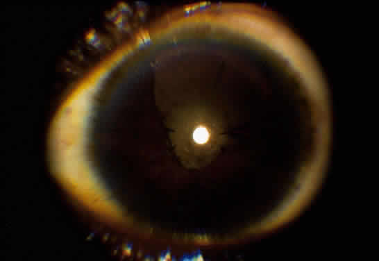
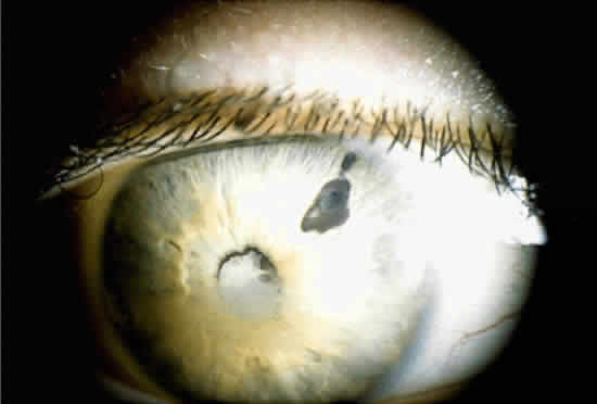
When a cataract develops in a uveitis patient, the management is more complex than in the nonuveitis patient. The presence of posterior synechiae, pupillary membranes, and inflammation may make the surgery more difficult, and the postoperative course is often stormy (Fig. 1). Much controversy still exists regarding the best method of managing the uveitis patient with a cataract. Ideally, absolute control of inflammation should be obtained for at least 3 months preceding surgery; however, there are exceptions to this rule, as in the case of lensinduced uveitis.9

Fig. 1. Posterior synechiae may make cataract extraction difficult in this patient with chronic iridocyclitis. Note the previous laser iridotomy for pupillary block. Calcific band keratopathy is also present.

The choice of surgical technique is also controversial, but it is probably not as important as the perioperative management. The patient should be treated preoperatively with topical steroid drops (e.g., prednisolone acetate or phosphate) at least four times a day for at least 3 days before surgery. Also, we often pretreat with an oral steroid (usually prednisone 1 mg/kg/day) for 3 days, if the patient's medical status permits.

Adequate pupillary dilation is often difficult to achieve preoperatively because of posterior synechiae and pupillary membranes. If this is the case, a laser peripheral iridotomy can be performed preoperatively, or a surgical peripheral iridectomy can be performed at the time of surgery. The posterior synechiae can then be lysed with the aid of a cyclodialysis spatula; access is gained to the superior ones through the peripheral iridotomy. If adequate pupillary dilation is still not achieved, straight or curved long-handled retinal scissors can be used to fashion small membranotomies or sphincterotomies (1 to 2 per clock hour), and viscoelastic material can then be used to dilate the pupil. This technique has an advantage over a single large sphincterotomy in that the pupil usually remains functional afterward. If adequate pupillary dilation is still not achieved, iris hooks can be used to further dilate the pupil. In some cases, iris hooks provide sufficient dilation without the need for sphincterotomies. The lens is then removed with the use of either phacoemulsification or standard extracapsular techniques.

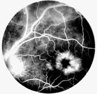
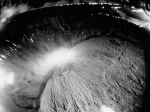
Implantation of an intraocular lens (IOL) is still controversial. There is much evidence that an IOL can be safely implanted in cases of Fuchs' iridocyclitis10–12 and pars planitis.8,13,14 In contrast, most evidence in cases of children with JRA is in favor of lensectomy/vitrectomy with no IOL (Fig. 2).4,15–17 The use of an IOL in uveitis of other etiologies is still uncertain, but an IOL should never be inserted in a patient with inflammation that could not be adequately controlled preoperatively. If an IOL is to be inserted, we prefer an all polymethyl-methacrylate lens to one with polypropylene haptics, because this may, at least in theory, lessen the chance of postoperative inflammation.12,17 Recently, in selected cases, we had good postoperative results using foldable acrylic lenses. Placement of the IOL in the capsular bag rather than the ciliary sulcus is also preferred because this may lessen the risk of inflammation secondary to iris-haptic contact.

Fig. 2. Patient with chronic iridocyclitis secondary to juvenile rheumatoid arthritis (JRA). Patient had cataract surgery with an intraocular lens (IOL) and developed an inflammatory membrane around the IOL. The membrane has dragged the iris over the IOL.

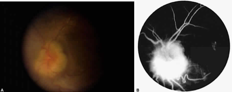
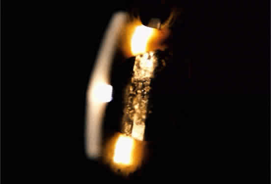
Occasionally, even in eyes with little or no preoperative inflammation, significant membranes form around an IOL (Fig. 3). This can occur even in the absence of significant anterior chamber cell and flare in the postoperative period.18 Although these cocoonlike membranes can be lysed with the Nd:YAG laser, they tend to reform. Therefore, it is imperative to be vigilant in the first 6 to 12 weeks after surgery, aggressively treating any signs of inflammatory deposits on the IOL, even in the absence of anterior chamber reaction. If laser membranectomy is required, vigorous anti-inflammatory treatment is necessary to prevent re-formation of the membrane. In severe cases, removal of the IOL may be required.

Fig. 3. Patient with pars planitis with giant-cell precipitates and inflammatory membrane encasement of IOL. Note peripheral iridectomy (these may not always be necessary in patients with uveitis but should be considered when synechiae may be a problem).

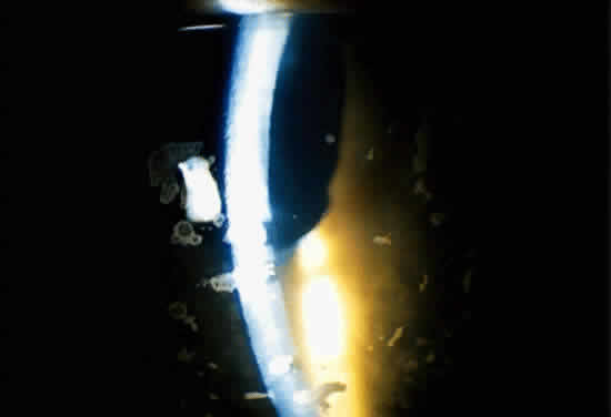
It has been suggested that pars plana vitrectomy may be warranted when substantial vitreous cell and debris might preclude good postoperative visual acuity.4,8,12,19 Although we do not routinely perform pars plana vitrectomy on all uveitis patients with cataract, this procedure is considered for eyes with significant vitreous debris or inflammation.

The postoperative management is similar to that of nonuveitis patients, except that inflammation is usually more severe and prolonged, often requiring depot steroid injections and systemic anti-inflammatory treatment.

The results of cataract surgery in uveitis patients vary according to the preoperative diagnosis. Patients with Fuchs' iridocyclitis generally do well, with a visual acuity of 20/40 or better being the rule.10–12 Patients with pars planitis also do well, with 60% to 82% achieving a visual acuity greater than 20/40.8,18 Most pars planitis patients who fail to achieve good visual acuity do so because of cystoid macular edema.4,8,9 In these patients, perhaps more aggressive anti-inflammatory treatment, both preoperatively and postoperatively, will improve visual outcome.

Patients with JRA do not tend to have as good an outcome as those with Fuchs' or pars planitis; a visual acuity greater than 20/40 is generally achieved in only about 60% of patients.15 However, one study using aggressive preoperative control of inflammation has reported a visual acuity greater than 20/40 in 75% of JRA patients.20 Finally, those patients with idiopathic and other forms of nongranulomatous anterior uveitis tend to do well postoperatively, with almost 80% achieving a visual acuity of 20/40 or better, again provided inflammation is well controlled preoperatively.9

Back to Top
GLAUCOMA
Glaucoma is a frequent complication of uveitis, occurring in up to one fourth of patients with chronic inflammation.21–23 Therefore, it is essential to measure the intraocular pressure (IOP) of the uveitis patient at each visit. Elevated IOP can be seen in any type of uveitis, but is most common in Fuchs' iridocyclitis, Vogt-Koyanagi-Harada syndrome, JRA-associated iridocyclitis, and iritis secondary to herpes simplex and herpes zoster.5,6,24 The glaucoma occurring with chronic uveitis can be either secondary open angle or closed angle, or a combination of both types. Careful gonioscopy of the uveitis patient with elevated IOP is therefore critical.

Open-angle glaucoma is the type most often seen in patients with chronic uveitis.25 The chamber angle may become blocked by debris and inflammatory cells. Alternatively, the trabecular meshwork may itself become inflamed, reducing outflow facility. This is the proposed mechanism of elevated IOP in Possner-Schlossman syndrome (glaucomatocyclitic crisis). It has also been postulated that the endothelial cells of the trabecular meshwork are capable of phagocytosis and that during periods of inflammation these cells ingest debris, migrate off the trabecular beams, and eventually may be lost. This loss of endothelial cells would then be reflected in a decreased outflow facility and an increased IOP.25

Another mechanism that may be responsible for increased IOP in uveitis patients is the so-called steroid response. This elevated IOP can occur with any route of steroid administration, but it is seen most often after topical use; it is least likely with systemic treatment. The IOP rise seems to be dependent on the type, frequency, and duration of treatment, as well as on patient characteristics. It is seen in up to 30% of the general population after 3 to 4 weeks of treatment with topical steroids, and in a much higher percentage of glaucoma patients. The IOP rise in glaucoma patients tends to be faster and of a greater magnitude than in nonglaucoma patients. The incidence is also high in patients with a family history of glaucoma, suggesting a genetic basis for the response.26–31

The mechanism responsible for steroid-induced ocular hypertension is not yet clear but does seem to result from a decrease in outflow facility.26–30,32 This may result from a stabilization of lysosomal membranes, which in turn could cause an increased deposition of glycosaminoglycans in the trabecular meshwork, because lysosomal enzymes are, at least in part, responsible for glycosaminoglycan breakdown.33

Not all IOP increases seen with steroid use constitute a true “steroid response.” During episodes of acute anterior uveitis, the IOP is often low secondary to hyposecretion by an inflamed ciliary body. With a reduction of this inflammation, aqueous production may return to normal, resulting in an elevation of IOP.

Angle-closure glaucoma is another possible sequela of chronic uveitis. Inflammatory peripheral anterior synechiae may close off the chamber angle, resulting in an elevation of IOP. The angle may also be closed by rubeosis iridis, which can occur as a consequence of chronic inflammation. Angle closure can also result from pupillary block, which can arise when extensive posterior synechiae prevent the flow of aqueous from the posterior to the anterior chamber. This results in iris bombe, because aqueous trapped in the posterior chamber causes the iris to be displaced anteriorly, mechanically closing the angle. One final cause of angle-closure glaucoma in the uveitis patient is anterior rotation of the ciliary body. This can occur secondary to ciliary body swelling or extensive exudative retinal detachment.

The treatment of uveitic glaucoma depends on its etiology. Steroid-induced pressure rises usually respond to withdrawal of, or a reduction in dose of, the offending agent.26–28,32 In general, those steroids with better anti-inflammatory activity result in a greater pressure rise.34 Patients with steroid-induced glaucoma may therefore benefit from a change in medication from a more potent steroid, such as prednisolone acetate, to one with a lesser effect on IOP, such as fluorometholone alcohol or acetate. There are also newer topical steroid preparations, such as rimexolone, that are purported to have less of an effect on IOP.35 In addition, a decrease in drug concentration or frequency of administration may suffice to lower IOP.

The medical management of uveitic glaucoma is similar to that of other types of glaucoma, except that miotics and epinephrine-based compounds should be avoided because they may increase inflammation and the risk of synechia formation. In addition, the glaucoma seen with certain types of uveitis, such as Fuchs' heterochromic iridocyclitis, may be refractory to medical treatment.22

If the glaucoma cannot be medically managed, argon laser trabeculoplasty should probably be avoided. It has a low success rate in uveitic glaucoma and may cause a significant increase in IOP and inflammation.36 The success rate of trabeculectomy is lower in uveitic patients than in patients with primary open-angle glaucoma,23 but it may be increased with the use of antimetabolites.25 Both postoperatively administered subconjunctival 5-fluorouracil and intraoperatively administered mitomycin C have been demonstrated to increase the success rate of filtration surgery in uveitic glaucoma.37–40 Another surgical alternative for the treatment of uveitic glaucoma is placement of a seton, such as the Molteno implant.39

Like other forms of pupillary block, uveitic pupil-block glaucoma is treated with a laser peripheral iridotomy. In uveitis patients, however, there is a tendency for the iridotomy to close because of the inflammatory reaction. We therefore tend to fashion multiple, large iridotomies, often using the argon as well as the Nd:YAG laser. In heavily pigmented eyes, which often have a more marked inflammatory reaction, a surgical peripheral iridectomy may be required (Fig. 4).

Fig. 4. Sector iridectomy in patient with chronic iridocyclitis. In patients with uveitis, laser iridotomies frequently close or are initially unsuccessful . Surgical iridectomy is then required. Sector iridectomy may be preferable to peripheral iridectomy in that the sector iridectomy will permit visualization of the fundus and may delay the need for cataract surgery by allowing the patient to see around a posterior subcapsular cataract.

Back to Top
CYSTOID MACULAR EDEMA
Cystoid macular edema (CME) is by far the most common macular sequela of chronic inflammation, although retinal and subretinal neovascularization, preretinal membranes, macular ischemia, and macular holes can also occur.41–43 Chronic CME and its sequelae are a significant cause of visual morbidity and probably account for the majority of cases of low visual acuity (20/40) in uveitis patients (Fig. 5).44–46 The main symptoms experienced by patients with CME are decreased vision and metamorphopsia, often with marked fluctuations in acuity. The diagnosis of CME is often difficult and may require careful stereoscopic examination at the slit lamp with a contact, Hruby, 78 or 90 D lens. Frank cystoid changes may not be present, and often only a loss of the normal foveal reflex is seen. For cases in which the diagnosis is not clear, fluorescein angiography is a helpful tool. This demonstrates leakage from perifoveal capillaries, with or without the characteristic petaloid hyperfluorescence in the late phases of the angiogram.

Fig. 5. Typical cystoid macular edema (CME) on fluorescein angiography in patient with chronic iridocyclitis. Note petaloid pattern of leakage around macula. Staining of the disc is often present.

The pathogenesis of uveitic CME is not completely understood, but it seems to be primarily related to a disturbance in the blood-retinal barrier; however, other factors such as vitreoretinal traction may also be involved.46 The cause of the breakdown of the blood-retinal barrier is unclear, but it may be the result of action by inflammatory mediators, such as cytokines and arachidonic acid metabolites. Also, a role has been postulated for oxygen free radicals within the vitreous cavity.47,48

The mainstay of treatment for uveitic CME thus far consists of steroids, nonsteroidal anti-inflammatory drugs (NSAIDs), and carbonic anhydrase inhibitors. Vitrectomy, cryotherapy, and immunosuppressive agents may also be indicated in selected cases.

Because prostaglandins have been implicated in the development of uveitic CME, the use of systemic NSAIDs in its treatment is theoretically appealing. However, data supporting their use in uveitic CME are not yet available.46 Topical NSAIDs have shown much promise in aphakic and pseudophakic CME49,50 but have not proved to be of much benefit in CME secondary to chronic inflammation.51

Topical steroids are of little benefit in uveitic CME, although a short course may be indicated to assess their therapeutic effect and, more importantly, to determine if the patient is a “steroid responder.” If there is no significant rise in IOP with topical steroids, posterior subtenon injections of steroid (e.g., triamcinolone diacetate or triamcinolone acetonide) are probably the best choice of treatment. The injections are performed using the technique described by Smith and Nozik,52 in which the injection is placed in the posterosuperior subtenon space with a short 25-gauge needle. This placement facilitates delivery of the drug (which is believed to be absorbed transsclerally) to the macular area.53 The injections are repeated every 3 weeks until a response is obtained, or until three injections have not resulted in any improvement. Thereafter, the injections are repeated every few months as needed.

Patients who cannot tolerate or do not respond to steroid injections may benefit from systemic steroids, although their use in most cases should be limited to a maximum of 6 months because of their numerous side effects. Some patients do, however, seem to tolerate long-term, low-dose oral steroid treatment (e.g., prednisone 20 mg qod). These patients, like all patients on systemic treatment, must be warned of the possible side effects and followed appropriately.

Some studies have shown a reduction in uveitic CME in patients treated with systemic carbonic anhydrase inhibitors (CAIs),54,55 although others have failed to demonstrate this effect.51,56 Therefore, these agents should be considered for patients in whom steroid injections are not an option, or as an adjunct to therapy in patients receiving steroids.

In some patients, standard pharmacologic therapy is not effective in decreasing CME or improving visual acuity. Many of these patients may benefit from more aggressive therapy, especially if the visual loss is severe.

There is evidence that cryotherapy of the pars plana results in a decrease in vitritis and presumably CME in patients with pars planitis who have neovascularization of the vitreous base and pars plana.57,58 Although this treatment may be beneficial in selected patients, one must remember that it carries the risk of complications such as retinal detachment and proliferative vitreoretinopathy.59 Peripheral laser may be as effective as cryotherapy with a lower rate of complications.

Pars plana vitrectomy has been advocated as a treatment option for refractory uveitic CME because it has been postulated that the vitreous may harbor antigen-antibody complexes and immunocompetent cells that may be responsible for the development or persistence of CME.60–63 In addition, vitrectomy serves to relieve vitreous traction, which may play a role in the development of CME. Although the subgroup of patients who would most benefit from this treatment has yet to be defined, the initial results of pars plana vitrectomy for uveitic patients intolerant of or unresponsive to steroid treatment have been promising.60,63

Finally, immunosuppressive therapy remains an option for selected patients with severe, bilateral, sight-threatening uveitis. Cyclosporin (CSA), a specific modulator of T-cell function, has been used to treat a variety of forms of uveitis, often with good results; however, visual acuity may worsen when the drug is stopped.64–66 The most significant side effect of CSA is nephrotoxicity; renal function must therefore be closely monitored.67 Other drugs that have been used to treat uveitis are antimetabolites, such as methotrexate and azathioprine, and alkylating agents, such as cyclophosphamide and chlorambucil. All of these agents have serious side effects, and their use should be reserved for specific patients with severe, bilateral, blinding disease who are nonresponsive to or intolerant of conventional therapy.

When following uveitis patients with CME, it is important to realize that an improvement in visual acuity may occur before there is a decrease in capillary leakage on fluorescein angiography or a change in blood-retinal barrier permeability as measured with vitreous fluorophotometry.63,64,68 In fact, only retinal thickness has proved to correlate well with visual acuity.69 Thus, although fluorescein angiography is very helpful in making the diagnosis of CME, stereophotographs of the macula coupled with a good clinical examination may be a better way to follow a patient's response to treatment.

Back to Top
HYPOTONY
The definition of ocular hypotony is not as exact as that of glaucoma, but it can probably be said to exist when repeated tonometry reveals an IOP 4 mmHg, or when the IOP is low enough that complications ensue. The complications of hypotony include keratopathy with Descemet's folds, optic disc edema, maculopathy, and, eventually, phthisis bulbi (Fig. 6).

Fig. 6. A. Patient with chronic iridocyclitis and hypotony. Note disc hyperemia and exudation of white cells around disc. B. Fluorescein angiogram of patient shows disc leakage and CME.

Hypotony may be the end result of a number of mechanisms, including decreased aqueous production because of ciliary body inflammation, ciliary body detachment by a cyclitic membrane, or secretory failure by an atrophic or fibrotic ciliary body.

Hypotony secondary to inflammation usually responds to steroid treatment. In these cases, the IOP may be increased because of an increased aqueous production by the ciliary body and because of a decreased outflow facility as a result of the steroids. Hypotony due to ciliary body detachment by cyclitic membranes may respond to pars plana vitrectomy with membranectomy and reattachment of the ciliary body. Unfortunately, the hypotony caused by ciliary body fibrosis or atrophy has no specific treatment. Often, the IOP can be maintained for a short while with aggressive steroid therapy, but eventually this treatment may fail and aqueous production completely cease. Pars plana vitrectomy with silicone oil injection has been used in a number of patients with intractable hypotony, with improvement in both IOP and visual acuity.70 Repeated intravitreal injection of viscoelastic material has also been reported in one patient with chronic hypotony in whom visual acuity and IOP were maintained over a period of 6 years. The patient received injections through the limbus under general anesthesia every 4 to 5 months, as the viscoelastic cleared from the eye.71 Viscoelastic injection through the pars plana under local anesthesia has also been used successfully in two patients with chronic hypotony, again maintaining IOP and visual acuity.72 The use of viscoelastic material instead of silicone oil eliminates the corneal decompensation seen with oil but has the disadvantage of requiring repeated injections, with each injection carrying the risk of infection and retinal detachment. Despite these drawbacks, both of these techniques can serve as last-ditch efforts to save eyes that might otherwise be lost to phthisis bulbi.

Back to Top
BAND KERATOPATHY
Calcific band keratopathy results from the deposition of calcium hydroxyapatite in the cornea at the level of Bowman's membrane. Histologically, there is basophilia and calcification of Bowman's layer. Eventually, there is fragmentation of Bowman's layer, with deposition of hyaline material beneath the epithelium.73,74 The differential diagnosis of band keratopathy is long, reflecting systemic and local ocular disease. It is probably most commonly associated with JRA, where it is found in 30% to 55% of patients.1,2,75

Band keratopathy usually occurs in the interpalpebral area. It typically begins at the periphery as grayish white opacities. The opacification may spread centrally and in time may form a complete band in the interpalpebral zone. A lucid interval is noted between the band and the limbus, because Bowman's layer does not extend to the absolute limbus.75

Small lucid areas are noted in the opacity, representing regions where corneal nerves penetrate Bowman's layer. The presence of these holes imparts a “Swiss cheese” appearance to band keratopathy when examined at the slit lamp.

The reason for the deposition of calcium in the interpalpebral area is unclear, but it may result from an elevation of pH in this area because of tear evaporation.74,76

Band keratopathy does not need to be treated unless there is a decrease in visual acuity or significant foreign body sensation or discomfort. If treatment is desired, it can be accomplished with disodium ethylenediamine tetraacetate (EDTA). Calcium EDTA should not be used. The corneal epithelium must first be removed with a blade or a spatula. A 2% to 3% solution of EDTA is then applied to the area of band keratopathy. This is most easily accomplished by rubbing the affected corneal surface with a cellulose sponge dipped in the EDTA solution. The cornea is repetitively wiped in this fashion, removing the abnormal deposits. Care must be taken to avoid application to uninvolved areas of cornea or to the conjunctiva, because EDTA is very irritating to the ocular surface. This procedure can be repeated as often as is necessary to maintain vision and prevent discomfort (Fig. 7).

Fig. 7. Patient with chronic iridocyclitis and aphakia secondary to JRA. Residual white calcium deposits are seen on the cornea after chelation with sodium EDTA.

The treatment of patients with uveitis can be both very rewarding and very frustrating. The disease itself, its complications, and the complications of treatment make each patient a unique and interesting challenge. A knowledge of the potential complications of uveitis and the treatment options available enable the physician to best manage the patient and preserve vision in a potentially blinding disease.

Back to Top
REFERENCES

1. Key SN, Kimura SJ: Iridocyclitis associated with juvenile rheumatoid arthritis. Am J Ophthalmol 80(3):425, 1975

2. Wolf MD, Lichter PR, Ragsdale CG: Prognostic factors in the uveitis of juvenile rheumatoid arthritis. Ophthalmology 94(10):1242, 1987

3. Dugel PU, Rao NA, Smith RE: Cataract extraction in patients with uveitis. In Dernouchamps JP, Verougstraete C, Caspers-Velu L, Tassignon MJ (eds): Recent Advances in Uveitis, pp 587–592. New York, Kugler, 1993

4. Hooper PL, Rao NA, Smith RE: Cataract extraction in uveitis patients. Surv Ophthalmol 35(2):120, 1990

5. Jones NP: Fuchs' heterochromic uveitis: a reappraisal of the clinical spectrum. Eye 5:649, 1991

6. Rao NA, Forster DJ, Spalton DJ: Complications of uveitis: medical and surgical management. In Rao NA, Forster DJ, Augsburger JJ (eds): The Uvea: Uveitis and Intraocular Neoplasms. New York, Gower Medical Publishing, 1992

7. Grant WM, Schuman JS: Toxicology of the Eye, 4th ed, pp 454–456. Springfield, IL, Charles C Thomas, 1993

8. Foster CS: Cataract surgery and intraocular lens implantation in patients with pars planitis. In Dernouchamps JP, Verougstraete C, Caspers-Velu L, Tassignon MJ (eds): Recent Advances in Uveitis, pp 593–595. New York, Kugler, 1993

9. Foster CS, Vitale AT: Cataract surgery in uveitis. Ophthalmol Clin North Am 6(1):139, 1993

10. Gee SS, Tabbara KF: Extracapsular cataract extraction in Fuchs' heterochromic iridocyclitis. Am J Ophthalmol 108: 310, 1989

11. Seamone CD, Deschênes J, Jackson WB: Cataract extraction in uveitis: comparison of aphakia and posterior chamber lens implantation. Can J Ophthalmol 27(3):120, 1992

12. Foster RE, Lowder CY, Meisler DM, Zakov ZN: Extracapsular cataract extraction and posterior chamber intraocular lens implantation in uveitis patients. Ophthalmology 99(8):1234, 1992

13. Kaufman AH, Foster CS: Cataract extraction in pars planitis patients. Ophthalmology 100(8):1210, 1993

14. Tessler HH, Farber MD: Intraocular lens implantation versus no intraocular lens implantation in patients with chronic iridocyclitis and pars planitis. Ophthalmology 100(8):1206, 1993

15. Nussenblatt RB, Palestine AG: Uveitis: Fundamentals and Clinical Practice. Chicago, Yearbook Medical Publishers, 1989

16. Kanski JJ: Juvenile arthritis and uveitis. Surv Ophthalmol 34:253, 1993

17. Foster CS, Fong LP, Singh G: Cataract surgery and intraocular lens implantation in patients with uveitis. Ophthalmology 96(3):281, 1993

18. Michelson JB, Friedlaender MH, Nozik RA: Lens implant surgery in pars planitis. Ophthalmology 97(8):1023, 1990

19. Heiligenhaus A, Bornfield N, Foerster MH, Wessing A: Long term results of pars plana vitrectomy in the management of complicated uveitis. Br J Ophthalmol 78:549, 1994

20. Foster CS, Barrett F: Cataract development and cataract surgery in patients with juvenile rheumatoid arthritis-associated iridocyclitis. Ophthalmology 100(6):809, 1993

21. Panek WC, Holland GN, Lee DA, Christensen RE: Glaucoma in patients with uveitis. Br J Ophthalmol 74:223, 1990

22. LaHey E, de Vries J, Langerhorst CT et al: Treatment and prognosis of secondary glaucoma in Fuchs' heterochromic iridocyclitis. Am J Ophthalmol 116:327, 1993

23. Jones NP: Glaucoma in Fuchs' heterochromic uveitis: aetiology, management and outcome. Eye 5:662, 1991

24. Forster DJ, Rao NA, Hill RA et al: Incidence and management of glaucoma in Vogt-Koyanagi-Harada syndrome. Ophthalmology 97(Sept suppl):143, 1990

25. Yaldo MK, Lieberman MF: The management of secondary glaucoma in the uveitis patient. Ophthalmol Clin North Am 6(1):147, 1993

26. Armaly MF: Effect of corticosteroids on intraocular pressure and fluid dynamics: I. The effect of dexamethasone in the normal eye. Arch Ophthalmol 70:482, 1963

27. Armaly MF: Effect of corticosteroids on intraocular pressure and fluid dynamics: II. The effect of dexamethasone in the glaucomatous eye. Arch Ophthalmol 70:492, 1963

28. Becker B, Mills DW: Corticosteroids and intraocular pressure. Arch Ophthalmol 70:500, 1963

29. Becker B, Hahn K: Topical corticosteroids in primary open angle glaucoma. Am J Ophthalmol 57:543, 1964

30. Becker B: Intraocular pressure response to topical corticosteroids. Invest Ophthalmol 4:198, 1965

31. Armaly MF: The heritable nature of dexamethasoneinduced ocular hypertension. Arch Ophthalmol 75:32, 1966

32. Miller D, Peczon JD, Whitworth CG: Corticosteroids and functions in the anterior segment of the eye. Am J Ophthalmol 59:31, 1965

33. Francois J: The importance of the mucopolysaccharides in intraocular pressure regulation. Invest Ophthalmol 14(3): 173, 1975

34. Shields MB: Textbook of Glaucoma, 3rd ed, p 377. Baltimore, Williams & Wilkins, 1992

35. Leibowitz HM, Bartlett JD, Rich R et al: Intraocular pressure-raising potential of 1. 0% rimexolone in patients responding to corticosteroids. Arch Ophthalmol 114:933, 1996

36. Lieberman MF, Hoskins HD, Hetherington J: Laser trabeculoplasty and the glaucomas. Ophthalmology 90(7):790, 1983

37. Jampel HD, Jabs DA, Quigley HA: Trabeculectomy with 5-fluorouracil for adult inflammatory glaucoma. Am J Ophthalmol 109:168, 1990

38. Patitsas CJ, Rockwood EJ, Meisler DM, Lowder CY: Glaucoma filtering surgery with postoperative 5-fluorouracil in patients with intraocular inflammatory disease. Ophthalmology 99(4):594, 1992

39. Hill RA, Nguyen QH, Baerveldt G et al: Trabeculectomy and Molteno implantation for glaucomas associated with uveitis. Ophthalmology 100(6):903, 1993

40. Kitazawa Y, Kawase K, Matsushita H, Minobe M: Trabeculectomy with mitomycin: a comparative study with fluorouracil. Arch Ophthalmol 109:1693, 1991

41. Nussenblatt RB: Macular alterations secondary to intraocular inflammatory disease. Ophthalmology 93(7):984, 1986

42. Bentley CR, Stanford MR, Shilling JS et al: Macular ischemia in posterior uveitis. Eye 7:411, 1993

43. Malinowski SM, Pulido JS, Folk JC: Long-term visual outcome and complications associated with pars planitis. Ophthalmology 100(6):818, 1993

44. Smith RE, Godfrey WA, Kimura SJ: Complications of chronic cyclitis. Am J Ophthalmol 82(2):277, 1976

45. Fox GM, Flynn HW, Davis JL, Culbertson W: Causes of reduced visual acuity on long term follow-up after cataract extraction in patients with uveitis and juvenile rheumatoid arthritis. Am J Ophthalmol 114(6):708, 1992

46. Dick AD: The treatment of chronic uveitic macular edema (editorial). Br J Ophthalmol 78:1, 1994

47. Rao NA, Romero JL, Fernandez MAS et al: Role of free radicals in uveitis. Surv Ophthalmol 32(3):209, 1987

48. Rao NA: Role of oxygen free radicals in retinal damage associated with experimental uveitis. Trans Am Ophthalmol Soc 88:797, 1990

49. Jampol LM: Pharmacologic therapy of aphakic and pseudophakic cystoid macular edema. Ophthalmology 92(6): 807, 1985

50. Flach AJ, Stegman RC, Graham J, Kruger LP: Prophylaxis of aphakic cystoid macular edema without steroids. Ophthalmology 97:1253, 1990

51. Guex-Crosier Y, Othenin-Girard P, Herbort CP: Traitement differencie de l'oedeme maculaire cystoide inflammatoire postoperatoire et secondaire aux uveitis. Klin Monatsbl Augenheilkd 200:367, 1992

52. Smith RE, Nozik R: Uveitis: A clinical approach to diagnosis and management, 2nd ed. Baltimore, Williams & Wilkins, 1986

53. McCartney HJ, Drysdale IO, Gornall AG, Basu PK: An autoradiographic study of the penetration of subconjunctivally injected hydrocortisone into the normal and inflamed rabbit eye. Invest Ophthalmol 4(3):297, 1965

54. Cox SN, Hay E, Bird AC: Treatment of chronic macular edema with acetazolamide. Arch Ophthalmol 106:1190, 1988

55. Farber MD, Lam S, Tessler HH et al: Reduction of macular oedema by acetazolamide in patients with chronic iridocyclitis: a randomized prospective crossover study. Br J Ophthalmol 78:4, 1994

56. Guex-Crosier Y, Othenin-Girard P, Herbert P: Acetazolamide (Diamox) may be sufficient for pseudophakic cystoid macular edema (CME) but not for uveitis-induced CME. In Dernouchamps JP, Verougstraete C, Caspers-Velu L, Tassignon MJ (eds): Recent Advances in Uveitis, pp 573–575. New York, Kugler, 1993

57. Aaberg TM, Cesarz TG, Flickinger RR: Treatment of peripheral uveoretinitis by cryotherapy. Am J Ophthalmol 75: 635, 1973

58. Aaberg TM, Cesarz TG, Flickinger RR: Treatment of pars planitis: I. Cryotherapy. Surv Ophthalmol 22:120, 1977

59. Dugel PU, Smith RE: Intermediate uveitis (pars planitis). Ophthalmol Clin North Am 6(1):29, 1993

60. Diamond JG, Kaplan HJ: Uveitis: effect of vitrectomy combined with lensectomy. Ophthalmology 86:1320, 1979

61. Dugel PU, Rao NA, Smith RE: Stepwise approach to management of pars planitis. In Dernouchamps JP, Verougstraete C, Caspers-Velu L, Tassignon MJ (eds): Recent Advances in Uveitis, pp 567–572. New York, Kugler, 1993

62. Hikichi T, Trempe CL: Role of the vitreous in the prognosis of peripheral uveitis. Am J Ophthalmol 116(4):401, 1993

63. Dugel PU, Rao NA, Ozler S et al: Pars plana vitrectomy for intraocular inflammation-related cystoid macular edema unresponsive to corticosteroids: a preliminary study. Ophthalmology 99(10):1535, 1992

64. Nussenblatt RB, Palestine AG, Chan C: Cyclosporin: a therapy in the treatment of intraocular inflammatory disease resistant to systemic corticosteroids and cytotoxic agents. Am J Ophthalmol 96(3):275, 1983

65. Towler HMA, Whiting DH, Forrester JV: Combination low dose cyclosporin and steroid therapy in chronic intraocular inflammation. Eye 4:514, 1990

66. deVries J, Baarsma GS, Zaal MJW et al: Cyclosporin in the treatment of severe chronic idiopathic uveitis. Br J Ophthalmol 74:344, 1990

67. de Smet MD, Nussenblatt RB: Clinical use of cyclosporine in ocular disease. Int Ophthalmol Clin 33(4):31, 1993

68. Jennings T, Rusin MM, Tessler HH, Cunha-Vaz JG: Posterior sub-tenon's injections of corticosteroids in uveitis patients with cystoid macular edema. Jpn J Ophthalmol 32(4):385, 1988

69. Nussenblatt RB, Kaufman SC, Palestine AG et al: Macular thickening in patients with cystoid macular edema. Ophthalmology 94(9):1134, 1987

70. Morse LS, McCuen BW II: The use of silicone oil in uveitis and hypotony. Retina 11(4):399, 1991

71. Cadera W, Harding PW, Gonder JR, Hooper PL: Management of severe hypotony with intravitreal injection of Healon. Can J Ophthalmol 28(5):236, 1993

72. Deschênes JG, Goldstein DA: Unpublished data, 1994

73. Spencer WH: Ophthalmic Pathology: An Atlas and Textbook, 3rd ed, pp 251–253. Philadelphia, WB Saunders, 1985

74. Sugar A: Corneal and conjunctival degenerations. In Kaufman HE, Barron BA, McDonald MB, Waltman SR: The Cornea, pp 445–447. New York, Churchill Livingstone, 1988

75. Smiley WK: The eye in juvenile rheumatoid arthritis. Trans Ophthalmol Soc UK 94:817, 1974

76. Arffa RC: Grayson's Diseases of the Cornea, 3rd ed, pp 346–349. Chicago, Mosby-Yearbook, 1991

Back to Top