|
|
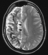
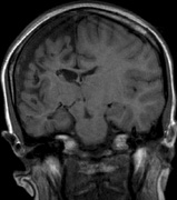
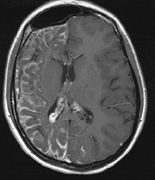
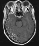
| Fig. 22. Axial T2-weighted (a) and coronal T1-weighted (b) images of a 16-year-old boy with a port-wine lesion over the right side of his face. The right hemisphere is markedly atrophic and abnormal draining veins are seen within the right lateral ventricle (arrowheads). (c, d) The entire right hemisphere is covered by an enhancing pial angioma and the choroid plexi are enlarged. Enhancing retinal angiomas (arrows), typical of Sturge-Weber syndrome, are seen in (d). |