|
|
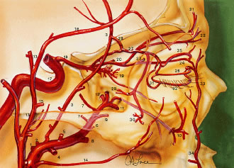
| Fig. 27. Lateral view demonstrating the relationship of the internal and the external carotid arterial systems to the orbit. Internal maxillary artery (O): (1) deep auricular; (2) anterior tympanic; (3) middle meningeal; (4) inferior alveolar; (5) masseteric; (6) pterygoid; (7) deep temporal; (8) buccal; (9) posterior superior alveolar; (10) infraorbital; (11) sphenopalatine; (12) artery of the pterygoid canal; (13) superficial temporal artery; (14) transverse facial; (15) zygomatico-orbital; (16) frontal branch; (17) internal carotid artery; (18) ophthalmic; (19) intraconal branches of ophthalmic artery; (20) posterior ethmoidal branch of ophthalmic; (21) supraorbital artery; (22) supratrochlear; (23) anterior ethmoidal branch of ophthalmic; (24) infratrochlear; (25) peripheral arcade (superior); (26) marginal arcade (superior); (27) lacrimal; (28) recurrent meningeal; (29) zygomaticotemporal; (30) zygomaticofacial; (31) lateral palpebral; (32) inferior marginal arcade; (33) angular; (34) facial; (35) central retinal; (36) lateral posterior ciliary; (37) muscular branches to superior rectus, to levator palpebrae, and to superior oblique; (38) medial posterior ciliary; (39) short ciliary; (40) long ciliary; (41) anterior ciliary; (42) greater circle of iris; (43) lesser circle of iris; (44) episcleral; (45) subconjunctival; (46) conjunctival; (47) marginal arcade; (48) vortex vein; (49) medial palpebral; (50) dorsal nasal. (From Zide BM, Jelkes GW. Surgical Anatomy of the Orbit. New York: Raven Press, 1985.) |