
|
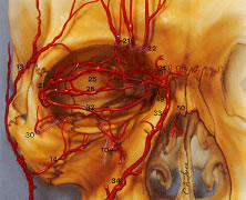
| Fig. 29. Superficial blood supply to the eyelids. From the external carotid artery, three branches of the facial vascular system ultimately supply the eyelid: the facial artery, the superficial temporal artery, and the infraorbital artery. The main points here are the following: First, most branches of the ophthalmic artery system from the posterior third of the orbit travel forward. Second, the ophthalmic artery is tethered to the medial orbital wall by the ethmoid arteries. Third, the collateralization that occurs among internal carotid artery (ICA) (17), the recurrent meningeal (28), and the facial arterial tree33,34 accounts for the reversal of flow when the ICA is obstructed. Finally, the central retinal artery enters the optic nerve in the posterior third. Internal maxillary artery (O): (1) deep auricular; (2) anterior tympanic; (3) middle meningeal; (4) inferior alveolar; (5) masseteric; (6) pterygoid; (7) deep temporal; (8) buccal; (9) posterior superior alveolar; (10) infraorbital; (11) sphenopalatine; (12) artery of the pterygoid canal; (13) superficial temporal artery; (14) transverse facial; (15) zygomatico-orbital; (16) frontal branch; (17) internal carotid artery; (18) ophthalmic; (19) intraconal branches of' ophthalmic artery; (20) posterior ethmoidal branch of ophthalmic; (21) supraorbital artery; (22) supratrochlear; (23) anterior ethmoidal branch of ophthalmic; (24) infratrochlear; (25) peripheral arcade (superior); (26) marginal arcade (superior); (27) lacrimal; (28) recurrent meningeal; (29) zygomaticotemporal; (30) zygomaticofacial; (31) lateral palpebral; (32) inferior marginal arcade; (33) angular; (34) facial; (35) central retinal; (36) lateral posterior ciliary; (37) muscular branches to superior rectus, to levator palpebrae, and to superior oblique; (38) medial posterior ciliary; (39) short ciliary; (40) long ciliary; (41) anterior ciliary; (42) greater circle of iris; (43) lesser circle of iris; (44) episcleral; (45) subconjunctival; (46) conjunctival; (47) marginal arcade; (48) vortex vein; (49) medial palpebral; (50) dorsal nasal. (From Zide BM, Jelkes G. Surgical Anatomy of the Orbit. New York: Raven Press, 1985.) |