|
|
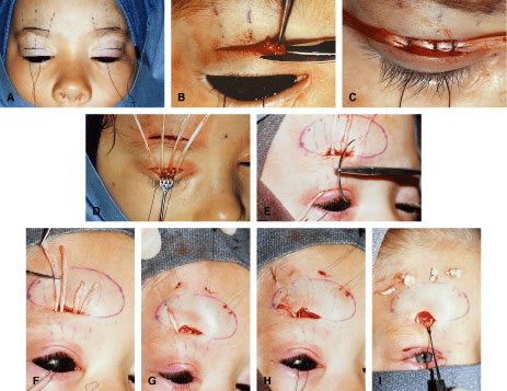
| Fig. 11. A. Corneal protective shields are placed. The planned incisions are marked at the proposed eyelid crease and above the eyebrow. B. The eyelid incision is carried out from skin down to the superior border of tarsus. Redundant orbicularis and tissue is cleaned off from the anterior surface of tarsus. C. Two strips of prepared fascia lata measuring approximately 5 mm by 7 cm are sutured to the upper tarsal border with interrupted sutures of 7-0 silk, one placed medially and one laterally. D. The superior brow incision is made. A forehead pocket anterior to the frontalis muscle is dissected above the brow. E. Each of the ends of the fascial strips is passed through the hub of a large curved needle and brought through the eyelid wound through orbital septum and exiting the superior brow wound. F and G. The fascial ends are trimmed and tagged with a double-armed Prolene suture. H. The suture is passed in a radial fashion from the brow incision out through the skin at the level of the hairline. I. The sutures are temporarily tied over bolsters, and the wounds are closed. Sutures are then adjusted and permanently tied over bolsters, then covered with Steri-strips. |