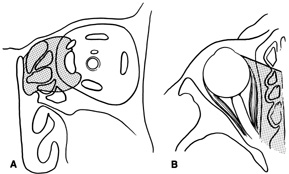
Fig. 17.
Schematic demonstration of areas amenable to frontoethmoidal orbitotomy. Coronal
(A)
and axial
(B)
views. This approach can be used for exposure of the medial orbit, ethmoid and sphenoid sinuses, and optic canal.