

|
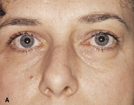
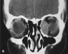
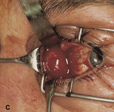
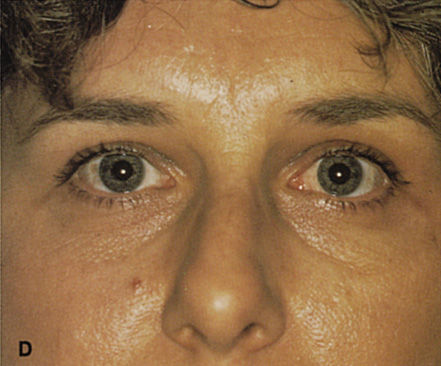
| Fig. 28. A. Patient with a medial orbital mass resulting in lateral displacement of the left globe. B. Coronal CT scan reveals the mass to be in the medial peripheral surgical space. C. Transconjunctival orbitotomy approach with conjunctival incision and dissection medial to the globe. In this case, the medial rectus muscle was not disinserted because the lesion lay in the peripheral surgical space outside the muscle cone. D. Postoperative appearance of the patient. Note that the lateral displacement of the globe has resolved. The conjunctival incision leaves no visible scar. |