Chapter 19
Metabolic Diseases and the Eye
SELWA AL-HAZZAA, PINAR OZAND, and ELIAS I. TRABOULSI
Main Menu   Table Of Contents

Search

LYSOSOMAL STORAGE DISEASES
DISORDERS OF LYSOSOMAL ENZYME PHOSPHORYLATION
DISORDERS OF GLYCOPROTEIN DEGRADATION AND STRUCTURE
SCHINDLER DISEASE
DISORDERS OF LIPID METABOLISM
GANGLIOSIDOSES
DISORDERS OF CARBOHYDRATE METABOLISM
LIPOPROTEIN AND LIPID DISORDERS
AMINO ACID DISORDERS
DISORDER OF ORGANIC ACID METABOLISM
ABNORMALITIES OF VITAMIN METABOLISM
MISCELLANEOUS DISORDERS
REFERENCES

A landmark in the history of human biochemical genetics was set at the turn of the 20th century with Sir Archibald Garrod's monographs on inborn errors of metabolism.

Metabolic and genetic disorders that affect the eye may cause significant visual disturbances and sometimes blindness. Furthermore, metabolic disorders may have characteristic ocular findings that assist in their diagnosis, making the ophthalmologist an invaluable member of the team that cares for these patients.

Metabolic disorders generally are inherited in an autosomal recessive fashion. There is reduced or absent function of one or more enzymes in a biochemical pathway that is critical to normal cellular function, growth, and development. Accurate biochemical diagnosis is essential for treatment, for genetic counseling, and for monitoring future pregnancies and children.

Clinically, enzyme deficiencies produce systemic and ophthalmic signs and symptoms by several mechanisms: accumulation of undegraded products, lack of production of an essential substrate, blockage of the normal conversion of one product to the other, or activation of alternate metabolic pathways that are deleterious to cellular integrity.

Inborn errors of metabolism can be divided into two broad categories4:

  Category 1 consists of diseases that involve only one functional or anatomic system or affect only one organ. The presenting symptoms usually are uniform, and diagnosis is easy, even when the basic biochemical lesion gives rise to systemic consequences. Included in this category are bleeding disorders that result from coagulation factor defects or hemolytic anemia from defects of glycolysis.
  Category 2 consists of diseases in which the basic biochemical lesion either affects a metabolic pathway common to a large number of cells or organs or is restricted to one organ but gives rise to humoral and systemic consequences. The presenting symptoms are diverse. This category includes most inborn errors of intermediary metabolism, diseases of intracellular trafficking, and lysosomal disorders.

Pathologic changes in the eye sometimes are characteristic of the underlying metabolic disease process. The detection of these ocular abnormalities depends on their prominence, the severity and ease of diagnosis of the systemic illness, and the familiarity of the ophthalmologist with their nature and significance. In many instances, the patient is referred with a diagnosed or suspected systemic disease, and a search for the known ocular complications of the illness is undertaken. In other instances, a metabolic disorder is suspected but no diagnosis is offered, making the ophthalmologic findings, if present, even more valuable in the diagnostic process.

Ophthalmologic findings such as corneal opacities, cataracts, cherry-red spot, and retinal degeneration may be the earliest signs of many metabolic disorders. Prompt and accurate diagnosis of the systemic disease aids in determining the prognosis and clinical expectations regarding career and life planning for the affected individual. It allows the early institution of treatment, when available, and the provision of genetic counseling on the risk of recurrence in siblings or in children. Measurement of enzyme levels or mutation analysis of DNA from fetal cells obtained through amniocentesis allows the prenatal diagnosis of many of these diseases.

Advances in molecular biology, biochemistry, and enzymology have allowed a better understanding of these diseases and their chemical defects. Currently, a biochemically based terminology is used instead of eponyms, and almost all disorders can be diagnosed by enzyme or gene analysis. Serum, leukocytes, or cultured skin fibroblasts can be assayed for enzyme activity if the molecular genetic defect is known, circumventing the need for biopsy of the liver, brain, or other tissues.

More than 300 human diseases that result from inborn errors of metabolism currently are recognized. Their true incidence may be underestimated because of failure of diagnosis. The detection of metabolic diseases relies only in part on screening programs and primarily depends on a high index of clinical suspicion and coordinated access to expert laboratory services.

Corneal opacities frequently occur in the mucopolysaccharidoses, mucolipidosis III and IV, α-mannosidosis, Fabry's disease, multiple sulfatase deficiency (Austin disease), Farber disease, LCAT (lecithin: cholesterol acyltransferase) deficiency, cystinosis, tyrosinosis type II, and Tangier disease.

Cataracts are some of the more conspicuous ocular signs of metabolic disorders.5 At birth and in infancy, cataracts are prominent findings in Lowe syndrome. They can be the only presenting sign of sorbitol dehydrogenase deficiency. Cataracts commonly occur in peroxisomal biogenesis disorders and in Cockayne syndrome. They are the only signs of galactokinase deficiency. They also are present in various galactitol or sorbitol accumulation states of unknown origin. Cataracts can be associated with other typical systemic signs and symptoms in galactosemia, mannosidosis, sialidosis, respiratory chain defects, hypoglycemia due to galactosemia, the severe form of mevalonic aciduria, aspartylglucosaminuria, multiple sulfatase deficiency, and Fabry's disease. In childhood, cataracts are present in one fourth of patients with untreated Wilson's disease. They also are signs of hypoparathyroidism and pseudohypoparathyroidism. In adults, isolated cataracts can be a sign of Lowe syndrome or of the heterozygous state for inborn errors of galactose metabolism. They frequently are present in patients with cerebrotendinous xanthomatosis. Posterior subcapsular cataracts with onset in the second decade of life are nearly constant complications of gyrate atrophy of the choroid and retina.

A macular cherry-red spot is a characteristic finding in some lysosomal disorders. The ganglion cells filled with storage material in the macula are opaque and give rise to a white ring that encircles the red, ganglion cell-free fovea. The diseases associated with a cherry-red spot are GM1 gangliosidosis (Landing disease), GM2 gangliosidosis Type 1 (Tay-Sachs disease), GM2 gangliosidosis Type 2 (Sandhoff's disease), Niemann-Pick Types A and B, sialidosis (cherry-red spot myoclonus), mucolipidosis I, metachromatic leukodystrophy, and galactosialidosis.

Retinal degeneration, with or without pigmentary retinopathy, occurs commonly in inherited metabolic disorders. Although its pathophysiology is not known, retinal dysfunction in metabolic disorders may be induced by toxic effects of certain metabolites, errors of synthetic pathways, or deficient energy metabolism. Table 1 lists the disorders associated with a pigmentary retinopathy.


This chapter summarizes the systemic and ophthalmologic manifestations of metabolic disorders in which the enzyme deficiency results in visually significant or diagnostic ocular manifestations. Because the diseases of interest have etiologies and manifestations in complex overlapping, noncategorical biologic systems, the authors are faced with the classic dilemma of balancing the practicality of categorical thinking against the reality of biologic variation.

Back to Top
LYSOSOMAL STORAGE DISEASES

THE MUCOPOLYSACCHARIDOSES

The mucopolysaccharidoses (MPSs) are transmitted as autosomal recessive traits, with the exception of Hunter syndrome (MPS II), which is inherited in an X-linked recessive fashion.6,7 They are caused by deficiency of lysosomal enzymes needed for the degradation of mucopolysaccharides and glycos-aminoglycans.8,9 The catabolism of dermatan sulfate, heparan sulfate, keratan sulfate, and chondroitin sulfate may be blocked singly or in combination, depending on the enzymatic deficiency. The storage process eventually results in cell, tissue, and organ dysfunction. Glycosaminoglycan fragments are excreted in the urine.

A defect in 1 of 10 enzymes gives rise to each of the seven distinct clinical forms of MPS and their subtypes. Table 2 presents the classification, major systemic features, and ophthalmologic findings of the MPSs.


The MPSs share several clinical features that are variable in severity in individual diseases. These findings include skeletal abnormalities, coarse facial features, mental deficiency, cardiac disease, hepatosplenomegaly, ocular abnormalities, and deafness. A chronic and progressive course is typical. Ocular manifestations include progressive corneal clouding, retinal pigmentary degeneration, optic nerve head swelling, optic atrophy, and glaucoma.

Tissue-specific differences in the structure of mucopolysaccharides account for the variability in clinical manifestations. Excess dermatan and keratan sulfates appear in the cornea, whereas heparan sulfate accumulates in the retina and central nervous system (CNS). The accumulation of these substances is the result of the faulty catabolism of mucopolysaccharides in lysosomes due to defective lysosomal acid hydrolases. The large storage vacuoles in histiocytes, lymphocytes, or leukocytes show metachromasia.

In patients whose disease leads to the storage of heparan sulfate in the retina, a retinal pigmentary degeneration associated with night blindness develops. Early in the course of their disease, patients with MPS IH, IS, IH-S, IV and VI report moderately severe photophobia. Papilledema is a frequent finding, occurring in one third or more of patients with certain types of MPS, such as Hunter syndrome.10 Optic nerve head swelling has been attributed to the hydrocephalus that results from meningeal thickening with the storage material. Collins et al10 postulated that it could be caused by narrowing of the scleral canal at the optic nerve head, as a result of posterior scleral thickening with mucopolysaccharide accumulation. Acute and chronic glaucoma may be seen in MPS IH, IS, and IH-S.

Conjunctival biopsy, a simple and applicable procedure, is a reliable screening test for patients with suspected lysosomal storage diseases.11 The diagnosis of MPS is based primarily on the characteristic clinical findings and on the detection of mucopolysaccharides in the urine. For definite diagnosis and for further categorization of the types and subtype of MPSs, specific enzymatic assays or gene analysis should be performed.

HURLER SYNDROME (MPS I-H)

Infants with Hurler syndrome appear healthy at birth. Signs of progressive organ dysfunction and dysostosis multiplex appear in the first year of life. Hurler syndrome has been the prototype for the description of the MPS and is the most severe form, with unfortunate death of all patients in the first decade of life. Clinical features include dwarfism, coarse facial features (Fig. 1), severe mental retardation, hirsutism, cardiovascular disease, skeletal abnormalities, and hepatomegaly.12 Acute cardiomyopathy has been described as the presenting feature in some infants younger than 1 year of age.13 Respiratory infection and cardiac failure are the usual causes of death.

Fig. 1. Coarse facial features of infant with Hurler syndrome (MPS I-H).

Progressive diffuse punctate stromal corneal opacities occur in all patients. They may conceal retinal degeneration and result in progressive visual loss.14,15 When the retina can be evaluated, a retinitis pigmentosa-like picture indistinguishable from other forms of heredofamilial retinal pigmentary dystrophies usually is present. The electroretinogram is diminished or nonrecordable.16 Glaucoma is relatively rare17 but has been reported in some patients.14,18 Optic nerve head swelling and optic atrophy are common10 (Fig. 2). Congenital cataracts occasionally have been observed.19

Fig. 2. Optic nerve head and peripapillary elevation in a patient with Hurler syndrome (MPS I-H).

Hurler syndrome is caused by the absence of α-L-iduronidase, which catalyzes the cleavage of iduronic acid residues from polysaccharide chains. As a result, heparan sulfate and dermatan sulfate accumulate and are excreted in the urine.20 The gene maps to 4p16.3.21 Residual α-L-iduronidase activity in Hurler fibroblasts is heat stable, whereas that in Scheie fibroblasts is heat labile. The enzyme from Hurler-Scheie compound fibroblasts is of inter—mediate activity between Hurler and Scheie-syndromes.

Bone marrow transplantation was performed in two patients homozygous for the relatively common W402X α-L-iduronidase mutation at the ages of 14 and 11 months, respectively. As opposed to untreated patients homozygous for this mutation who have a very severe clinical phenotype with rapid clinical deterioration and death before 6 years of age, the transplanted children were alive at 12 and 14 years of age, respectively. One showed limited mobility but was coping well at school; the patient was wheelchair-bound with severe disability and attended a school for the physically handicapped.22 Transfer and expression of the normal gene in autologous bone marrow may become an alternative method of treatment in the future.23

SCHEIE SYNDROME (MPS I-S; FORMERLY MPS V)

The clinical features of Scheie syndrome24 aremilder than those of Hurler syndrome. Signs and symptoms of the disease usually appear after the age of 5 years, leading to the diagnosis at about 15 years of age. α-L-Iduronidase is deficient, and the disease is allelic with Hurler syndrome.20 Patients have claw-hand deformities, joint stiffness, aortic valve insufficiency, hernias, and deafness. They are of normal height and intelligence and have a relatively normal life span. Facial features are only slightly coarse.

The predominant ocular feature is a peripheral corneal opacification that progresses centrally with age, eventually resulting in visual loss.25 Mucopolysaccharides accumulate in all cellular components of the cornea, with profound alteration of Bowman's layer and of the corneal lamellae. Ophthalmologic manifestations include glaucoma and pigmentary retinal degeneration that occurs in the first decade of life and is accompanied by night blindness and visual field constriction in the teens and 20s. If vision is reduced markedly, penetrating keratoplasty may be considered even though poor results have been reported, presumably because of the accompanying retinal degeneration.24

As in MPS I-H, dermatan sulfate and heparan sulfate are present in the urine. Scheie syndrome should be differentiated from mild MPS VI-B.

HURLER-SCHEIE COMPOUND (MPS I H-S)

Patients with MPS IH-S have a phenotype of intermediate severity between Hurler and Scheie syndromes.8 The disease is a genetic compound of the two alleles, H and S. Most patients have severe bone involvement, with little or no intellectual impairment. They have a longer life expectancy than those with Hurler syndrome. Micrognathia is a prominent clinical feature and leads to a characteristic facial appearance (Fig. 3). Arachnoid cysts with spinal rhinorrhea are characteristic and may lead to the enlargement of the sella turcica. The cervical cord may be compressed as a result of MPS accumulation in the dura. Symptoms appear at about 5 years of age, and survival to adulthood is common.

Fig. 3. Facial features of patient with Hurler-Scheie compound (MPS I -HS).

There is progressive corneal clouding, chronic disc edema, retinal degeneration, and a diminished or extinguished electroretinogram. When there is advanced vision loss, penetrating keratoplasty can be very helpful. Glaucoma is rare. Biochemical and enzymatic assays are identical to those in MPS I-H and I-S.

The residual α-L-iduronidase activity in Hurler fibroblasts is heat stable; it is labile in Scheie fibroblasts. The enzyme activity in MPS IH-S is intermediate between the two. Four novel mutations underlying mild or intermediate forms of α-L-iduronidase deficiency have been discovered, providing insight into genotype-phenotype correlation in this group of patients.26

HUNTER SYNDROME (MPS II)

There are mild (type B) and severe (type A) forms of Hunter syndrome. They are distinguished on clinical grounds only and have a wide spectrum of clinical severity. The two types are allelic and are caused by mutations at the X-linked locus for the enzyme iduronate sulfate sulfatase. Wilson et al27 localized the gene to Xq28, distal to the fragile X site. Characteristic pebbly, ivory-colored skin lesions over the back, neck, scapula, and thigh are present in patients with MPS II (Fig. 4).

Fig. 4. Nodular skin lesions in the scapular area of a patient with Hunter syndrome.

Individuals with the severe form have many features of Hurler syndrome, but of lesser severity and with slower progression of somatic and CNS disease. Only males are affected. Death usually occurs before the age of 15 years.

In the milder type B, survival may extend past the age of 45 years.28 The longest known survival is to 87 years of age.29 Intelligence is normal and skeletal involvement moderate, with minimal or no CNS involvement. Corneal clouding usually is absent in type B,28 although deposits of acid mucopolysaccharides are found histologically, even in clinically clear corneas.30 Pigmentary retinopathy with an abnormal or extinguished electroretinogram may lead to visual impairment of varying degrees in all patients.31 In patients with mild Hunter syndrome without raised intracranial pressure, elevated and blurred disc margins lead to the diagnosis of chronic-papilledema, which may culminate into optic-atrophy.10,28,32,33

Dermatan sulfate and heparan sulfate are present in the urine. Definitive diagnosis is made by assaying for the activity of sulfoiduronate sulfatase in fibroblasts. The enzyme defect can be corrected in vitro using an amphotropic retroviral vector containing the human gene.34

SANFILIPPO SYNDROME (MPS III)

Patients with this autosomal recessive syndrome have severe CNS degeneration but only mild somatic disease. Onset of clinical features occurs between 2 and 6 years of age. Significant delay in diagnosis is caused by the mild somatic (Fig. 5) and radiographic features. There may be moderate dwarfism, minimal skeletal dysostosis, and moderate hepatosplenomegaly. The presenting problem may be that of marked hyperactivity, destructive tendencies, and other behavioral aberrations. Four biochemically distinct types of MPS III are difficult to differentiate clinically. Type A is most severe, with an earlier onset, more rapid progression of symptoms, and earlier death than types B, C, or D.35 Types B and D are heterogeneous.36,37. Type C is intermediate between A and B. Excessive heparan sulfate, but not dermatan sulfate, is excreted in the urine. N-sulfated glucosamine residues are removed during the degradation of heparan sulfate through the sequential action of four enzymes that are defective in Sanfilippo syndrome. In Type A, there is a deficiency of heparan N-sulfatase or sulfamidase; in type B, α-N-acetyl-glucosaminidase (NAG) is lacking; in type C, acetyl CoA: α-glucosaminidase is deficient; and in type D, the defect is in N-acetyl glucosamine 6-sulfatase.

Fig. 5. Relatively normal facial features in a patient with Sanfilippo syndrome type A (MPS IIIA).

The corneas are clear clinically but show similar but less severe ultrastructural abnormalities as other forms of MPS.15 A retinitis pigmentosa-like fundus picture frequently is present.38 Optic atrophy may be present.

The MPS urine spot test is positive, and the urine contains excessive amounts of heparan sulfate. The four types of Sanfilippo syndrome can be differentiated by enzymatic assays. Type A has been mapped to 17q25.3,39 type B to 17q21,40 type C to chromosome 14, and type D to 12q14.41

MORQUIO SYNDROME (MPS IV)

Two types of Morquio syndrome are recognized with a wide spectrum of clinical manifestations. Type A is caused by deficiency of N-acetyl galactosamine 6-sulfatase,42 and type B is caused by deficiency of β-galactosidase.43 As with most MPSs, no clinical abnormalities are apparent at birth. An awkward gait, retarded growth, knock knees, sternal bulging, and flaring of the rib cage become evident in the second year of life. Patients with Morquio syndrome have short trunk dwarfism (Fig. 6). They are of normal intelligence, and their distinctive skeletal abnormalities become prominent in the first 10 years of life. The predominant clinical features are related to the effect of the disease process on the spinal cord.44 The joints are loose and hyperextensible, the wrists enlarged, and the hands misshapen,8 and there is genu valgum and kyphosis. The facies is characteristic with a broad mouth, prominent maxilla, short nose, and widely spaced teeth.7 Odontoid hypoplasia may lead to atlantoaxial subluxation and spinal cord compression later in the course of the disease. Cervical myelopathy develops early. Death occurs late in childhood from respiratory paralysis secondary to spinal cord compression or from recurrent pneumonia.

Fig. 6. Child with Morquio syndrome (MPS IV).

Corneal clouding, the most common ocular feature, generally is not present before 10 years of age45 (Fig. 7), but one of us (EIT) has observed it in three Lebanese siblings younger than 10 years of age. The corneal epithelium and Bowman's layer appear normal under the slit lamp, and there is a homogeneous clouding of the corneal stroma. There is no retinal dystrophy.46

Fig. 7. Corneal opacification in a child with Morquio syndrome (MPS IV).

The gene for type A maps to 16q24.3.47

MAROTEAUX-LAMY SYNDROME (MPS VI)

This autosomal recessive disease was first recognized in 1963.48 Severe and mild phenotypes have been described.7 Type A, or classic form, is more severe than its allelic form, type B. The deficient enzyme is arylsulfatase B (N-acetylgalactosamine 4-sulfatase).49 Heparan sulfate and dermatan sulfate are excreted excessively in the urine. The extent of corneal clouding is the same in both types, but skeletal dysplasia is less pronounced in the latter. The clinical features, except for normal intelligence, are reminiscent of those of Hurler syndrome, especially in the severe form (Fig. 8).

Fig. 8. Facial and skeletal abnormalities in a child with Maroteaux-Lamy syndrome (MPS VI).

Growth retardation is first noted at 2 or 3 years of age. Restriction of joint movement develops in the first year of life. Significant cardiomyopathy may arise, and hepatosplenomegaly may be present. Hypoplasia of the odontoid process can cause spinal cord compression and spastic paraplegia.50 Umbilical and inguinal hernias are common. Patients with type A Maroteaux-Lamy syndrome die in their teens from hydrocephalus secondary to meningeal infiltration with mucopolysaccharides. Radiographic findings are similar to those of patients with Hurler syndrome and are striking examples of dysostosis multiplex.

Diffuse corneal stromal opacification develops early in life. The cornea increases in thickness, especially in its periphery, where the clouding is most dense.11 Papilledema and optic atrophy occur.10,51 Retinal degeneration with electroretinographic abnormalities is a feature of the disease. Corneal transplantation has been performed in patients with-Maroteaux-Lamy syndrome, but the long-term visual outcome is poor.52,53 Vision may be poor despite clear corneal grafts because of associated retinal or optic nerve disease.

The gene maps to 5q11–q13.54

SLY SYNDROME (MPS VII)

β-Glucuronidase is deficient in this type of mucopolysaccharidosis.55 There may be two allelic forms leading to two clinical phenotypes with a wide range of severity of signs and symptoms.

The gene map for β-glucuronidase was refined to 7q21.11 by Ward et al56 using dosage analysis of chromosomal aberrations. There is excessive urinary excretion of glycosaminoglycans and coarse granulocyte inclusions in a variety of tissues.

The severe form of the disease is characterized by rapidly progressive mental, motor, and growth retardation. Patients have hepatosplenomegaly, massive ascites, inguinal hernias, thoracolumbar gibbus, and skeletal radiographic changes similar to those of the other mucopolysaccharidoses. Individuals with the milder phenotype exhibit hepatosplenomegaly, skeletal abnormalities, and an unusual facies. Mental retardation is not present at birth but develops with aging. Corneal clouding is variable and usually mild.57 A mild pigmentary retinopathy develops later in life.

Back to Top
DISORDERS OF LYSOSOMAL ENZYME PHOSPHORYLATION
Spranger and Wiedemann coined the term mucolipidoses for this group of disorders.58 The mucolipidoses have phenotypic and biochemical features of both the mucopolysaccharidoses and the sphingolipidoses without excessive urinary excretion of mucopolysaccharides. Ultrastructurally, the affected cells resemble those of the MPSs, with cytoplasmic vacuoles filled with fine fibrillogranular-material. The vacuoles also contain membranous lamellar inclusions similar to those present in the lipidoses.15

Four diseases have been included in this category: 1) mucolipidosis I, or sialidosis; 2) mucolipidosis II, or inclusion cell disease (I-cell disease); 3) mucolipidosis III, or pseudo-Hurler polydystrophy; and 4) mucolipidosis IV, or Berman disease.

MUCOLIPIDOSIS I (MLS I) (SIALIDOSIS TYPE 1; CHERRY-RED SPOT MYOCLONUS SYNDROME)

In MLS I, there is deficiency of the enzyme sialidase that leads to a syndrome characterized by a retinal cherry-red spot and myoclonus. MLS I was reclassified as sialidosis type 159 and is discussed later in this chapter.

MUCOLIPIDOSIS II (MLS II) (I-CELL DISEASE)

Patients with this rare and severe autosomal recessive mucolipidosis display many of the clinical and radiographic features of Hurler syndrome.60 There are striking fibroblast inclusions, hence the name I-cell disease.61 There is no mucopolysacchariduria.62,63 The disease results from the absence of the enzyme that attaches a recognition phosphate group onto a mannose residue in hydrolases. There is abnormal lysosomal enzyme transport in cells of mesenchymal origin. Severe progressive psychomotor retardation occurs. MLS II is differentiated from Hurler syndrome by the earlier onset of signs and symptoms, the absence of mucopolysacchariduria, the early and striking gingival hyperplasia, and the rapidly progressive course leading to death in the first decade.64

Early in development, congenital dislocation of the hips, thoracic deformities, hernia, and gingival hyperplasia are evident.65,66 Radiographic studiesshow bony changes of dysostosis multiplex more severe than in MPS I. Patients have coarse facial features, craniofacial abnormalities, and restricted joint mobility.

Early in the course of the disease, the corneas are clear. Late corneal clouding is common; it correlates positively with survival and occurs in 40% of cases.67 Corneal opacities are evident on slit-lamp examination as diffuse stromal granules. Glaucoma occurs in 6% of patients.68 No cherry-red macula is present. Conjunctival biopsy is diagnostic.68

The diagnosis can be made biochemically by demonstration of elevated serum and urine lysosomal enzyme levels and by measuring the UDP-N-acetylglucosamine lysosomal enzyme N-acetylglucosamine phosphotransferase (GNPTA) in fibroblasts. The gene for this enzyme maps to 4q21–q23.69 Prenatal diagnosis is reliable and carrier detection is possible. There is no definitive treatment.

MUCOLIPIDOSIS III (MLS III) (PSEUDO-HURLER POLYDYSTROPHY)

Mucolipidosis III results from the same enzyme deficiency as MLS II. N-acetylglucosamine 1-phosphotransferase is composed of at least two distinct polypeptides: a recognition subunit that is defective in the MLS III variant and a catalytic subunit that is deficient or altered in the classic forms of MLS II and III as well as in the MLS II variant.70

Onset of clinical signs and symptoms is later than in MLS II. The clinical course progresses more slowly, and survival into adulthood is possible.71,72 Most patients have been Ashkenazi Jews. Children usually present at about 3 years of age with joint stiffness, coarse facial features, and short stature.73 They have many of the features of Hurler syndrome but with a much slower clinical evolution and no mucopolysacchariduria. The arms and hands are involved most markedly, and carpal tunnel syndrome with wasting of the thenar eminence is common (Fig. 9). Aortic valve disease occurs in most cases. The pathology of MLS III is not well documented. Cultured fibroblasts have inclusion bodies similar to, but not as prominent as, MLS II cells.

Fig. 9. Child with mucolipidosis III. Note mild coarse facial features and joint contractures, most prominent in the hands.

Nearly 50% of reported patients have some learning disability or mental retardation.74 Life expectancy and the natural course of the illness beyond the third decade are unknown.

Corneal clouding (Fig. 10), hyperopic astigmatism, and a mild retinopathy with surface-wrinkling maculopathy (Fig. 11) appear to be the constant ocular triad.75 Some patients have retinal vascular tortuosity, optic nerve head swelling (Fig. 12), visual field defects, and abnormalities in color vision. Visual complaints are uncommon.

Fig. 10. Mild corneal clouding in a child with mucolipidosis III. (Traboulsi E, Maumenee I: Ophthalmologic findings in mucolipidosis III. Am J Ophthalmol 102:529, 1986)

Fig. 11. Surface-wrinkling maculopathy in a patient with mucolipidosis III. (Traboulsi E, Maumenee I: Ophthalmologic findings in mucolipidosis III. Am J Ophthalmol 102:529, 1986)

Fig. 12. Optic nerve head swelling in a patient with mucolipidosis III. (Traboulsi E, Maumenee I: Ophthalmologic findings in mucolipidosis III. Am J Ophthalmol 102:529, 1986)

Prenatal diagnosis by means of amniocentesis is possible. There is no specific or definitive treatment.

MUCOLIPIDOSIS IV (MLS IV) (BERMAN DISEASE)

Mucolipidosis IV was first described by Berman.12 It is characterized by psychomotor retardation, lack of skeletal deformities and organomegaly, and variable ophthalmologic abnormalities in the form of corneal opacities and retinal degeneration. Patients have been described with onset of clinical findings from 1 year of age to the mid 20s. Life span and prognosis beyond this age remain to be determined. The disease is seen mainly among Ashkenazi Jews.76 MLS IV differs from most other lysosomal storage disorders in the apparent lack of progression in some patients. In most, however, there is marked psychomotor and physical retardation. Biochemically, MLS IV is characterized by the accumulation of gangliosides,77 phospholipids, 78 and acidic mucopolysaccharides.79 Deficiency of ganglioside sialidase was reported as the possible metabolic defect causing this disorder, and partial reduction in activity of this enzyme was noted in obligate heterozygotes.80 Conjunctival biopsy shows characteristic-intracellular inclusions.81

There is progressive severe visual impairment from corneal opacification12,82 and from progressive retinal degeneration.83,84 The age of appearance and the extent and severity of the clinical course of these abnormalities are variable. The superficial corneal opacification is a characteristic feature of MLS IV and leads to the clinical diagnosis in most children. The opacities are not congenital in all patients, as suggested previously.12 With time, the corneal opacities may increase in density, with poor fundus visualization leading to the erroneous diagnosis of cataracts in some patients. Epithelial edema also hasbeen noted. Corneal clearing may occur after epithelial removal, but clouding returns after a few days or weeks. A progressive rod-cone dystrophy is present with a subnormal or extinguished electroretinogram (ERG).82,84 The static nature and, at times, improvement in the degree of corneal opacification in more than 50% of cases is characteristic of MLS IV. In other lysosomal storage disorders, corneal clouding and visual impairment are progressive. Deterioration of visual function in MLS IV correlates with retinal degeneration rather than with worsening of corneal opacities. Prenatal diagnosis is possible.81,82

Back to Top
DISORDERS OF GLYCOPROTEIN DEGRADATION AND STRUCTURE
Glycoproteins are synthesized via two pathways: (1) the glycosyltransferase pathway that synthesizes oligosaccharides linked to serine or threonine and (2) the dolichol, lipid-linked pathway that synthesizes oligosaccharides linked to asparagine. Specific enzyme deficiencies cause individual glycoprotein storage diseases.

MANNOSIDOSIS

There are α and β types of mannosidosis.85 We restrict our comments to α-mannosidosis because of the associated ophthalmologic abnormalities. α-mannosidosis results from a deficiency of the lysosomal enzyme α-mannosidase. There are elevated serum levels and excretion of small mannose-richoligosaccharides. Patients with α-mannosidosis excrete a unique pattern of increased amounts of several oligosaccharides.86,87 The gene for α-mannosidase was mapped to chromosome 19p13.2-q12.88 In severe infantile type I mannosidosis, there is rapidly progressive mental retardation, hepatosplenomegaly, severe dysostosis multiplex, and often death between the ages of 3 and 10 years.89 The milder juvenile and adult type II mannosidosis is characterized by a more slowly progressive course with survival into adulthood.90 Hearing loss is a prominent feature of this disease, but clinical heterogeneity is evident.91 Recurrent bacterial infections and hernias are seen.92,93 Types I and II are not clearly separated. Ophthalmologic manifestations are present in both types and consist of superficial corneal opacities and posterior spoke-like lens opacities.94

The diagnosis of mannosidosis can be confirmed by direct measurement of α-mannosidase in leukocytes, fibroblasts, or cultured amniotic fluid cells. There are several reports of successful prenatal diagnosis.95,96

FUCOSIDOSIS

Fucosidosis is caused by deficiency of the lysosomal enzyme α-fucosidase.97 This results in accumulation and excretion of a variety of glycoproteins, glycolipids, and oligosaccharides containing fucoside moieties. The disorder is panethnic, with a higher incidence in Italy and in the Southwestern parts of the United States.98 There is faulty degradation of both sphingolipids and polysaccharides.99 Urine samples from individuals with fucosidosis contain excessive amounts of several fucoglycoconjugates.100 The most precise way of diagnosing fucosidosis is based on enzymatic assay of α-L-fucosidase in cells of any type.

There is a spectrum of severity of the clinical findings with mild to severe phenotypes. The more severely affected patients exhibit psychomotor retardation, coarse facies, growth retardation, dysostosis multiplex, and neurologic retardation in the first year of life. The milder phenotype is characterized by angiokeratomas (Fig. 13) and by a longer survival. The gene for fucosidosis maps to chromosome 1p24.101

Fig. 13. Cutaneous lesions in fucosidosis.

The ocular features of fucosidosis are not prominent. There may be tortuosity of conjunctival vessels and a mild pigmentary retinopathy.102–104

SIALIDOSIS

Sialidosis is a rare lysosomal storage disease with clinical features resembling those of Hurler syn-drome, but milder. The two forms of the disease, sialidosis type 1 and type 2, result from deficiency of neuraminidase. The gene defects of the two types are not allelic. Complementation studies between sialidosis type 1 and type 2 fibroblasts result in restoration of both β-galactosidase and sialidase activities in fused cells. Moderate but progressive mental retardation, cerebellar signs, peripheral neuropathy, myoclonic jerks, tremor, dysostosis multiplex, and peculiar inclusions are present in cultured fibroblasts,8,105,106 but there is no mucopolysacchariduria. There is accumulation of syalyloligosaccharides in various tissues and organs of the body and deficiency of the enzyme α-N-acetylneuraminidase in cultured fibroblasts.107

Diagnosis is based on electron microscopy. Fibroblasts in conjunctival biopsy specimens contain small membrane-bound vacuoles containing fibrillogranular and membranous lamellar bodies detectable by electron microscopy.15 The differential-diagnosis must include other diseases causing a cherry-red macula. There is no known treatment, but prenatal diagnosis is possible.

Sialidosis Type 1

Sialidosis type 1 is characterized by the cherry-red spot myoclonus phenotype (cherry-red spot myo-clonus syndrome) and is the milder form of sialidosis. There are at least 15 confirmed patients.108,109 The disease results from a defect in the structural gene for sialidase, which maps to 10pter-q23.110 Retinopathy and myoclonus occur simultaneously at the onset of the disease, which tends to be in early adolescence but can be variable. The striking neurologic manifestation is a stimulus-sensitive myoclonus that limits daily activities. Ataxia and generalized grand-mal seizures also may occur. Intellect is preserved, and patients with this rare disorder can survive beyond 30 years of age.

The presenting symptom may be a reduction in visual acuity; visual loss is progressive and often severe. It may be associated with impaired color vision and night blindness.111 The macular cherry-red spot is consistent but can be atypical. Punctate lens opacities can occur.109,112,113 Nystagmus, optic-atrophy, and visual field constriction have been-described.

An important diagnostic screening test in this disorder is the examination of the urine for excessive excretion of sialic acid-containing compounds. Urine thin-layer chromatography stained with resorcinol gives a blue color, suggesting the presence of sialylated oligosaccharides. The definitive test is the demonstration of deficient sialidase in fresh cultured skin fibroblasts.

Sialidosis Type 2

Sialidosis type 2 is distinguished from sialidosis type 1 by the early onset of a severe progressive muco-polysaccharidosis-like phenotype with visceromegaly, dysostosis multiplex, and mental retardation. The disease may be the result of a defect in the gene for a protective protein on chromosome 20.110 There are several subtypes with marked variability in clinical manifestations. Recognized are a congenital, or hydropic form, an infantile form, and a juvenile form, the latter of which is the most common and which is described in this chapter under the title of galactosialidosis.

Congenital sialidosis type 2 patients all have been stillborn infants with hydrops fetalis, ascites, hepatosplenomegaly, stippling of the epiphyses, and periosteal cloaking of the long bones. There are no documented ophthalmologic findings.

Patients with infantile-onset sialidosis type 2 are relatively healthy at birth.114 Later, a progressive severe MPS-like phenotype develops. Development is slow, with mental retardation and gait ataxia. Myoclonus is present. Grandmal seizures, deafness, and a peripheral neuropathy may occur. Skeletal abnormalities are prominent with dysostosis multiplex. Vision is retained despite the presence of a macular cherry-red spot and punctate lens opacities.

Peripheral blood lymphocytes are vacuolated, and foam cells are present in the bone marrow. Sialidase is deficient in cultured fibroblasts.

Juvenile sialidosis type 2 is described under β-galactosialidosis.

ASPARTYLGLUCOSAMINURIA

Aspartylglucosaminuria is an autosomal recessive disorder that occurs primarily in Finland.115 Rare, isolated cases are reported from other countries. The accumulation of aspartylglucosamine results from a deficiency of the lysosomal enzyme, aspar—tylglucosaminidase, which maps to chromosome 4q32–q33.116

Patients are healthy for the first few months of life. Recurrent infections, diarrhea, and hernias are noted during the first year of life. Coarsening of facial features and saggy skin folds occur in the first decade. Mental deterioration begins between the ages of 6 and 15 years. Crystal-like lens opacities are observed.117 Aspartylglucosamine is the major storage compound found in tissues and fluids of patients with this disease.118 There may be vacuolated lymphocytes and neutropenia. The urine contains large amounts of aspartylglucosamine. No treatment is known.

Back to Top
SCHINDLER DISEASE
Schindler disease is caused by deficient activity of the lysosomal hydrolase enzymes α-N-acetylgalactosaminidase and α-galactosidase B.119 The gene has been localized to 22q13.1–q13.2.120 The disease is clinically heterogeneous, and there are two major phenotypes. Only type 1 is described because of its ophthalmologic features.

In type 1 Schindler disease, there is infantile onset of severe neural degeneration and neuroaxonal dystrophy. The disorder originally was described in two German brothers who were the products of a consanguineous marriage.119,121 Development is normal in the first 9 to 15 months of life. This is followed by rapid neural degeneration and by loss of developmental milestones, resulting in severe psychomotor retardation, myoclonic seizures, and cortical blindness. Limited information is available on pathologic and biochemical abnormalities because all known patients are still living.

Ophthalmologic findings include strabismus, optic atrophy, nystagmus, and eventually, cortical blindness. Neuroimaging studies demonstrate generalized atrophy of the brainstem, cerebellum, and cortex. Visual evoked potentials have low amplitude, delayed responses, or both.121,122

Diagnosis of affected homozygote and heterozygote carriers can be made only by determination of α-N-acetylgalactosaminidase activity in various tissues. Prenatal diagnosis is possible. There is no specific treatment, and appropriate supportive care should be implemented as needed.

Back to Top
DISORDERS OF LIPID METABOLISM

NIEMANN-PICK DISEASE

Niemann-Pick disease (NPD) results from impaired sphingomyelin metabolism. The disease was first reported by Niemann in 1914.123 Thirteen years later, Pick described the characteristic vacuolated or foam cells in many body tissues.124 NPD results from deficient activity of acid sphingomyelinase (ASM) that maps to 18q11–q12.125

Crocker126 categorized the NPD phenotypes into four clinical entities designated A to D. The broad classification of NPD into five phenotypic variants (A through E) followed and was based on the age of onset, the severity and type of neurologic involvement, and the evolution of the disease. Hepatosplenomegaly and foam cells in the bone marrow are constant features in all variants. Type A is the infantile neurodegenerative form. Type B is distinguished from the severe neuronopathic type A by the absence of neurologic involvement, by the presence of hepatosplenomegaly, and by survival into adulthood. Patients with type C NPD are healthy for the first 1 or 2 years of life and then experience a slowly progressive and variable neurodegenerative course, with less severe hepatosplenomegaly and survival into adulthood; they often present with prolonged neonatal jaundice. Type D patients share a common ancestry traceable to Acadians from Nova Scotia. Neurologic symptoms develop later in childhood, and these patients have a slower neurodegenerative course than those with type C. Type E is the adult form with a mild degree of visceral sphingomyelin storage, mild splenomegaly, foam cells in the bone marrow and absence of neurologic signs.

ASM is deficient in types A and B, which are believed to be allelic.127 In type A NPD, ASM is reduced dramatically,128,129 whereas in type B, there is residual ASM activity.130 In type C, sphingomyelinase activity usually is normal.131 The metabolic block in type C is in the intracellular trafficking of cholesterol.132 The biochemical defects in type D and type E NPD remain to be determined.

The pathologic hallmark of types A and B is the Niemann-Pick cell, which is a lipid-laden foam cell133 (Fig. 14). Sphingomyelin accumulates in the brain and autonomic ganglia. The neurons become swollen and have a pale cytoplasm. Ultrastructurally, the cells contain concentric lamellated bodies representing storage cytosomes. Inclusion profiles in the viscera, lymph nodes, and foam cells also correlate with an increase in sphingomyelin content. Diagnosis can be made readily by enzymatic determination of ASM activity in cells and tissues. More than 300 cases of type A and B NPD have been reported. Prenatal diagnosis has been accomplished by enzyme assays of cultured amniotic fluid cells in types A and B.

Fig. 14. Foam cells in bone marrow aspirate of patient with Niemann-Pick type B. Giemsa stain.

Niemann-Pick Disease Type A

Type A NPD is fatal in infancy. It is characterized by failure to thrive, hepatosplenomegaly, and severe CNS involvement, with loss of motor function leading to death by 2 to 3 years of age. A higher incidence is seen in the offspring of Ashkenazi Jews compared with the general population.134

A cherry-red macula is present in 50% of infants in the first and second year of life.135 There is no distinction between the appearance of the cherry-red spot in infantile NPD and in Tay-Sachs disease.136,137 Occasionally, a macular halo syndrome with a gray, granular-appearing macula is observed.138 Optic atrophy develops with time. Subtle lens opacities and corneal clouding can occur.137 ERG is abnormal. The stored lipid is localized to the ganglion and amacrine cells of the retina, most conspicuously in the parafoveal region. The other retinal layers appear unaffected.135

Niemann-Pick Disease Type B

Type B NPD has a variable phenotype, with only visceral involvement. It is diagnosed in childhood between 3 and 11 years of age, or in adult life with hepatosplenomegaly and progressive pulmonary infiltrates that cause the major disease complications.139 Most patients have a normal intellect and survive into adulthood. Patients with type B NPD are of mixed ethnic backgrounds. Harzer and associates140 were first to demonstrate a low sphingomyelinase level in this disease.

A unique retinal abnormality, the macular halo syndrome, has been reported in type B NPD by Cogan and Kuwabara135 and consists of a ring of opacities around the macula that causes no visual impairment (Fig. 15). This abnormality has been reported by several authors.141–143The crystalloid halo which measures about half the disc diameter occurs at the outer edge of the retina mainly in Henle's fiber layer causing only minor obstruction of the overlying vessels. Matthews and associates144 proposed that the macular halo represents the mildest form of a cherry-red spot in the ganglion cell layer of the retina. Their findings are in conflict with those of Cogan et al.138 The precise location of the opacities in the retina remains uncertain because of the lack of histopathology. The available clinical data suggest that such opacities are permanent.

Fig. 15. Macular halo in a patient with Niemann-Pick type B.

Bone marrow transplantation has been successful in type B NPD. Enzyme replacement and somatic gene therapy using macrophage-targeted recombinant enzyme may be available in the future.

Niemann-Pick Disease Type C

Type C NPD is a panethnic autosomal recessive lipidosis associated with lysosomal accumulation of unesterified cholesterol. It was first recognized in 1958145 and is biochemically distinct from type A and type B NPD.146 It is as common as types A and type B NPD combined. This distinction was supported by somatic cell hybridization studies.147 Genetic isolates have been described with the Nova Scotia form, formerly type D NPD,126 and in southern Colorado.148 There is no evidence of genetic heterogeneity. Sea-blue histiocytes in the bone marrow are common but nonspecific.149 The metabolic block results from defective intracellular cholesterol trafficking.132 The disease is diagnosed by filipin staining and by demonstration of fluorescence around the nucleus after low-density lipoprotein (LDL)-cholesterol loading.150 Cultured fibroblasts from patients and carriers have high levels of unesterified cholesterol. There is no macular cherry-red spot or a macular halo in type C NPD. The differentiating clinical features of type C NPD (ophthalmoplegic neurovisceral lipidosis, vertical supra's disease) are its three main clinical features, which led Cogan et al151 to recommend the acronym DAF syndrome to denote downgaze paralysis, ataxic athetosis, and foam cells in the spleen, liver, and bone marrow. There is extensive infiltration of bone marrow, spleen, liver, and other tissues with foam cells. Sphingomyelinase activity in leukocytes and cultured fibroblasts is decreased or normal.152,153

Type C NPD has a subacute clinical course and presents in infancy with neonatal hepatitis or later in childhood with moderate splenomegaly and gradual neurologic deterioration. Most patients have seizures and limitation of vertical gaze. The presence of downgaze paresis is characteristic154 and has been noted in all juvenile and adult cases.155 Subtle slowing of vertical saccades begins in late infancy. Voluntary vertical gaze is completely paralyzed in late stages of the illness.156 Horizontal eye movements may be affected with a total supranuclear ophthalmoplegia. Patients usually die before the end of the second decade.

Niemann-Pick Disease Types D, E, and F

Type D NPD is caused by a defect in cholesterol metabolism, and although few biochemical studies have been published, it may be allelic with type C.157,158 The underlying metabolic defect in type D and type E NPD still is not understood, but these two variants cannot be diagnosed by a single laboratory test. Increase in organ sphingomyelin content or presence of foam cells in the bone marrow helps to corroborate clinical suspicions. Neurologic symptoms develop later in childhood in patients with type D disease, and they have a slower neurodegenerative course than type C patients.159 They share a common ancestry traceable to Acadians. There are no ocular abnormalities in type D. Patients with the type E variant may have a macular cherry-red spot. In 1978, Schneider et al160 described two patients with a heat-labile form of ASM and classified it as type F NPD.

FABRY'S DISEASE (ANGIOKERATOMA CORPORIS DIFFUSUM UNIVERSALE)

Fabry's disease results from a defect in glycosphingolipid catabolism. The disease has an incidence of approximately 1 in 40,000.122 The gene is X-linked recessive and has been localized to Xq22.161 It codes for the lysosomal hydrolase α-galactosidase A.162 Most heterozygous female carriers have an intermediate level of enzymatic activity.163,164 There is progressive and systematic accumulation of the glycosphingolipid ceramide trihexaside, particularly in vascular endothelial cells.165 Carrier females in a pedigree can be examined clinically and biochemically for heterozygote identification.

The disease usually has its onset during childhood or adolescence. Classically, affected hemizygotes have pain and paresthesia of the extremities (acroparesthesia) around the time of puberty166 ; vascular cutaneous lesions (angiokeratomas) of the scalp, mucous membranes, skin, and inguinal and umbilical regions; hypohidrosis; and the characteristic corneal and lens opacities. Severe renal impairment leads to hypertension and uremia. Death occurs from renal failure or from cardiac or cerebrovascular disease.167

The ocular deposition of glycosphingolipids results in unique and diagnostic eye findings in severely affected hemizygous males and minimally affected heterozygous carrier females.168 The ocular findings have been recognized as one of the distinctive hallmarks of this disease and among its earliest clinical manifestations.169 The corneal opacities appear as whorled streaks from a central vortex and have been called cornea verticillata170,171 (Fig. 16). Bilateral inferior granular anterior capsular or posterior subcapsular lens opacities occur in one third of hemizygous males but rarely in heterozygous females. Mild to severe conjunctival (Fig. 17) and retinal vessel tortuosity are present early in life. Visual acuity is not impaired. However, acute visual loss has occurred in hemizygotes as a result of unilateral central retinal vascular occlusion.171 Other ocular findings include lid edema, myelinated nerve fibers, mild optic atrophy, papilledema, nystagmus, and internuclear ophthalmoplegia.171,172 (Fig. 18). Confirmation of the clinical diagnosis in hemizygotes and heterozygotes requires the demonstration of deficient α-galactosidase A activity in plasma, leukocytes, or tears or increased levels of ceramide trihexaside in plasma or urinary sediment. The diagnosis in female heterozygotes can be established by linkage analysis.163,174 These carriers may have some of the systemic manifestations of the disease, but they usually are milder than in affected males. The most frequent clinical finding in females is the characteristic whorl-like corneal epithelial dystrophy. Corneal opacification in female carriers may be more prominent than in male patients because of the admixture of corneal epithelial cell lines with the normal gene and those with the abnormal gene (lyonization). Abnormal glycosphingolipid deposits are found in all ocular and orbital vessels, in smooth muscle of iris and ciliary body, and in perineural cells and connective tissue of the lids and cornea.175

Fig. 16. Cornea verticillata in a carrier female of Fabry disease. (Courtesy of Irene H. Maumenee, MD)

Fig. 17. Corkscrew-like tortuosity and dilatation of conjunctival vessels in Fabry's disease. (Courtesy of George Spaeth, MD)

Fig. 18. Spoke-like lens changes in Fabry's disease. (Courtesy of George Spaeth, MD)

Prenatal diagnosis is possible by demonstration of the specific α-galactosidase A mutation in chorionic villi or cultured amniotic cells.176

GAUCHER DISEASE

Gaucher disease is an autosomal recessive lysosomal glycolipid storage disorder characterized by the accumulation of glucocerebroside (glucosylceramide) in reticuloendothelial cells.177,178 The gene coding for the deficient enzyme glucocerebrosidase (acid beta-glucosidase) is located on chromosome 1q21.179 The disease is common in the Ashkenazi Jewish population.178 Three phenotypes are recognized based on the absence (type 1) or presence and severity (types 2 and 3) of primary central nervous involvement.180

Type 1 is most common. It is a chronic, non-neuronopathic disorder of adult onset and it accounts for 90% of cases. Splenomegaly, anemia thrombocytopathic, pathologic bone fractures, bleeding episodes, and a yellow skin pigmentatipon and features the disease. The absence of cerebral involvement ditinguishes it from types 2 and 3. Brownish piguecula-like masses containing Gaucher cells are the only significant ocular feature.181 These lesions enlarge and assume a yellow color. The nasal and temporal bulbar conjunctiva are involved with equal frequency in only one fourth of patients. Their presence and significance in this disease has been questioned by Chu et al,182 who did not find any pingueculae in a group of 10 patients.

Type 2 is the acute neuronopathic infantile form. It has an early onset, with severe CNS involvement, failure to thrive, progressive hepatomegaly, splenomegaly, and dysphagia. Later, persistent retroflexion of the head and signs of pseudobulbar palsy develop. Early and late onset varieties have been delineated.183 The classic Gaucher triad consists of trismus, strabismus, and opisthotonus. Oculomotor abnormalities often are the first manifestation of the disease, with strabismus or oculomotor apraxia.183 Most infants with type 2 disease die within the first 2 years of life.184

Type 3 is the subacute, juvenile neuronopathic form, with onset in the teenage years and a chronic course. The severity of type 3 is intermediate between types 1 and 2. The neurologic features are milder and have a later onset. The first symptoms are the result of massive visceral involvement.185 The disease is common in Norrbottnian Swedes. As in type 2, disorders of eye movements are the usual presenting signs.

An additional variant with cardiac valvular calcifications, abnormal eye movements, and corneal opacification that does not interfere with vision is associated with homozygosity for the D409H mutation in the glucocerebrosidase gene.186,187

A tentative diagnosis is based on the detection of Gaucher cells (storage cells) in the bone marrow. Decreased tissue levels of glucocerebroside activity confirm the diagnosis.188 Mutation analysis of the glucocerebrosidase gene also can be performed. The quality of life of patients with Gaucher disease can be improved by a variety of medical and surgical procedures. The clinical abnormalities can be ameliorated and reversed by repeated infusions of modified acid β-glucosidase (ceredase). Cure theoretically is possible using bone marrow transplantation189

METACHROMATIC LEUKODYSTROPHY

Metachromatic leukodystrophy (MLD), known for many years as “diffuse brain sclerosis,” is an autosomal recessive disorder of myelin metabolism.190,191 It is characterized by accumulation of cerebroside sulfate in the CNS and peripheral nerves. Late infantile, juvenile, and adult forms are recognized, based on the age of onset. Heterogeneity exists within each group. The MLD group of diseases also is classified according to the individual biochemical defect. The more common forms are associated with deficiency of arylsulfatase A. The enzyme is absent in all tissues.192,193 This results in abnormal sulfatide metabolism with demyelination and storage of sulfatides in the central and peripheral nervous systems and increased excretion of sulfatides in the urine. The gene for arylsulfatase A maps to 22q13.31-qter.194 The prevention of MLD mainly relies on identifying carriers in known MLD families and on providing genetic counseling, with the possibility of prenatal diagnosis.

Late-Infantile Metachromatic Leukodystrophy

Late-infantile metachromatic leukodystrophy is the most common type of MLD. Clinical signs develop in the second year of life, and patients die within a few years. The earliest clinical finding is a gait disorder characterized by flaccid paraparesis, hypotonia, and absent tendon reflexes secondary to a severe demyelinating peripheral neuropathy.195 Occasionally, ataxia and weakness occur. Hagberg196 divided the clinical course into four stages. In stage 1, which lasts up to 1 year, patients are hypotonic and unsteady. In the following 6 months that make up stage 2, mental retardation develops and speech deteriorates as the disease progresses. Twenty percent of patients have abnormal subtle grayness of the fovea, and 50% have optic atrophy. Nystagmus is present. Macular changes can be detected before disturbances of vision are apparent.197 The general appearance of the cherry-red spot is similar to that in Tay-Sachs disease, but the perifoveal region is faint gray instead of white, and therefore, the cherry-red spot is much less obvious.198 By the onset of the stage 3, the child is bedridden, with quadriplegia combined with bulbar and pseudobulbar palsy and optic atrophy. Decorticate, decerebrate, or dystonic postures and flexor spasms occur. In the fourth and final stage, patients have lost all contact with their surroundings. They are mute, blind, unresponsive to stimuli, and without volitional movements. The terminal stage is very protracted, often lasting as long as 7 years. Death usually occurs from bronchopneumonia.

Histochemical and ultrastructural studies of the eye in MLD have been reported.199 The retinal ganglion cells contain a metachromatic complex that, on electron microscopy, appears to be formed of inclusion bodies with a laminated structure. Electron microscopic studies of the eyes of patients with different clinical and genetic variants of MLD have revealed profound demyelination and loss of axons in the optic nerve in all cases.198 A variety of membrane-bound inclusions are present in the cytoplasm of glial cells; some appear whorled, homogenous, and granular, whereas others have a lamellar or prismatic configuration.198,200 The varied ultrastructural appearance of the storage inclusions is explained by the progressive transformation of their content because the nonmetabolized sulfatide moiety of myelin becomes relatively more concentrated as the molecule undergoes catabolism. The final stage of this process is represented by the prismatic inclusions, which are believed to consist of sulfatide. Demyelination with intact axons is found in corneal and conjunctival nerves.

Prenatal diagnosis has been achieved successfully using cultured amniotic fluid cells.

Juvenile Metachromatic Leukodystrophy

Symptoms develop at about 5 years of age in patients with juvenile MLD. In some, the disease begins later in childhood, suggesting the possibility of two subgroups. Death usually occurs before the 20 years of age. Patients present with motor dysfunction and decline in school performance.201 Frequently, there is an unexplained progressive dystonia. Signs of extrapyramidal dysfunction also may be present.202

Slight visual disturbance may be the initial or early symptom of juvenile MLD. Fundus changes are similar to those of the late-infantile form of the disease; however, optic atrophy is more common than macular changes.

Microscopic studies of the eye in juvenile and adult MLD have revealed no abnormalities of the retina. Inclusions were limited to the conjunctival, corneal, ciliary, and optic nerves.198 Optic atrophy may be present. Only isolated reports are available on visual evoked responses in MLD.203,204 In the late-infantile or juvenile forms, flash-evoked visual potentials were reported variably as normal, poorly formed, or absent.

Adult Metachromatic Leukodystrophy

Adult MLD has its onset from the mid teens205 to middle age or, rarely, late life.206 Psychological disturbances, sometimes simulating schizophrenia and progressive dementia, predominate at the onset.207 As the disease progresses, a frontal lobe syndrome develops. The course is variable, with some patients reaching a plateau after a period of decline. Others continue to deteriorate, with a terminal stage resembling the vegetative state of late-infantile disease.208

In strictly adult cases with onset after 21 years of age, visual symptoms and blindness are considered to be most exceptional. Austin and associates209 -observed two adults with progressive disease for-30 years without noting retinal changes or optic-atrophy.

Tissues from patients with MLD show a striking pink metachromasia when stained with toluidine blue and a brownish color when stained with cresyl violet in acetic acid. This latter phenomenon is specific for MLD and useful for clinical diagnosis.210,211 The most useful clinical tests are elevated cere-brospinal fluid protein levels,212 delayed nerve con-duction velocity, brain white-matter changes oncomputed tomography or magnetic resonance imag-ing 207 and evoked potential studies. However, diagnosis relies on the demonstration of deficient arylsulfatase A or sulfatidase in leukocytes213 or cultured fibroblasts214 and demonstration of excessive excretion of sulfatide in the urine. The latter test is important because it uncovers cases of MLD with activator protein deficiency and normal arylsulfatase A activity.215

GLOBOID CELL LEUKODYSTROPHY (KRABBE'S DISEASE)

Globoid cell leukodystrophy, or Krabbe's disease, is an autosomal recessive, rapidly progressive, invariably fatal disease in infants that begins between the ages of 3 and 6 months. There is deficiency of the enzyme galactosyl-ceramidase or galactocerebroside-β-galactosidase,216 which degrades galactocerebroside to ceramide and galactose. The accumulation of psychosine (galactosylsphingosine) results in the destruction of myelin-producing oligodendroglia, causing demyelination and a widespread leukodystrophy, despite a normal brain-content of galactocerebroside. Infantile and late-infantile forms are differentiated on the basis of age of onset. The gene has been mapped to chromosome 14.217 The clinical course is characterized by irritability, hypersensitivity, hypertonicity with hyperactive reflexes progressing to severe mental and motor deterioration, flaccidity, and hypotonicity.

Krabbe first described the disease in 1916.218 Typical phagocytic cells of the central white matter were termed globoid by Collier and Greenfield219 in 1924. Similar changes occur in the myelin of peripheral nerves. Numerous multinucleated macrophages, globoid cells, total loss of myelin and oligodendroglia, and astrocytic gliosis of the white matter are the basis for diagnosis.

Infantile Globoid-Cell Leukodystrophy

Infantile globoid-cell leukodystrophy is panethnic, but it may have a higher prevalence in Scandinavian countries.220 Symptoms develop before 6 months of age. There are instances of very early typical signs and others with later onset and atypical clinical manifestations. Neurologic and nonneurologic signs may be present in the neonatal period. There may be stiffness with clenched fists and extended extremities. Irritability and vomiting are prominent signs. Feeding difficulty, seizures, and episodic fever are common.196,221

Development is normal in the first few months after birth. Psychomotor deterioration follows. Cerebrospinal fluid protein level is elevated and nerve conduction velocities are slowed. Computed tomography and magnetic resonance imaging show symmetrical diffuse cerebral atrophy, very rarely accompanied by calcification. Death occurs by the second to fifth years of life. Head and brain size often are small,222 but hydrocephalus has been observed.223 The white mater is gliotic and hypercellular because of astrocytosis. Optic atrophy and sluggish pupillary reactions to light are common. Macular cherry-red spots have been reported.224 Globoid cells are present and are identifiable as giant multinucleated cells containing lamellar inclusions, some enclosed by a membrane. The sarcoplasmic reticulum contains dilated sacs and tubular angular inclusions. Other inclusions are crystalline and octagonal. Inclusions also are seen in pericytes and endothelial cells. The peripheral nerves show segmental demyelination. Globoid cells are present in areas of myelin breakdown. The cytoplasm of Schwann cells frequently contains inclusions similar to those seen in CNS white matter.

Diagnosis is based on the measurement of galactocerebroside-β-galactosidase activity in leukocytes or cultured fibroblasts.216 Prenatal diagnosis has been achieved using cultured amniotic fluid cells and chorionic villus samples.

Juvenile Globoid-Cell Leukodystrophy

Cases of late-onset globoid-cell leukodystrophy have been reported with increasing frequency.225 These patients often were diagnosed as diffuse sclerosis, and the correct diagnosis was made histopathologically.226 Clinical manifestations in these patients are highly variable and significantly different from those with the typical infantile form.227 The disease has its onset at about 10 years of age. Spasticity, developmental delay, and optic atrophy always are present. Dystonia and cerebellar ataxia are variable.225 The cerebrospinal fluid protein level is mildly elevated but may be normal.225 Nerve conduction velocities frequently are normal. Computed tomographic and neuropathologic findings are identical to those of the infantile form. The enzyme galactocerebroside-β-galactosidase is deficient.216 Cell complementation studies have shown the infantile and juvenile forms to be allelic.

Assay of galactosylceramidase in fibroblasts and leukocytes can establish a definitive diagnosis. There is no effective therapy. Prenatal diagnosis is available using enzyme assays on amniotic fluid cells or biopsies from chorionic villi.

MULTIPLE SULFATASE DEFICIENCY (AUSTIN DISEASE)

Multiple sulfatase deficiency (MSD) is a rare form of late-infantile MLD. There is deficiency of several sulfatases, including steroid sulfatase and the various mucopolysaccharide sulfatases, with lysosomal storage of sulfatides, glycosaminoglycans, glycolipids, and sulfated steroids. At least nine sulfatases are known to be involved.228–230 The defect in MSD is not allelic with the usual forms of MLD, and its exact biochemical basis remains unclear.

The clinical features of patients with MSD are a combination of those of diseases that result from the individual enzyme defects.228 There is an arrest in growth, and patients usually die before or during the teenage years. MPS-like features may be evident early in the course of the disease,231 or they may not be appreciated until later. Mild coarsening of the facial features, hepatosplenomegaly,232 joint stiffness, growth retardation, and skeletal anomalies are seen (Fig. 19). Ichthyosis develops at 2 to 3 years of age.

Fig. 19. Saudi child with Austin disease. Note coarse facial features. The skeletal abnormalities are reminiscent of Morquio syndrome.

Ophthalmologic features include skew deviation,233,234 optic atrophy, retinal degeneration, and occasional cherry-red macula.232 Corneal clouding usually is not present. A variant of Austin disease has been reported from Saudi Arabia and is characterized by corneal clouding, pseudoproptosis, macrocephaly, severe dysostosis multiplex, cervical cord compression, severe deafness, and absence of ichthyosis235,236 (see Fig. 19). Tissues from patients with MSD contain an excess of sulfatide. Both dermatan sulfate and heparan sulfate have been detected in tissues. The disease is diagnosed by demonstrating deficient activity of multiple sulfatases in fibroblasts.

FARBER LIPOGRANULOMATOSIS

Farber disease is characterized by tissue accumulation of ceramide caused by lack of lysosomal acid ceramidase.237,238 There are seven subtypes.239 The characteristic pathologic lesion in Farber disease is granulomatous infiltration of the subcutaneous tissues, joints, and other organs by foam macrophages filled with periodic acid-Schiff (PAS)-positive material extractable with lipid solvents. Ultrastructurally, these cells have irregular cytoplasmic vacuoles limited by a single membrane and containing curvilinear tubular structures referred to as Farber bodies. The vacuoles are acid phosphatase positive and probably represent lysosomes. Elevated levels of ceramide have been found in subcutaneous nodules, in the liver, kidney, and brain, and in the lungs.

The disease begins in the first few weeks of life and leads to death within the first few years, although a few cases of intermediate severity and longer survival have been reported. Pulmonary disease is the usual cause of demise. The recognized clinical manifestations are irritability, intermittent fever, hoarseness, failure to thrive, painful, progressively deformed swollen joints, and subcutaneous periarticular nodules near the joints and pressure areas. Swallowing difficulties with episodic bouts of fever and pulmonary consolidation are common. Systolic cardiac murmurs related to granulomatous valvular involvement develop in some infants. Most have a moderate generalized lymphadenopathy with occasional moderate hepatomegaly and rare splenomegaly. Characteristically, intellect is unaffected, and involvement of the nervous system is related moderately to the accumulation of ceramide and gangliosides in neurons.

Ophthalmologic findings are common. The retinal changes in Farber disease are subtle and easily overlooked. Cogan et al240 reported a diffuse grayish opacification of the retina about the fovea, producing a mild cherry-red macula with no effect on vision. The appearance of the macula in Farber disease differs from that of Tay-Sachs disease because the former has a subtle appearance and no pallor of the optic disc. It resembles the cherry-red macula of MLD. The retinal vessels are normal. Visual function is unaffected. Granulomatous nodules have been observed in the conjunctiva. Subepithelial corneal opacities and lens changes also have been documented. Microscopic examination of conjunctival granulomas shows a histologic picture similar to that of the subcutaneous granulomas, with groups of irregular large foam cells that have a granular cytoplasm weakly positive for fat stains. Histochemical studies of paraffin-embedded ocular tissues show no histologic abnormalities, but frozen, unstained sections reveal accumulation of lipid in the parafoveal region, where ganglion cells are abundant. The intracellular deposit in ganglion cells is likely to be a complex lipid rather than neutral fat or cholesterol. The optic nerve is not atrophic.

Specific diagnosis depends on demonstration of a deficiency of acid ceramidase in cultured fibroblasts241 and leukocytes.214 Acid ceramidase activity is reduced in heterozygotes.242 Prenatal diagnosis has been performed using cultured amniotic fluid cells. At this time, there is no specific therapy.

STEROID SULFATASE DEFICIENCY

Steroid sulfatase (STS) deficiency is an X-linked inborn error of metabolism causing ichthyosis.243 The gene has been mapped to Xp22.3. Patients have a dark scaly skin of their upper and lower limbs and trunk (Fig. 20). This is present at birth or up to 4 months of age. Characteristic corneal opacities occur in one fourth of patients but have no effect on vision. These opacities are found in Descemet's membrane or in the deep stroma.244–246 An increase in cholesterol sulfate in plasma and stratum corneum is the cause of the ichthyotic changes. Management of STS deficiency focuses on the treatment of ichthyosis by the topical application of ammonium lactate.

Fig. 20. Scaly skin lesions in steroid sulfatase deficiency.

Back to Top
GANGLIOSIDOSES
The gangliosidoses are autosomal recessive neuronal lipid storage disorders characterized by progressive mental and motor deterioration due to the storage of GM1 or GM2 gangliosides in neurons.

GENERALIZED GANGLIOSIDOSIS (GM1 GANGLIOSIDOSIS)

GM1 gangliosidosis is caused by deficiency of acid β-galactosidase.247 Most patients have disease onset in early infancy. Patients with a later onset of clinical abnormalities have been described with progressive neurologic symptoms in childhood. Developmental arrest is followed by progressive neurologic deterioration and generalized spasticity with sensorimotor and intellectual dysfunction. Facial dysmorphism, hepatosplenomegaly, and generalized skeletal dysplasia are present. There is diffuse atrophy of the brain. Neurons are filled with membranous cytoplasmic bodies. β-galactosidase activity is reduced markedly or almost completely deficient in cells and body fluids. The gene has been mapped to chromosome 3p21.33. 248 GM1 is divided into infantile (type I), late-infantile or juvenile (type II), and adult or chronic (type III) types.

Infantile (Type I) GM1 Gangliosidosis

In the infantile type, the clinical features are present at birth or shortly thereafter.249 In most patients, developmental arrest or delay is observed at 3 to 6 months. Midfacial structures appear prominent because of a protuberant maxilla. Facultative findings include telangiectatic skin lesions, hepatosplenomegaly, psychomotor retardation, failure to thrive, and heart disease. Death ensues within a few years. There is absence of all fractions of β-galactosidase. Two substances are excessively stored: a ganglioside that is stored in the brain, visceral organs and skeleton, and a keratan sulfate-like mucopolysaccharide that accumulates in the abdominal organs.

A cherry-red spot is present in a least 50% of patients and is characteristic. Nystagmus, decreased visual acuity, retinal hemorrhages, and optic atrophy may be noted. Mild, diffuse corneal clouding has been reported. Conjunctival vascular tortuosity may be a feature. GM1 gangliosides accumulate in-the cytoplasm of corneal epithelium and kerato—cytes.

β-galactosidase deficiency can be demonstrated in leukocytes. The urine has increased keratan sulfate levels. High levels of GM1 ganglioside are found in erythrocytes, and foamy storage cells are present in the bone marrow.

Late Infantile/Juvenile Type II GM1 Gangliosidosis

GM1 gangliosidosis type II has a later onset and longer survival rate than type I.250 Skeletal and visceral changes also are milder. Patients have a variable phenotype.251 Progressive neurologic deterioration begins during the first year of life, with severe motor and mental retardation and generalized atrophy of the brain.252,253 GM1 gangliosides accumulate in the brain but not in the viscera; however, the viscera show excessive amounts of an undersulfated keratan sulfate-like mucopolysaccharide. Whereas all β-galactosidase isoenzymes are deficient in the classic (infantile) type, only isoenzymes B and C are deficient in type II. Normal levels of isoenzyme A are found in the viscera. Visceromegaly and dysmorphism are absent.

No cherry-red spot has been detected, but patients may have optic atrophy. Diagnosis is based on the biochemical demonstration of deficient levels of β-galactosidase in leukocytes.251

Adult/Chronic Form (Type III) GM1 Gangliosidosis

Suzuki et al254 reported six Japanese patients with hereditary β-galactosidase deficiency. These patients had cerebellar ataxia, action myoclonus dystonia, mild dysmorphism, and vertebral dysplasia. Corneal clouding and cherry-red spots were present. Biochemical screening detected a specific deficiency of β-galactosidase in leukocytes and serum.

GALACTOSIALIDOSIS

Galactosialidosis is a lysosomal storage disease associated with combined deficiency of neuraminidase and β-galactosidase.255 These two enzymatic abnormalities are secondary to a defect of another lysosomal protein called the protective protein.256 Galactosialidosis has been classified into three categories based on age of onset and severity of clinical manifestations. Patients with the early-infantile type have fetal hydrops, neonatal edema, proteinuria, coarse facies, inguinal hernias, telangiectasia, vis-ceromegaly, psychomotor delay, and skeletal spinal changes.257 Ocular abnormalities include corneal clouding and cherry-red maculae.258 Death occurs at an average age of 8 months. Patients with the late-infantile type present in the first months of life with coarse facies, hepatosplenomegaly, spinal dysostosis multiplex, cherry-red spots, or corneal clouding.259,260 Cardiac valvular involvement is common.

The juvenile form of sialidosis is described here in the section on galactosialidosis because no documented cases of juvenile disease due to isolated neuraminidase deficiency have been described. The gene for this disease has been localized to chromosome 20q13.1. Most reported patients are Japanese. Clinical manifestations begin between the ages of 8 and 15 years, but the disease can present later in adult life. Patients have mild coarse facies, vertebral changes, myoclonus, ataxia, angiokeratoma, mental retardation, neurologic deterioration, absence of visceromegaly, and long survival.254,255,261 The disease progresses slowly, and intellect is impaired only minimally. Typical ophthalmologic findings consist of macular cherry-red spots and mild corneal clouding, with loss of visual acuity in the second decade of life. Punctate lens opacities may be present. Some patients may have color vision defects.262 Foam cells are observed in the bone marrow, and vacuolated lymphocytes are detected in blood smears. Leukocytes are deficient in β-galactosidase, and cultured fibroblasts are deficient in both β-galactosidase and sialidase. Prenatal diagnosis has been established in cultured amniotic fluid cells.263 No specific therapy is available.

THE GM2 GANGLIOSIDOSES

The GM2 gangliosidoses are a class of disorders caused by excessive intralysosomal accumulation of ganglioside GM2 and related glycolipids. They result from the deficiency of β-hexosaminidase A (Hex A) or β-hexosaminidase B (Hex B).

Hex A activity requires three separate gene products: an α-subunit, a β-subunit, and an activator protein encoded by genes located on two different chromosomes. HEXA maps to chromosome 15q23–q24;264 HEXB maps to 5q13;265 and the GM2A gene has been mapped to chromosome 5q32–33.266 Three forms of GM2 gangliosidosis have been described: 1) Tay-Sachs disease and its variants result from mutations of the Hex A gene; 2) Sandhoff disease and its variants result from mutations of the Hex B gene and deficient activity of both Hex A and Hex B; and 3) GM2 activator deficiency results from mutation of the GM2A gene. There is massive accumulation of GM2 ganglioside in neurons, where they form characteristic inclusions. The gross pathology is similar in Tay-Sachs disease, Sandhoff disease, and GM2 activator deficiency, except that visceral organ involvement sometimes is evident in Sandhoff disease.

Specific therapy for GM2 gangliosides is not available. However, all Hex A deficiency disorders and their variants can be diagnosed by analysis of amniotic fluid or cultured amniotic fluid cells or from chorionic villus biopsies.

The extent of the deficiency of hexosaminidase activity determines the rate of ganglioside accumulation and, hence, the time of onset and the clinical severity of the disease. Tay-Sachs disease is the most severe form of GM2 gangliosidosis and is characterized by an almost complete absence of Hex A activity with preservation of Hex B; thus, it is called the B variant. Less severe forms are characterized by the presence of residual but insufficient levels of Hex A activity. These milder variants present as juvenile, chronic, or adult forms of GM2 gangliosidosis. Another variant of GM2 gangliosidosis is caused by the deficiency of an activator protein, the GM2 activator protein. The function of this protein is to bind and attach the ganglioside substrate to hexosaminidase.266

GM2 GANGLIOSIDOSIS TYPE I (TAY-SACHS DISEASE)

GM2 gangliosidosis type I (infantile) is the most common sphingolipid storage disease. Tay, a British ophthalmologist, was the first to recognize the macular cherry-red spot in 1881.267 In 1896, Sachs, an American neurologist, emphasized the association of this ocular manifestation with signs of progressive CNS deterioration, such as dementia, blindness, convulsions, and early death.268 The carrier frequency of mutations in this gene is very high among individuals of Ashkenazi Jewish or French Canadian descent.268 Tay-Sachs disease screening programs primarily target these two populations. Children with Tay-Sachs disease show neurologic signs early in infancy. Observant parents notice an increased startle reaction to sound and hypotonia at 2 to 3 months of age.

The macular cherry-red spot is a major diagnostic criterion of Tay-Sachs disease (Fig. 21). It is caused by the accumulation of intracytoplasmic membranous bodies in retinal ganglion cells. Tay-Sachs disease is the most common storage disease causing macular cherry-red spots.269 The circular appearance of the fundus lesion reflects the anatomy of the macula. No ganglion cells are present at the very center of the macular region, the foveola, and the central red spot simply represents the normal choroidal background color. The ganglion cell layer surrounding the foveola is constituted of several layers of neurons. The loading of these neurons by storage products results in loss of retinal transparency and in a white parafoveal halo. Peripheral to the macular region, the ganglion cell layer is only one layer thick, and therefore, lipid accumulation in these cells is less conspicuous.

Fig. 21. Posterior pole view of a patient with Tay-Sachs disease. The optic nerve head is atrophic, and there is opacification of the retina surrounding the foveal area, giving rise to a cherry-red spot.

A dynamic process of development of the macular cherry-red spot parallels the infant's progressive neurologic disorder. The cherry-red spot can be observed as early as 2 months of age and is conspicuous at 4 to 6 months. Loss of visual acuity may occur without noticeable change in the circular halo. In time, the ganglion cells drop out and optic atrophy and loss of the nerve fiber layer occur. At this stage, blindness coincides with disappearance of the macular lesion. The visual evoked response (VER) is no longer elicited, but the flash ERG remains intact. The frequency of disappearance of the cherry-red spot is unknown.269 The diagnosis of Tay-Sachs disease should be entertained in the neurologically impaired infant even in the absence of a cherry-red spot, and appropriate biochemical screening should be performed.

Collins270 performed the first histologic study of the eye in Tay-Sachs disease. There is loss of ganglion cells and extravasation of storage material.271 In Tay-Sachs disease, unlike most other retinal-storage diseases, all ganglion cells die early.

Tay-Sachs disease is diagnosed by assaying for Hex A in serum or leukocytes, cultured skin fibroblasts, cultured amniotic fluid cells obtained by amniocentesis at 16 weeks gestation, or on fresh and cultured chorionic villus cells aspirated between 8 and 11 weeks. Heterozygous carriers also can be detected by serum and leukocyte assay. Genetic counseling of carriers permits reduction in the incidence of the disease through the use of early prenatal monitoring of a pregnancy.

GM2 GANGLIOSIDOSIS (SANDHOFF DISEASE)

In 1968, Sandhoff and colleagues272 described this phenotype variant of classic Tay-Sachs disease. The disorder is panethnic. It is characterized by the neuronal and visceral deposition of GM2 gangliosides. The hexosaminidase deficiency is caused by a defect in the locus on chromosome 5 that codes for the β-subunit of this enzyme. Patients with defects of the β-subunit have deficiencies of both isoenzyme A and B activities.272,273 Thus, this variant has been designated variant “0,” indicating zero hexosaminidase activity.

The clinical and neurologic features of Sandhoff disease and its variants are clinically indistinguishable from classic Tay-Sachs disease except for, in some cases, the presence of moderate hepatosplenomegaly and mild skeletal dysostosis. Death occurs before 10 years of age, usually from pneumonia.

The macular cherry-red spot in Sandhoff disease is identical to that of Tay-Sachs disease.251 Blindness and optic atrophy occur. The cornea in infants with Sandhoff disease is clear.

In Sandhoff disease, there is accumulation of GM2 ganglioside and of asialo-GM2 and GA2 globoside. The presence of occasional foamy histiocytes in bone marrow and the occurrence of N-acetylglucosamine-containing oligosaccharides in urine274 also may distinguish variants of Sandhoff disease from those of Tay-Sachs disease. Ultrastructurally, the metabolic products are stored in almost every tissue as cytoplasmic bodies or cytosomes. Histologic evidence of lipid storage in the viscera is present.

There are few reports in the literature describing the ocular pathology of Sandhoff disease, and the findings hardly differ from those in Tay-Sachs disease.275,276

Absence of Hex A and Hex B in the serum and leukocytes confirms the diagnosis. Prenatal diagnosis has been performed successfully.

GM2 GANGLIOSIDOSIS ACTIVATOR DEFICIENCY

This is a rare disease caused by a deficiency of the GM2 activator protein required for the hydrolysis of ganglioside GM2 and some related glycolipids by Hex A.277 The GM2A gene is located on chromosome 5.278 The complete absence of GM2 activator causes a pronounced neuronal storage of ganglioside GM2. In this AB variant of GM2 gangliosidosis, the clinical course and pathologic findings closely resemble those of Tay-Sachs disease. The onset of the disease is at 3 to 8 months of age, when motor and cognitive abilities start to be impaired. Nystagmus and a startle response are noted. Cherry-red maculae and optic atrophy are present.279 Myoclonus to sound, epileptic fits, or generalized seizures commence at about 1 year of age, and psychomotor regression and blindness become apparent. It is distinguished from classic Tay-Sachs disease and Sandhoff disease by the presence of normal Hex A and Hex B levels in the serum if artificial substrates are used, but by absence of the enzymes with natural GM2 as the-substrate.

Back to Top
DISORDERS OF CARBOHYDRATE METABOLISM

GLYCOGEN STORAGE DISEASE TYPE I (VON GIERKE'S DISEASE)

Von Gierke280 described glycogen storage disease type I in 1929, and the disease still is widely referred to as von Gierke's disease. It is caused by a deficiency of glucose-6-phosphatase activity in the liver, kidney, and intestinal mucosa, with excessive accumulation of glycogen in these organs.281 The gene for G6PT maps to 17q21.282 Delayed growth, feeding difficulties, massive hepatomegaly, hypoglycemia, lactic acidemia, hyperuricemia, hyperlipidemia, and upper respiratory infections are noted in infancy. The developing child has short stature and poor muscle tone. There is a low fasting blood sugar, high blood lipids, high uric acid, and increased platelet count. Death frequently is caused by ketoacidosis. Ten percent of these patients have xanthomas.

Faint brown cloudy infiltration of the corneal periphery has been described. Multiple yellowish discrete perimacular lesions may be present.283 These changes appear to correlate with the degree of hyperlipidemia. Recently, increased bilateral subcutaneous fat in the lower eyelids and inverted eyelashes have been described in three adult Japanese patients with von Gierke's disease.284

Definitive diagnosis requires a liver biopsy to demonstrate a deficiency of glucose-6-phosphatase activity. In the past, many patients died and prognosis was guarded. With maintenance of normal blood glucose levels after early diagnosis and initiation of treatment, the prognosis has improved dramatically. Prenatal diagnosis has been accomplished by fetal liver biopsy.285

GALACTOSEMIA

The name galactosemia has been given to a toxicity syndrome associated with administration of galactose to patients with an inherited disorder of-galactose utilization. Three inherited disorders-of galactose metabolism result in galactosemia: 1) galactose-1-phosphate uridyl-transferase (GALT) deficiency that maps to 9p13286 ; 2) galactokinase deficiency, which maps to 17q24287 ; and 3) galactose epimerase deficiency, which maps to 1p36–p35.288 Cataracts are associated with the first two types.

The clinical manifestations in infants with GALT deficiency result from exposure to galactose and become apparent within a few days after milk ingestion. Elevated blood galactose and galactosuria are invariable laboratory findings. Tissue damage in galactosemia results from the toxic accumulation of galactose and its reduced conjugate, galactitol (dulcitol). Galactitol is a sugar alcohol to which the plasma membranes are relatively impermeable. Thus, galactitol induces a hypertonic environment at its site of accumulation, draining fluid away from neighboring tissues. The appearance of galactitol in the lens is followed by an influx of water, and by swelling of the lens fibers. Ultimately, the basic-biochemical metabolism of the lens is impaired, resulting in opacification.

Mason and Turner289 gave the first comprehensive description of galactosemia in 1935. The most common clinical sign is failure to thrive in all patients with GALT or with epimerase deficiency. Vomiting and diarrhea start within a few days of milk ingestion.290 Jaundice occurs in the first few weeks. Hepatomegaly and mental retardation are present in infancy. Cataracts have been observed within a few days of birth, but they may appear later in the disease process and are best detected by slit-lamp examination (Fig. 22). Bilateral cataracts occur in approximately 75% of patients within a few days or weeks after birth. They progress rapidly and have an oil-droplet appearance.

Fig. 22. Concentric lamellar opacities in the lens of a patient with galactosemia.

Galactokinase deficiency also is characterized by elevated blood galactose and galactosuria levels but is clinically much less severe, with cataracts as the only significant functional abnormality.291–294 The diagnosis is established by specific enzyme assays in erythrocytes.

Galactosemia is a treatable inborn error of metabolism if diagnosed early. The elimination of lactose and galactose from the newborn's diet is critical for prevention of toxicity. Regression of established deficits, including mild cataracts, has been observed.295 Unfortunately, advanced cataracts and mental retardation are not reversed if dietary restriction is begun late.

Galactosemia can be diagnosed in utero using transabdominal amniocentesis and tissue culture. It has been suggested that dietary restriction should begin with the pregnant mother of the galactosemic fetus because even heterozygous mothers may transmit excessive toxic sugar across the placenta. Indeed, early cataractous changes have been demonstrated by light and electron microscopy in the lenses of a 5-month aborted galactosemic fetus. Ovarian dysfunction and sterility occur in a high percentage In females with controlled galactosemia.296

Back to Top
LIPOPROTEIN AND LIPID DISORDERS

LECITHIN CHOLESTEROL ACYLTRANSFERASE DEFICIENCY

This rare metabolic disorder is characterized by absence or near-absence of the enzyme lecithin cholesterol acyltransferase (LCAT) from the plasma.297 The LCAT gene is situated on chromosome 16 in proximity to the α-haptoglobin locus. This disease is not restricted geographically to Scandinavia, as previously reported. LCAT circulates in the plasma and catalyzes the formation of plasma lipoprotein cholesterol esters that transform fatty acid from lecithin to cholesterol. This results in high serum levels of free cholesterol and lecithin. The plasma may be turbid or milky. The erythrocytes show abnormalities in morphology and in lipid composition,298 with target cell formation, anemia, and reduced erythrocyte life span. Phagocytosis of excess lipid results in the sea-blue histiocytes and foam cells in the bone marrow and spleen.299

The clinical abnormalities in familial LCAT deficiency include anemia and proteinuria.300 Premature atherosclerosis may develop, as well as renal failure secondary to lipid deposition within glomeruli. Renal failure can be a life-threatening complication.300

Corneal opacities are present in all patients from early childhood and are easily detectable. They consist of numerous, minute, grayish dots in the corneal stroma. An arcus resembling arcus senilis is present near the limbal area298 (Fig. 23).

Fig. 23. Severe corneal arcus in a patient with LCAT deficiency.

Corneal grafting has been performed successfully.301 Blood transfusions have been employed, and dietary treatment has been tried. Kidney transplantation has been performed successfully in several patients.

ABETALIPOPROTEINEMIA (BASSEN-KORNZWEIG SYNDROME)

Abetalipoproteinemia is a rare disorder of lipid metabolism characterized by the absence of very low-density lipoproteins (VLDLs) and LDLs from plasma. The first description of this syndrome was in 1950.302 It is inherited as an autosomal recessive disorder and can affect persons of any race. There are five characteristic features: abetalipoproteinemia, malabsorption of fat, acanthocytosis (crenated erythrocytes with spiny excrescences), ataxic neuropathy, and retinitis pigmentosa. Of the four major families of lipoproteins: chylomicrons, VLDLs, LDLs, and high-density lipoproteins (HDLs),303 LDLs (also known as beta lipoproteins), VLDLs, and chylomicrons are absent. Plasma cholesterol and triglycerides are low. Defects of transport of tocopherol in the blood result in spinocerebellar ataxia, peripheral neuropathy, ceroid myopathy, and degenerative pigmentary retinopathy.

The molecular defect is in the microsomal triglyceride transfer protein, the gene of which maps to 4q22–q24.304,305 The first obvious abnormalities are steatorrhea and abdominal distention during infancy in a child who appeared healthy at birth. Growth and weight gain are retarded. Between 5 and 10 years of age, a neurologic picture resembling Friedreich's ataxia develops. Disability becomes progressively severe, and death may occur in childhood or within the first 3 decades of life. Diagnosis is made by examining peripheral blood smears for acanthocytosis.306 The erythrocytes apparently assume the acantholytic form because of abnormal distribution of lipids between the bilayered plasma membrane.307 The lipid abnormalities can be detected by electrophoretic and ultracentrifugal studies.

Atypical retinitis pigmentosa is the most prominent ophthalmic abnormality in abetalipoproteinemia.302 The vision and retinal appearance apparently are normal at birth and through early childhood. Retinal degeneration has been noted as early as 18 months of age but generally occurs between 5 and 10 years of age. By adolescence or early adulthood, most patients have decreased visual acuity, field defects, loss of night vision,302 loss of color vision,308 and retinal pigmentary disturbance. Major pathologic features are loss of photoreceptors and pigment epithelium in the fundus periphery. The first abnormality may be the development of fine pigment granules in the macula and later in the periphery. The presence of angioid streaks in some patients indicates the involvement of Bruch's membrane.309 Bright colloid bodies similar to those seen in retinitis punctata albescens also may become evident. Electroretinography and dark adaptometry reveal diminished rod function. Presumably, the retinal degeneration is secondary to absence of plasma carotenoids and low concentrations of vitamin A. Ophthalmoplegia due to primary aberrant regeneration of the oculomotor nerve,310,311 lens opacities, choroiditis, anisocoria, and ptosis also have been reported. Parenteral vitamin A and vitamin E supplements benefit these patients and result in the reversal of some of the clinical abnormalities.312

TANGIER DISEASE (FAMILIAL HIGH-DENSITY LIPOPROTEIN DEFICIENCY)

In this rare disorder of lipid metabolism, there is absence or severe deficiency of HDLs and storage of cholesterol esters in many body tissues.313 Two features are pathognomonic when found together: a low plasma cholesterol concentration and a normal or elevated triglyceride level. The condition is inherited as an autosomal recessive trait, with heterozygotes having abnormally low concentrations of HDLs. Peripheral neuropathy, lymphadenopathy, splenomegaly,314 and an orange-yellow-discoloration of the tonsils are the most common clinical manifestations.

Hazy infiltration of the entire corneal stroma, which could resolve into multiple, fine, “equidistant” dots, is observed by slit-lamp examination.314 There is no corneal arcus. Vision is not affected. Transient diplopia and ptosis also have been-recorded.

CEREBROTENDINOUS XANTHOMATOSIS

Cerebrotendinous xanthomatosis is an autosomal recessive sterol storage disease. There is a defect of bile acid biosynthesis caused by a deficiency of hepatic C24 cholesterol hydroxylation, with accumulation of cholestanol and cholesterol in most tissues. The disease was first reported by van Bogaert et al in 1937.315 In 1991, Cali et al316 demonstrated mutations in the 27-hydroxylase gene located on 2q33-qter in two patients with this disorder. The clinical findings include dementia, spinal cord paresis, cerebellar ataxia, premature atherosclerosis, tuberous tendon xanthomas, and cataracts. Patients usually present in the second decade of life with neurologic symptoms such as paresis, dementia, ataxia, and neuropathy. Endocrine and pulmonary dysfunction may be present. Cataracts are common and may appear by 5 or 6 years of age.315–317 The cataractous lens contains elevated levels of cholestanol.318 The clinical diagnosis is confirmed by the demonstration of increased concentrations of plasma cholestanol. Replacement therapy with oral chenodeoxycholic acid arrests the progression of the disease and may reverse some of its manifestations.

FAMILIAL HYPERCHOLESTEROLEMIA

Familial hypercholesterolemia (FH) is the most common inborn error of metabolism. It is characterized by an elevated LDL level,319 hypercholesterolemia,320 and deposition of LDLs in tendons, skin, arteries, and the cornea. Familial hypercholesterolemia is an autosomal dominant disorder.308,321 It was the first genetic disorder recognized to cause myocardial infarction.322 Patients with FH manifest two distinct syndromes, depending on whether the mutant LDL receptor gene is present in the heterozygous or homozygous form. The clinical picture in homozygotes is remarkably uniform and distinctly different from that of heterozygotes.308 Unique yellow-orange cutaneous xanthomas develop after 20 years of age in heterozygous individuals and within the first 4 years in homozygous individuals.308 Atherosclerosis develops after 30 years of age in the heterozygote and in childhood in the homozygote. Homozygotes have severe hypercholesterolemia at birth, with cutaneous xanthomas of the extremities and oral xanthomas in infancy and arcus in early childhood. Heterozygotes also have hypercholesterolemia at birth and develop arcus and xanthomas later in childhood. Symptomatic coronary artery disease develops in the fourth decade.

Prenatal diagnosis is possible by using functional assays for quantitative assessment of LDL receptor activity in cultured amniotic fluid cells.323

Treatment is directed at lowering plasma levels of LDLs. Effective treatment can lead to reduced rate of progression and, in some cases, an actual regression of clinical abnormalities.

Back to Top
AMINO ACID DISORDERS

CYSTINOSIS

The biochemical hallmark of cystinosis is a high intracellular content of free cystine, which results in cystine crystal deposition in the eye, bone marrow, lymph nodes, leukocytes, and internal organs, including the kidneys. The major clinical manifestation of cystinosis is renal failure at 9 to 10 years of age. Renal glomerular damage progresses inexorably, and patients require dialysis or renal transplantation by 12 years of age. Cystinosis is often considered a disease of fair skinned individuals but can occur in other races. The primary biochemical defect is probably a defective carrier mediated transport of the amino acid cystine across the lysosomal membrane. The disease has been mapped to 17p.324

Three cystinotic phenotypes have been described: an infantile form, an intermediate adolescent form, and a benign adult form. All three are inherited in an autosomal recessive pattern. Genetic heterogeneity exists. Children with infantile cystinosis are healthy at birth, but signs of the renal tubular Fanconi syndrome develop by 1 year of age. Dehydration, acidosis, vomiting, electrolyte imbalances,-hypophosphatemic rickets, failure to thrive,325 hypothyroidism, and decreased ability to sweat occur.

The clinical feature common to the three types of cystinosis is the pathognomonic deposition of cystine crystals in the cornea (Fig. 24) and conjunctiva.326 Severe photophobia often is the only pre-senting visual symptom.327 The symptoms result from the diffraction of light by the corneal crystals. Crystal deposits decrease with cystamine eye drop therapy.327 The fusiform crystals initially involve the anterior portion of the central cornea but occupy the full thickness of the peripheral cornea by 1 year of age.328,329 No visual impairment occurs in the early stages. By 7 years of age, most patients have crystals either within or on the endothelial surface of the cornea, with markedly decreased corneal sensitivity. Decreased tearing and painful corneal erosions occur.327 Corneal thickness is increased.330 The conjunctiva has a ground-glass appearance.329 Birefringent, hexagonal, polychromatic, polymorphic, rectangular, or rhomboidal crystals can be seen with the biomicroscope.328 The iris contains an abundance of polymorphous crystals.327 Clinically, these can be seen as glistening dots on the surface of the iris. Thickened iris stroma and posterior synechiae may occur.325 The entire uvea has polymorphic crystalline deposits, most heavily in the choroid. Pupillary block glaucoma has been reported.331 The crystals also deposit in sclera.329 The retinal findings consist of generalized depigmentation that may assume a patchy pattern.329 The pigmentary disturbance tends to be peripheral at first but progresses with age.332 Macular abnormalities have been observed. Intracellular crystals also have been observed within the retinal pigment epithelial cells on electron microscopy. The ERG is abnormal.327

Fig. 24. Crystalline corneal deposits in cystinosis.

The diagnosis is confirmed by the demonstration of elevated cystine content in polymorphonuclear leukocytes, cultured fibroblasts, or conjunctival tissue. The ocular findings of cystinosis are sufficiently unique and characteristic to form the basis for a diagnosis of this disease. Cystinosis can be diagnosed in utero by cystine measurements in amniocytes or chorionic villi.

The therapy of cystinosis centers on the treatment of Fanconi syndrome, the provision of thyroxine, insulin, pancreatic enzymes, and testosterone for deficient patients, and the use of cysteamine drops to chelate the cystine deposits from the cornea.327 More recently, treatment with oral phosphocysteamine was found to be useful in reducing the systemic storage of cystine.

HOMOCYSTINURIA

Homocystinuria is the second most common inborn error of amino acid metabolism. Phenylketonuria is the first. Homocystinuria is caused by a deficiency of the enzyme cystathionine β-synthase (CBS), which controls the synthesis of cystathionine, an intermediate in the degradation of homocysteine to cysteine.333 The disease first was reported in 1962.334 The block in this biochemical pathway causes the accumulation of homocysteine and methionine, with increased concentrations of these amino acids in blood and urine. Different mutations account for the pyridoxine responsive and unresponsive variants.335,336 Patients characteristically are lightly pigmented, with blond hair and blue eyes. They have a body habitus that resembles that of patients with Marfan syndrome.

The principal clinical features of homocystinuria involve the eye, skeletal, nervous, and vascular systems.337 Diffuse osteoporosis, genu valgum, kyphoscoliosis, and pectus excavatum often are present. Mental retardation and seizures occur in approximately one half of homocystinuric patients. Thromboembolic episodes of uncertain etiology and-involving both arteries and veins frequently oc—cur. Hence, premature deaths from myocardial-infarctions, pulmonary emboli, and cerebrovascu—lar accidents are not unusual. The diagnosis of-homocystinuria is established using amino acid-electrophoresis and chromatography of urine and-plasma.

The most common ocular sign of homocystinuria, and the one most likely to alert the examiner to this diagnosis, is ectopia lentis. The subluxation tends to be inferior and nasal (Fig. 25), in contrast to that encountered in Marfan syndrome, in which the lenses tend to move in a superior and temporal direction.338 Lens subluxation is present in about one third of homocystinuric patients by the 5 years of age and in almost all by 25 years of age.337 It has been observed as early as 4 weeks of age. Anterior lens subluxation or dislocation can lead to pupillary-block glaucoma. The most common primary defect is fraying and disruption of the zonular fibers that anchor the lens to the ciliary body. It has been demonstrated that these fibers are disorganized and retracted to the basement membrane of the nonpigmented ciliary epithelium. The fibers form a distinct thick PAS-positive amorphous layer on the equator of the lens capsule.339 However, zonular remnants have been observed on some lenses.340 The abnormal amino acid metabolism is presumed to cause defective formation or subsequent degeneration of the ciliary zonule. The subluxated lens initially may cause only myopia and mild visual impairment. In time, however, pupillary-block glaucoma or complete dislocation into the anterior chamber occurs in a significant number of patients and requires emergent treatment. The glaucoma is treated by dilation of the pupil, allowing the lens to fall back behind the iris; the patient then is placed on miotic agents, and a peripheral iridectomy is performed. Lens extraction becomes necessary if the lens dislocates frequently into the anterior chamber or if there is lenticulocorneal touch leading to corneal edema. Complete dislocation into the vitreous cavity also has been observed. Other frequent ocularfeatures of homocystinuria include myopia, stra—bismus, and retinal detachment. Less frequent are cataracts, peripheral retinal degeneration, optic atrophy, and central retinal artery occlusion.

Fig. 25. Inferonasal lens dislocation in a patient with homocystinuria.

Homocystinuria is one of the few metabolic errors for which therapy is available. Amelioration of the characteristic biochemical abnormalities has been achieved by the use of low methionine, by cystine- and betaine-supplemented diets for patients not responsive to pyridoxine administration, and by supplementation of pyridoxine (vitamin B6) for py—ridoxine-responsive patients. In countries in which neonatal screening is mandatory, initiation of dietary therapy at birth leads to the prevention of mental retardation and of lens subluxation. Surgeons must bear in mind that general anesthesia is particularly hazardous in patients with homocystinuria because of frequent intra- and postoperative thromboembolic episodes.337 Good hydration and the preoperative use of antiplatelet adhesion medications should be contemplated before surgery. Deferring lens extraction until it can be done using local anesthetic is advisable.

TYROSINEMIA TYPE II (RICHNER-HANHART SYNDROME)

Three types of tyrosinemia are recognized: neonatal, type I, and type II. Neonatal tyrosinemia is a transient disease that affects primarily premature infants and has no ocular manifestations. Tyrosinemia type I is rare and has no ocular manifestations.

Tyrosinemia type II (oculocutaneous tyrosinemia) results from deficiency of tyrosine aminotransferase (TAT).341 This leads to high serum levels of tyrosine and increased urinary excretion of tyrosine. The gene was mapped to 6q22.1–q22.3.342 The disease is characterized by the clinical triad of ocular lesions, cutaneous abnormalities, and mental retardation. Richner and Hanhart343,344 independently described this autosomal recessive disease. Skin lesions, which are limited to the palms and soles, may be preceded by the ocular manifestations,345 but the two abnormalities usually coexist. The cutaneous manifestations consist of painful, nonpruritic, hyperkeratotic, and erosive lesions of the palms and soles346 (Fig. 26). Mental retardation of varying severity occurs in less than 50% of patients.347

Fig. 26. Plantar erosive lesion in a patient with tyrosinemia II (Richner-Hanhart syndrome).

The corneal lesions usually appear in infancy, but a later onset has been documented. Tearing, photophobia, and pain are common symptoms. Dendritic-like corneal defects simulating herpetic keratitis are present; the lesions may stain poorly with fluorescein and can involve the epithelium or Bowman's layer and anterior stroma (Fig. 27). There are episodes of exacerbations and of partial remission.-Eye cultures are negative, despite purulent central corneal ulcers. Long-term consequences include corneal opacities (Fig. 28), decreased visual acuity, strabismus, amblyopia, and glaucoma.348 Other ocular findings include conjunctivitis, discrete conjunctival plaques, and papillary hypertrophy.

Fig. 27. Corneal epithelial lesion in a patient with tyrosinemia II (Richner-Hanhart syndrome).

Fig. 28. Corneal opacification in a patient with untreated tyrosinemia II (Richner-Hanhart syndrome).

Clinical diagnosis can be confirmed by amino acid analysis of the blood and urine, by the presence of hypertyrosinemia, and by the abnormal urinary excretion of tyrosine metabolites. However, the combination of the pseudodendritic corneal ulcers and skin lesions in the pediatric age group is almost pathognomonic of tyrosinemia. Some patients are treated for presumed herpes simplex keratitis before the correct diagnosis is established. The clinical features that aid in distinguishing the corneal lesions of tyrosinemia from herpes simplex keratitis include bilaterality and the stellate, plaque-like lesions thatlack club-shaped edges, stain minimally with rose bengal and fluorescein, and respond poorly to topical antiviral agents. The treatment of tyrosinemia consists of dietary restriction of tyrosine and-phenylalanine.

MAPLE SYRUP URINE DISEASE (MSUD)

Maple syrup urine disease, or branched chain keto-aciduria, is caused by a deficiency of the branched chain α-ketoacid dehydrogenase (BCKAD) complex. This metabolic block results in the accumulation of branched chain amino acids (BCCAs).349 The genetic heterogeneity in MSUD can be explained by the six loci that contribute to the human BCKAD complex. Based on clinical presentation and biochemical responses, MSUD can be divided into five phenotypes: classic, intermediate, intermittent, thiamine responsive, and dihydrolipoyl dehydrogenase deficient. Most untreated patients with classic MSUD die within the early months of life from recurrent metabolic crisis and neurologic deterioration.350

Ophthalmologic findings include ocular complications of pseudotumor cerebri,351 cortical blindness, and ophthalmoplegia during infancy.352

The age at diagnosis and the subsequent metabolic control are the most important determinants of long-term outcome. Patients in whom treatment is initiated after 10 days of age rarely achieve normal intellect. Treatment involves both long-term dietary management and aggressive intervention during acute metabolic decompensation. Tandem mass spectrometry has been used successfully to detect organic acid and amino acid abnormalities in blood and urine.353

Back to Top
DISORDER OF ORGANIC ACID METABOLISM

ALKAPTONURIA

Boedeker354 described the first patient with alkaptonuria in 1859. The enzyme homogentisic acid oxidase is deficient, with resulting accumulation of homogentisic acid and its excretion in urine. The gene locus is at 3q21–q23.355

The metabolic defect causes a characteristic triad of homogentisic aciduria, ochronosis, and arthritis.356 Homogentisic acid is an intermediary product in the metabolism of phenylalanine and tyrosine; hence, melanin is deposited in various tissues.

The cardinal features of alkaptonuria are signs due to the presence of homogentisic acid in the urine, causing it to be dark, pigmentation of cartilage and other connective tissue, and arthritis that appears consistently in old age. The metabolic defect does not appear to decrease the life span of affected subjects.357 Pigment is deposited both intracellularly and extracellularly and may be granular or homogenous.358 The pigmentation of the sclera is patchy and commonly involves the areas exposed to light leading to dark triangular sectors with their base at the limbus on the nasal and temporal aspects of the cornea (Fig. 29). The pigmentation increases with age. The triangle of pigmentation is found midway between the cornea and outer and inner canthi at the site of the insertion of the recti muscles. A more diffuse pigmentation may involve the conjunctiva and cornea.359 Treatment with ascorbic acid has been tried, but the long-term results are not known.360

Fig. 29. Pigmented scleral lesions in a patient with alkaptonuria. They are in the charateristic interpalpebral location.

ETHYLMALONIC ACIDURIA

Ethylmalonicaciduria (EMA) was considered to result from a defect in fatty acid oxidation, but extensive studies of cultured fibroblasts failed to reveal such a defect. It has been observed in patients with deficiency of short-chain acyl-CoA-dehydrogenase (SCAD).361–363 SCAD is one of three enzymes that catalyze the first step of each cycle of mitochondrial β-oxidation of saturated fatty acids. Three inherited defects have been described within the β-oxidation pathway, each due to a deficiency of one of the acyl-CoA dehydrogenase: long-chain (LCAD), medium-chain (MCAD), and short-chain (SCAD) acyl-CoA dehydrogenase. Each is inherited in an autosomal recessive manner. The human gene for SCAD has been assigned to chromosome 12q22 qter.364

Patients with SCAD deficiency excrete ethylmalonic acid.365 Deficiency of SCAD has been identified only recently in humans. The clinical presentation of this disease is heterogenous, ranging from hypotonia and recurrent hypoketotic hypoglycemia in the early infantile form with systemic involvement362,366,367 to lipid storage myopathy and cardiomyopathy in adults.368–370

As yet, there is not a common link among the few patients described in the literature except those reported by Burlina et al371,372 from Italy. These four Italian patients presented with a novel clinical phenotype characterized by acrocyanosis, relapsing petechiae, chronic diarrhea, progressive pyramidal signs and mental retardation. These patients are very similar to those described by Ozand et al373 and Al-Hazzaa et al.236 Convoluted retinal veins (Fig. 30) and an abnormal ERG are present.236,373 The infantile form of EMA is lethal in infancy or early childhood. There is no response to treatment with riboflavin, carnitine, glycine, ascorbic acid, or-vitamin B12.

Fig. 30. Retinal venous tortuosity in a patient with ethylmalonic aciduria.

Back to Top
ABNORMALITIES OF VITAMIN METABOLISM

METHYLMALONIC ACIDURIA WITH HOMOCYSTINURIA (COBALAMIN E AND G MUTANTS)

Congenital methylmalonic aciduria with homocystinuria is inherited in an autosomal recessive fashion and results from an incompletely defined abnormality of cobalamin metabolism. Patients with methylmalonic aciduria with homocystinuria (cobalamin E and G mutants) present with failure to thrive, seizures, megaloblastic anemia, hemolysis, poor feeding, and lethargy in the first 2 months of life. Neurologic manifestations are prominent. Most have hematologic abnormalities. Biochemical abnormalities include methylmalonic aciduria, homocystinuria, hypomethioninemia, and cystathioninuria. Most untreated patients die in the first months or year of life.

A large number of patients present with nystagmus, wandering eye movements, or abnormal lid movements.374,375 Lens dislocation, as in isolated homocystinuria, does not occur. Some patients present with a retinal degeneration that may be most marked in the posterior pole or that may take the form of salt-and-pepper pigmentary changes in the fundus periphery.375,376 The ERG is subnormal.375,377 Ocular histopathologic studies show photoreceptor atrophy.378

The diagnosis is made by the presence of methylmalonic aciduria, homocystinuria, and normal-serum cobalamin levels and is confirmed by cell-culture studies.379 Early diagnosis and prompt treatment with hydroxycobalamin improves the survival of these patients and may improve the retinal degeneration.379 Prenatal diagnosis of fetuses has been accomplished using cultured amniotic cells, amniotic fluid, or maternal urine.

Back to Top
MISCELLANEOUS DISORDERS

NEURONAL CEROID LIPOFUSCINOSIS

Zeman and Dyken380 were the first to use the term neuronal ceroid lipofuscinosis (NCL) to describe a group of disorders previously grouped under the familial amaurotic idiocies. The NCLs are inherited in an autosomal recessive fashion and are characterized by a progressive and uniformly blinding and fatal course, except for the adult form. They have an incidence of about 1 to 5 in 100,000 live births, with a carrier frequency of 1 in 150.381,382

Six major clinical types of NCLs have been described as well as several variants.383,384 The six types differ in age of onset, clinical course, and neuropathologic findings. The congenital,385 infantile (INCL, Santavuori-Haltia disease), late-infantile (LINCL, Jansky-Bielchowsky disease), LINCL variant, and juvenile (JNCL, Spielmeyer-Vogt disease) types have their onset in infancy and childhood.386,387 Adult NCL, or Kufs' disease, is not discussed in this chapter because of the absence of ocular involvement.388,389 There are atypical forms of NCLs that are rare and therefore difficult to classify.390 The clinical classification of the NCLs, which is based on the age at onset, clinical signs and symptoms, and type of intracellular inclusions, appears simple but raises problems with so-called variant forms.391 The most important clinical criteria for classifying patients into a specific subgroup are the pattern of the evolution of the disease and the magnitude of seizure activity; the age of onset of the disease and the fine structure of the storage material are less important. A more accurate classification is based on chromosomal localization and analysis of responsible mutant genes or specific biochemical abnormalities. The gene for JNCL was mapped to chromosome 16p12.392,393 INCL maps to chromosome 1p,394 and is caused by mutations in the palmitoyl-protein thioesterase gene.395 Classic LINCL is not an allelic form of the juvenile or infantile subtypes.396 The variant form of LINCL was assigned to chromosome 13q31–32.397

The NCLs are characterized by the accumulation of intracellular autofluorescent lipopigments (ceroid/lipofuscin) in various cell types, particularly in neurons and photoreceptors, with ensuing severe neurologic and visual defects.390,398,399 The storage material has been identified as subunit c of mitochondrial adenosine triphospate (ATP) synthase in LINCL and JNCL.400,401 The sphingolipid activator proteins saposins A and D constitute a major component of the proteins stored in INCL.402

In patients with the Haltia-Santavuori infantile form of NCL, decreasing vision develops before 2 years of age. They invariably have microcephaly, hypotonia, and ataxia. There is a rapid downhill clinical course, with mental retardation following a short period of normal psychomotor development. A generalized retinal degeneration consisting of mottling of the retinal pigment epithelium and a brownish-gray discoloration of the macula, hypo- and hyperpigmentation of the fundus periphery, visible choroidal vessels, narrow retinal vessels, and optic atrophy, which leads to blindness. The ERG becomes extinguished commonly before the onset of clinical findings.403 Death occurs between 5 and 7 years of age.

The Jansky-Bielschowsky or late-infantile type has its onset between the ages of 2 and 4 years. Behavioral changes, seizures, ataxia, and rapid systemic deterioration develop in patients. The ophthalmoscopic findings begin with a decreased foveolar reflex followed by a bulls-eye maculopathy and pigment mottling and clumping. Progressive optic atrophy and retinal vessel attenuation occur.404,405 The ERG is nonrecordable.406

The juvenile Vogt-Spielmeyer type407,408 has its onset at age 6 to 8 years, with an insidious intellectual deterioration of long course. Death occurs after 15 years of age. The ophthalmoscopic findings are those of a generalized retinal degeneration (Fig. 31), peripheral retinal pigment epithelial defects followed by retinitis pigmentosa-like picture, and a bulls-eye maculopathy.409 Loss of color vision occurs with progression of disease. The ERG is abolished in advanced cases.406

Fig. 31. Fundus photograph of an 8-year-old child with JNCL. There is optic atrophy, attenuation of the retinal blood vessels, and mild fine pigmentary changes in the macula and midperipheral fundus.

The ocular manifestations of the NCLs include night blindness, progressive loss of acuity and field of vision, retinal degeneration, and a reduced or nonrecordable ERG.410 Histologically, there is loss of ganglion and bipolar cells, with proliferation of glial elements. Pigment-laden macrophages are abundant. There is loss of rods and cones beginning in the macular area. The ganglion cells are distended with membrane-bound cytosomes containing curvilinear material; these also are present in the retinal pigment epithelium, rods and cones, Mueller cells, and retinal ganglion cells. The optic nerve is atrophic, and there is accumulation of curvilinear and occasional “fingerprint” bodies in the ganglion cells.410,411

Clinically, the diagnosis of NCL should be suspected in children with neurodegenerative diseases, seizure disorders, or visual loss from a retinal dystrophy. Patients with juvenile NCL characteristically present with visual loss first, and neurologic symptoms develop later. They often are misdiagnosed with retinitis pigmentosa if subtle neurologic deficits and mental changes are overlooked. Patients with LINCL already have neurologic problems by the time visual loss starts. In INCL, visual loss and neurologic deficits occur simultaneously. Laboratory investigations of blood, urine, and spinal fluid are important for ruling out metabolic disorders other than NCL. Ophthalmoscopy and neurophysiologic testing are indicated. Neuroimaging studies may show atrophic changes that appear early and progress rapidly. The atrophy seems to proceed in all areas of the CNS simultaneously. The changes are pronounced supratentorially and in the brainstem. White matter hypodense lesions may be seen in patients with advanced INCL; this finding can be used with other criteria to differentiate INCL from other clinical subtypes of NCL.412,413 The diagnostic step in the investigation of patients with suspected NCL is the electronmicroscopic demonstration of intracellular storage of lipopigments in biopsy specimens414 (Fig. 32). Demonstration of vacuolated lymphocytes in peripheral blood is a highly suggestive finding.415 The lymphocytes also may be examined electromicroscopically for the characteristic cytosomes found in all types of NCL (Fig. 33). The most accessible sites for tissue diagnosis of NCL are skin, conjunctiva, and rectal mucosa. Conjunctival biop-sies are obtained easily using topical anesthetic. Skin specimens offer the advantage of containing several types of tissue, such as sweat glands, blood vessels, smooth muscle, myelinated and nonmyelinated nerves with Schwann cells, and fibrocytes.

Fig. 32. Characteristic curvilinear inclusions (arrows) in conjunctival fibrocyte in a patient with JNCL. (Courtesy of W. Richard Green, MD)

Fig. 33. Abnormal inclusions (arrows) in lymphocyte of patient with neuronal ceroid lipofuscinosis.

Prenatal diagnosis based on electronmicroscopic search for inclusions has been possible from chorionic villus samples.416,417 There is a risk of sampling error involved in this technically very demanding analysis. Prenatal diagnosis has become possible with linkage of INCL and JNCL to informative DNA markers.418,419 A combination of DNA testing and EM analysis of chorionic villi biopsy specimen currently is used in Finland for prenatal diagnosis.420

Treatment with antioxidants such as sodium selenite and vitamin E, B2, and B6 has been tried with insignificant effects on the relentless clinical course of these disorders.

WILSON'S DISEASE (HEPATOLENTICULAR DEGENERATION)

Wilson's disease has an incidence of 1 in 100,000 live births. Biliary excretion of copper and incorporation into ceruloplasmin are severely impaired.421 The defective biliary excretion leads to excessive copper deposition in the liver and to cirrhosis. Copper deposition in the kidneys causes renal tubular damage; in the brain, it causes widespread failure of motor but not sensory function. As copper deposits in Descemet's membrane (Fig. 34), it forms the characteristic Kayser-Fleischer ring (Fig. 35). Deposition of copper in other sites leads to osteoporosis, arthropathy, cardiomyopathy, and hypoparathyroidism. The gene for Wilson's disease has been mapped to chromosome 13q14. The defect is in a copper adenosine triphosphatase (ATPase)422 and in the biliary excretion of ceruloplasmin.423 In the first few years of life, increasing amounts of copper are stored in the liver, leading to necrosis of liver cells and release of copper into the blood stream and secondary deposition in other tissues.

Fig. 34. Copper deposits at the level of Descemet's membrane in patient with Wilson's disease.

Fig. 35. Kayser-Fleischer ring in a patient with Wilson's disease.

Clinical manifestations rarely occur before 6 years of age and may be delayed until the fifth decade. Most patients are diagnosed between 8 and 16 years of age as they present with liver disease, neurologic symptoms, or both.424,425

The diagnostic Kayser-Fleischer corneal pigment ring eventually is present in all patients with Wilson's disease.426 Its absence, however, does not exclude the diagnosis. The deposits start as a golden brown, dull-copper-colored granular band of 1 to3 mm, which is observed initially at the upper and lower limbus at the level of Descemet's membrane. With time, the superior and inferior arcs of deposits meet. It sometimes is necessary to use a Goldmann three-mirror contact lens to detect the copper deposits on the inner layers of the cornea. Copper also can be deposited in the crystalline lens, leading to a peculiar sunflower-like cataract in 15% to 20% of patients. With effective penicillamine treatment, the ocular deposits may decrease. The ocular deposits in the cornea and in the lens capsule do not cause a reduction of visual acuity.

Treatment of Wilson's disease with penicillamine is very effective, and long-term results are excellent. Liver transplantation has been used for patients with irreversible liver damage.

Linkage analysis makes prenatal diagnosis possible. Mass screening of newborn babies has been suggested, and simple methods of measuring serum ceruloplasmin do exist.

OCULOCEREBRORENAL SYNDROME OF LOWE

The oculocerebrorenal syndrome of Lowe (OCRL) 427 has major abnormalities in the eyes, the nervous system, and the kidneys. It is X-linked and has been mapped to Xq25–q26.

The OCRL protein is 51% identical to inositol polyphosphate 5-phosphatase II from human platelets over a span of 744 amino acids. The enzyme converts phosphatidylinositol-4,5-bisphosphate to phosphatidylinositol-4-phosphate, suggesting that OCRL is mainly a lipid phosphatase that may control cellular levels of a critical metabolite, phosphatidylinositol-4,5-bisphosphate. Deficiency of this enzyme apparently causes the widespread clinical manifestations of Lowe syndrome.428

Patients have mental and growth retardation, hypotonia, aminoaciduria, reduced ammonia production by the kidneys, rickets, and ocular abnormali-ties, the most prominent of which are congenital cataracts in more than 90% of patients, congenital glaucoma in about 50% of cases, microphthalmos, and a miotic pupil. The prenatal development of cataracts is universal. The retina usually is normal.

This disorder should be considered in any male infant with both congenital cataracts and congenital glaucoma. Carrier females have punctate lens opacities that are white to gray and vary from being barely visible to a size of several millimeters (Fig. 36). The presence of lens opacities in heterozygotes assists in genetic counseling; the sensitivity of carrier detection by slit-lamp examination is more than 90%.

Fig. 36. Lens opacities in a female carrier of Lowe syndrome.

Biochemical findings are typical of metabolic acidosis and renal tubular acidosis. There is proteinuria, generalized aminoaciduria, organic aciduria, and occasional glycosuria. Rubella can cause both congenital cataracts and glaucoma but usually produces only one or the other. The treatment of patients with OCRL includes cataract extraction, refraction for aphakia, control of glaucoma, speech and physical therapy for developmental delay, the use of anticonvulsants, and replacement of urinary bicarbonate, water, and phosphate losses.

MENKES' DISEASE

Menkes' disease is inherited in an X-linked fashion. The clinical features and neuropathology were described in 1962.429 The clinical features of Menkes' disease are abnormal hair, abnormal facies, progressive cerebral degeneration, hypopigmentation, emphysema, bone changes, arterial rupture and thrombosis, and hypothermia.430 Premature delivery, neonatal hypothermia, and hyperbilirubinemia are very common. The baby may seem healthy during the first 2 to 4 months of age, although growth may be slow. At about 3 months of age, symptoms of developmental delay, loss of early developmental skills, and convulsions appear. Cerebral degeneration with various vascular complications, particularly subdural hematoma, may occur. The hair becomes tangled, lusterless, and grayish or ivory colored; broken hairs are palpable over the occiput and temporal regions, where the hair rubs on sheets. The facies are quite characteristic, with pudgy cheeks, sagging jaws, and abnormal eyebrows. The facial features are recognizable even in babies who have no hair. Several patients have lived in a decerebrate state for up to 12 years.431 In most cases, death occurs between 3 months and 3 years of age, most often at about 12 months. Abnormal sluggish pupillary response and retinal venous tortuosity, iris cysts, and retinal degeneration have been described. Scotopic ERG and VER were abnormal.432,433

A defect in copper transport explains the pleiotropic features of this disease.434 The hair abnormalities are similar to the wool defects in copper-deficient sheep. Menkes' disease is lethal at an early age because of the associated neurologic defects. The disease causes excessive accumulation of copper, accompanied by deficient activity of copper-dependent enzymes. Serum copper and ceruloplasmin levels are very low. The liver content of copper is diminished grossly, and duodenal or jejunal biopsy shows greatly increased copper content. No form of treatment has been proven to be truly effective. Any or all of the disturbances of copper metabolism in cultured cells can be used for prenatal diagnosis on cultured amniotic cells or cultured chorionic villus samples.

CANAVAN'S DISEASE (ASPARTOACYLASE DEFICIENCY)

Canavan's disease was first defined by van Bogaert et al in 1949.435 Their report was comprehensive and described the essential pathologic and clinical features of this disorder, as well as its occurrence in the Ashkenazim. Kaul et al436 isolated the cDNA for human aspartoacylase and identified the mutation in Jewish patients. The gene was later mapped to 17pter-p13.437 Matalon and associates438 and Divry and Mathieu439 are credited with the recognition that aspartoacyclase deficiency correlated with infantile spongy degeneration.

Infants appear virtually normal in the first few months of life. In the second to fourth month of life, they demonstrate poor head control, seizures, and abnormal muscle tone. Motor activity is abnormal early in life and is replaced by spasticity later. Increased head circumference and leukodystrophy are observed on neuroimaging studies. Optic atrophy and nystagmus occur.440 The disease is inherited in an autosomal recessive fashion,85 with consanguinity in 23's disease can be diagnosed on the basis of its clinical features and cranial imaging studies. The diagnosis can be confirmed by demonstrating increased amounts of N-acetyl-L-aspartic acid (NAA) in the urine or by-enzyme analysis of cultured fibroblasts.441,442

Back to Top
REFERENCES

1. Garrod A: The Croonian lectures on inborn errors of metabolism: Lectures I, II, III, IV. Lancet 2:1–7, 73–79, 142–148, 214–220, 1908

2. Garrod A: Inborn Errors of Metabolism. London: Oxford University Press, 1923

3. Bearn A: Archibald Garrod and the Individuality of Man. New York: Clarendon Press, 1993

4. Scriver C, Beaudet A, Sly W et al: The Metabolic and Molecular Bases of Inherited Disease. 7th ed. New York: McGraw-Hill, 1995

5. Endres W, Shin Y: Cataract and metabolic diseases. J Inherit Metab Dis 13:509, 1990

6. Sly W: The mucopolysaccharidoses in metabolic control and disease. In Bondy P, Rosenberg L (eds): Metabolic Control and Disease, p 545. Philadelphia: WB Saunders, 1980

7. McKusick V, Neufeld E: The mucopolysaccharide storage diseases. In Stanbury J, Wyngaarden J, Fredrickson D et al (eds): The Metabolic Basis of Inherited Disease, p 751. New York: McGraw-Hill, 1983

8. McKusick V: The mucopolysaccharidoses in heritable disorders of connective tissue. In McKusick V (ed): Hentable Disorders of Corrective Tissue, pp 521, 641, 646. St. Louis, Mosby, 1972

9. Spranger J: The systemic mucopolysaccharidoses. Ergeb Inn Med Kinderheilkd 32: 165, 1972

10. Collins M, Traboulsi E, Maumenee I: Optic nerve head swelling and optic atrophy in the mucopolysaccharidoses. Ophthalmology 97:1445, 1990

11. Kenyon K, Quigley H, Hussels IE: The systemic mucopolysaccharidosis: Ultrastructural and histochemical studies 
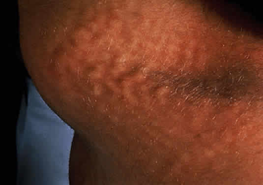
of conjunctiva and skin. Am J Ophthalmol 73:811, 1972

12. Berman E, Livni N, Shapira E et al: Congenital corneal clouding with abnormal systemic storage bodies: A new 
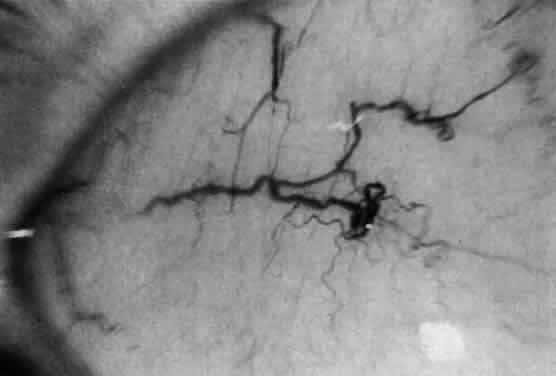
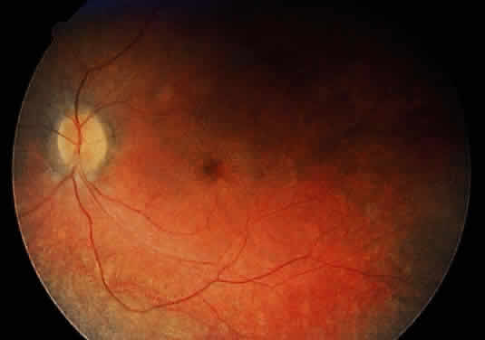
variant of mucolipidosis. J Pediatr 84:519, 1974

13. Donaldson M, Pennock C, Berry P et al: Syndrome with cardiomyopathy in infancy. J Pediatr 114:430, 1989

14. Rosen D, Haust M, Yamashita T et al: Keratoplasty and electron microscopy of the cornea in systemic mucopolysaccharidosis (Hurler's disease). Can J Ophthalmol 3:218, 1968

15. Kenyon K: Ocular ultrastructure of inherited metabolic disease. In Goldberg M (ed): Genetic and Metabolic Eye Disease, p 139. Boston: Little Brown, 1974

16. Gills P, Hobson R, Hanley B et al: Electroretinography and fundus oculi findings in Hurler's disease and allied mucopolysaccharidosis. Arch Ophthalmol 74:596, 1965

17. Nowaczyk M, Clarke J, Morin J: Glaucoma as an early complication of Hurler's disease. Arch Dis Child 63:1091, 1988

18. Spellacy E, Bankes J, Crow S et al: Glaucoma in a case of Hurler disease. Br J Ophthalmol 64:773, 1980

19. Duke-Elder S: Diseases of the Lens and Vitreous: System of Ophthalmology, p 172. London: Kimpton, 1969

20. Bach G, Friedman R, Weissmann B et al: The defect in the Hurler and Scheie syndromes: Deficiency of alpha-L-iduronidase. Proc Natl Acad Sci USA 69:2048, 1972

21. Scott H, Ashton L, Eyre H et al: Chromosomal localization of the human alpha-L-iduronidase gene (IDUA) to 4p16.3.Am J Hum Genet 47:802, 1990

22. Hopwood J, Vellodi A, Scott H et al: Long-term clinical progress in bone marrow transplanted mucopolysaccharidosis type I patients with a defined genotype. J Inherit Metab Dis 16:1024, 1993

23. Fairbairn L, Lashford L, Spooncer E et al: Long-term in vitro correction of alpha-L-iduronidase deficiency (Hurler syndrome) in human bone marrow. Proc Natl Acad Sci USA 93:2025, 1996

24. Scheie H, Hambrick GJ, Braner L: A newly recognized forme fruste of Hurler's disease (gargoylism): The Sanford R. Gifford lecture. Am J Ophthalmol 53:753, 1952

25. Quigley H, Goldberg M: Scheie syndrome and macular corneal dystrophy: An ultrasound comparison of conjunctiva and skin. Arch Ophthalmol 85:553, 1971

26. Tieu P, Bach G, Matynia A et al: Four novel mutations underlying mild or intermediate forms of alpha-L-iduronidase deficiency (MPS IS and MPS IH/S). Hum Mutat 6:55, 1995

27. Wilson P, Suthers G, Callen D et al: Frequent deletions at Xq28 indicate genetic heterogeneity in Hunter syndrome. Hum Genet 86:505, 1991

28. Al-Hazzaa S, Maumenee I, Traboulsi E: Ocular findings in adults with mucopolysaccharidosis II-B (Hunter syndrome mild). MEJO 1:15, 1993

29. Hobolth N, Pedersen C: Six cases of a mild form of the Hunter syndrome in five generations: Three affected males with progeny. Clin Genet 13:121, 1978

30. Spranger J, Cantz M, Gehler J et al: Mucopolysaccharidosis II (Hunter disease) with corneal opacities. Eur J Pediatr 129:11, 1978

31. Caruso R, Kaiser-Kupfer M, Muenzer J et al: Electroretinographic findings in the mucopolysaccharidoses. Ophthalmology 93:1612, 1986

32. Beck M: Papilloedema in association with Hunter syndrome. Br J Ophthalmol 67:174, 1983

33. Beck M, Cole G: Disc oedema in association with Hunters syndrome: Ocular histopatho-logical findings. Br J Ophthalmol 68:590, 1984

34. Braun S, Aronovich E, Anderson R et al: Metabolic correction and cross-correction of mucopolysaccharidosis type II (Hunter syndrome) by retroviral-mediated gene transfer and expression of human iduronate-2-sulfatase. Proc Natl Acad Sci USA 90:11830, 1993

35. Van de Kamp J, Niermeiyer M, Von Figura K, Giesberts M: Genetic heterogeneity and clinical variability in the Sanfilippo syndrome (types A, B, C). Clin Genet 20:152, 1981

36. Kaplan P, Wolfe L: Sanfilippo syndrome type D. J Pediatr 110:267, 1987

37. De Natale P: Sanfilippo B disease: A re-examination of a particular sibship after 12 years. J Inherit Metab Dis 14:23, 1991

38. Sanfilippo J, Podosin R, Langer L et al: Mental retardation associated with acid mucopolysacchariduria (heparitin sulfate type). J Pediatr 63:837, 1963

39. Scott H, Blanch L, Guo X-H et al: Cloning of the sulphamidase gene and identification of mutations in Sanfilippo A syndrome. Nature Genet 11:465, 1995

40. Zhao H, Li H, Bach G, Schmidtchen A et al: The molecular basis of Sanfilippo syndrome type B. Proc Natl Acad Sci USA 93:6101, 1996

41. Robertson D, Callen D, Baker E et al: Chromosomal localization of the gene for human glucosamine-6-sulphatase to 12q14. Hum Genet 79:175, 1988

42. Arbisser A, Donnelly K, Scott C Jr et al:Morquio-like syndrome with beta-galactosidase deficiency and normal hexosamine sulfatase activity: mucopolysaccharidosis IVB. Am J Med Genet 1:195, 1977

43. DiFerrante N, Ginsburg L, Donnelly P, Diferrante D, Caskey C: Deficiencies of glucosamine-6-sulfate or galactosamine-6-sulfate sulfatases are responsible for different mucopolysaccharidoses. Science 199:79, 1978

44. Holzgreve W, Grobe H, Von Figura K et al: Morquio syndrome: Clinical findings in 11 patients with MPS IV A and 2 patients with MPS IV B. Hum Genet 57:360, 1981

45. Sugar J: Corneal manifestations of systemic mucopolysaccharidoses. Ann Ophthalmol 11:531, 1979

46. Beck M, Glossl J, Grubisic A et al: Heterogeneity of Morquio disease. Clin Genet 29:325, 1986

47. Masuno M, Tomatsu S, Nakashima Y et al.Mucopolysaccharidosis IVA: assignment of the human N-acetylgalactosamine-6-sulfate sulfatase (GALNS) gene to chromosome 16q24. Genomics 16:777, 1996

48. Maroteaux P, Levesque B, Marie J, Lamy M: Une nouvelle dysotose avec elimination urinaire de chrondrotin-sulfate B. Presses Med 71:1849, 1963

49. O'Brien J, Cautz M, Spranger J: Maroteaux-Lamy disease (mucopolysaccharidosis VI): Subtype A: Deficiency of N-acetyl-galactosamine-4-sulfatase. Biochem Biophys Res Commun 60:1170, 1974

50. Young R, Kleinman G, Odemann R et al: Compressive myelopathy in Maroteaux-Lamy syndrome: Clinical and pathological findings. Ann Neurol 8:336, 1980

51. Goldberg M, Scott C, McKusick V: Hydrocephalus and papilledema in the Martoteaux-Lamy syndrome (MPS VI). Am J Ophthalmol 69:969, 1970

52. Schwartz MF, Werblin T, Green W: Occurrence of mucopolysaccharide in corneal grafts in Maroteaux-Lamy syndrome. Cornea 4:58,1985/1986

53. Naumann G: Clearing of cornea after perforating keratoplasty in mucopolysaccharidosis type VI (Maroteaux-Lamy syndrome). N Engl J Med 312:995, 1985

54. Litjens T, Baker E, Beckman K et al: Chromosomal localization of ARBS, the gene for human N-acetylgalactosamine-4-sulphatase. Hum Genet 82:67, 1989

55. Sly W, Quinton B, McAllister W, Rimoin D: B-glucoronidase deficiency: Report of clinical and radiological and biochemical features of a new MPS. J Pediatr 82:249, 1973

56. Ward J, Sharpe C, Luthardt F et al: Regional gene mapping of human beta-glucuronidase (GUSB) by dosage analysis: Assignment to region 7q11.23–7q21 (abstract). Am J Hum Genet 35:56A, 1983

57. Maumenee I: The eye in connective tissues diseases. In Daentl D (ed): Clinical, Structural and Biochemical Advances in Hereditary Eye Disorders, p 53. New York: Alan R. Liss, 1980

58. Spranger J, Wiedemann H: The genetic mucolipidoses. Diagnosis and differential diagnosis. Humangenetik 9:113, 1970

59. Kolodny E, Boustany R: Storage disease of reticular endothelial system. In Nathan D, Oski F (eds): Hematology of Infancy and Childhood, p 1212. Philadelphia: WB Saunders, 1987

60. Leroy J, Spranger J, Feingold M et al: I-Cell Disease: A Clinical Picture. J Pediatr 79:360, 1971

61. Tondeur M, Vamos-Hurwitz E, Mockel-Pohl S et al: Clinical biochemical and ultrastructured studies in a case of chondrodystrophy presenting the I-cell phenotype in tissue culture. J Pediatr 79:366, 1971

62. Leroy J, DeMars R: Mutant enzymatic and cytological phenotypes in cultured fibroblasts. Science 157:804, 1967

63. DeMars R, Leroy J: The remarkable cells cultured a human with Hurler's syndrome: An approach to visual selection for in vitro genetic studies. In Vitro 2:107, 1967

64. Okada S, Owada M, Sakiyama T et al: I-cell disease: Clinical studies of 21 Japanese cases. Clin Genet 28:207, 1985

65. Cipolloni C, Boldrini A, Donti E et al: Neonatal mucolipidosis II (I-cell disease): Clinical, radiological and biochemical studies in a case. Helv Paediatr Acta 35:85, 1980

66. Whelan D, Chang P, Cockshott P: Mucolipidosis II. The clinical, radiological and biochemical features in three cases. Clin Genet 24:90, 1983

67. Sprigz R, Doughty R, Spackman T et al: Neonatal presentation of I-cell disease. J Pediatr 93:954, 1978

68. Libert J, Van Hoof F, Farriaux J et al: Ocular findings in I-cell disease (mucolipidosis type II). Am J Ophthalmol 83:617, 1977

69. Mueller O, Wasmuth J, Murray J et al: Chromosomal assignment of N-acetylglucosaminylphosphotransferase, the lysosomal hydrolase targeting enzyme deficient in mucolipidosis II and III. Cytogenet Cell Genet 46:664, 1987

70. Ben-Yoseph Y, Mitchell D, Yager R et al: Mucolipidoses II and III variants with normal N-acetylglucosamine 1-phosphotransferase activity toward alpha-methylmannoside are due to nonallelic mutations. Am J Hum Genet 50:137, 1992

71. Freisinger P, Padovani JC, Maroteaux P: An atypical form of mucolipidosis III. J Med Genet 29:834, 1992

72. Taylor H, Thomas G, Miller C et al: Mucolipidosis III (Pseudo-Hurler polydystrophy): Cytological and ultrastructural observation of cultured fibroblast cells. Clin Genet 4:388, 1973

73. Spranger JW, Wiedemann HR. The genetic mucolipidoses. Diagnosis and differential diagnosis. Humangenetik 9:-113, 1970

74. Kelley T, Thomas G, Taylor H et al: Mucolipidosis III (Pseudo-Hurler polydystrophy): Clinical and laboratory studies in a series of 12 patients. Johns Hopkins Med J 137:156, 1975

75. Traboulsi E, Maumenee I: Ophthalmologic findings in mucolipidosis III. Am J Ophthalmol 102:529, 1986

76. Bach G: Mucolipidosis type IV. In Goodman R, Motulsky A (eds): Genetic Diseases Among Ashkenazi Jews, p 187. New York: Raven Press, 1979

77. Ben-Yoseph Y, Momoi T, Hahn L, Nadler H: Catalycally defective ganglioside neuraminidase in mucolipidosis IV. Clin Genet 21:374, 1982

78. Crandall B, Philippart M, Brown W et al: Mucolipidosis IV. Am J Med Genet 12:301, 1982

79. Bach G, Zeigler M, Kohn G: Mucopolysaccharide accumulation in cultured skin fibroblasts derived from patients with mucolipidosis IV. Am J Hum Genet 29:610, 1977

80. Bach G, Zeigler M, Schaap T et al: Mucolipidosis type IV: Ganglioside sialidase deficiency. Biochem Biophys Res Commun 9:1341, 1979

81. Kenyon K, Maumenee I, Green W et al: Mucolipidosis IV: Histopathology of conjunctiva, cornea and skin. Arch Ophthalmol 97:1106, 1979

82. Merin S, Livni N, Berman E et al: Mucolipidosis IV: Ocular systemic and ultrastructural findings. Invest Ophthalmol 14:437, 1975

83. Abraham F, Brand N, Blumenthal M et al: Retinal function in mucolipidosis IV. Ophthalmologica 191:210, 1985

84. Newell F, Matalon R, Meyer S: A new mucolipidosis with psychomotor retardation, corneal clouding and retinal degeneration. Am J Ophthalmol 80:440, 1975

85. McKusick V, Francomano C, Antonorakis S: Mendelian Inheritance in Man. Baltimore: Johns Hopkins University Press, 1994

86. Norden N, Lundblad A, Svensson S et al: A mannose-containing trisaccharide isolated from urines of three patients with mannosidosis. J Biol Chem 248:6210, 1973

87. Norden N, Lundblad A, Svensson S et al: Characterization of two mannose-containing oligosaccharides isolated from the urine of patients with mannosidosis. Biochemistry 13:871, 1974

88. Kaneda Y, Hayes H, Uchida T et al: Regional assignment of five genes on human chromosome 19. Chromosoma 95:8, 1987

89. Desnick R, Sharp H, Grabowski G et al: Mannosidosis: Clinical, morphologic, immunologic and biochemical studies. Pediatr Res 19:985, 1976

90. Warner T, Mock A, Nyhan W et al: Alpha-mannosidosis, analysis of urinary oligosaccharides with high performance liquid chromatography and diagnosis of a case with unusually mild presentation. Clin Genet 25:248, 1984

91. Chester M, Lundblad A, Ockerman P et al: Mannosidosis. In Durand P, O'Brien J (eds): Genetic Errors of Glycoprotein Metabolism, p 89. Berlin: Springer-Verlag, 1982

92. Spranger J, Gehler J, Cantz M: The radiographic features of mannosidosis. Radiology 119:401, 1976

93. Yunis J, Lewandowski R, Sanfilippo S et al: Clinical manifestations of mannosidosis—a longitudinal study. Am J Med 61:841, 1976

94. Murphree A, Beaudet A, Palmer E et al: Cataract in mannosidosis. Birth Defects 12:319, 1976

95. Maire I, Zabot M, Mathieu M et al: Mannosidosis tissue culture studies in relation to prenatal diagnosis. J Inherit Metab Dis 1:19, 1978

96. Fukuda M, Tanaka A, Isshiki G: Variation of lysosomal enzyme activity with gestational age in chorionic villi. J Inherit Metab Dis 13:862, 1990

97. Willems P, Gatti R, Darby J et al: Fucosidosis revisited: A review of 77 patients. Am J Med Genet 38:111, 1991

98. Willems P, Garcia C, De Smedt M et al: Intrafamilial variability in fucosidosis. Clin Genet 34:7, 1988

99. Durand P, Borrone G, Della Cella G: Fucosidosis. J Pediatr 75:665, 1969

100. Michalski J, Wieruszeski J, Alonson C et al: Characterization and 400-MHZ IH-NMR analysis of urinary fucosyl glycoasparagines in fucosidosis. Eur J Biochem 201:439, 1991

101. Cook P, Noades J, Newton M et al: On the orientation of the Rh:E1-1 linkage group. Ann Hum Genet 41:157, 1977

102. Snodgrass M: Ocular finding in a case of fucosidosis. Br J Ophthalmol 60:508, 1976

103. Smith E, Graham J, Ledman J et al: Fucosidosis. Cutis 19:195, 1977

104. Dvoretzky I, Fisher B: Fucosidosis. Intl J Dermatol 18:213, 1979

105. Spranger J, Cantz M. Mucolipidosis I, the cherry-red spot-myoclonus syndrome and neuraminidase deficiency. Birth Defects Orig Art Ser 14:105, 1978

106. Kelley T: The mucopolysaccharidosis and mucolipidosis. Clin Orthop 114:116, 1976

107. Cantz M, Gehler J, Spranger J: Mucolipidosis I: Increased sialic acid content and deficiency of an alpha-N-acetylneuraminidase in cultured fibroblasts. Biochem Biophys Res Commun 74:732, 1977

108. Federico A, Cecio A, Battini G et al: Macular cherry red spot and myoclonus syndrome: Juvenile form of sialidosis. J Neurol Sci 48:157, 1980

109. Rapin I, Goldfischer S, Katzman R et al: The cherry-red-spot-myoclonus syndrome. Ann Neurol 3:234, 1978

110. Mueller O, Henry W, Haley L et al: Sialidosis and galactosialidosis: chromosomal assignment of two genes associated with neuraminidase deficiency disorders. Proc Natl Acad Sci USA 83:1817, 1985

111. Thomas G, Tipton R, Ch'ien L et al: Sialidosis (alpha-N-acetylneuraminidase) deficiency: The enzyme defect in an adult with macular cherry-red spots and myoclonus without dementia. Clin Genet 13:369, 1978

112. Durand P, Gattlie R, Cavalier S et al: Sialidosis (mucolipidosis I). Helv Acta Paediatr 32:391, 1977

113. Thomas P, Abrams J, Swallow D et al: Sialidosis type 1: Cherry red spots myoclonus syndrome with sialidase deficiency and altered electrophoretic mobilities of some enzymes known to be glycoproteins. J Neurol Neurosurg Psychiatr 42:873, 1979

114. Spranger J, Cantz M: Mucolipidosis I, the cherry red spot myoclonus syndrome and neuraminidase deficiency. Birth Defects Orig Art Ser 14:105, 1978

115. Aula P, Autio S, Ravio K et al: Aspartylglucosaminuria. In Durand P, O'Brien J (eds): Genetic Errors of Glycoprotein Metabolism, p 123. Berlin: Springer-Verlag, 1982

116. Morris C, Heisterkamp N, Groffen J et al: Chrosomal localization of the human glycoasparaginase gene to 4q32–q33. Hum Genet 88:295, 1992

117. Autio S: Aspartylglycosaminuria: Analysis of thirty-four patients. J Men Def Res Monogr Ser 1:1, 1972

118. Maury C, Palo J: N-Acetylglucosamine-asparagine levels in tissues of patients with aspartyglycosaminuria. Clin Chim Acta 108:293, 1981

119. Desnick R, Wang A: Schindler disease: An inherited neuroaxional dystrophy due to alpha-N-acetylgalactosaminidase deficiency. J Inherit Metab Dis 13:549, 1990

120. deGrout P, Westerveld A, Khan P, Tager J: Localization of a gene for human α-galactosidase B on chromosome 22 (n-acetyl-alpha-d-glucosaminidase). Hum Genet 44:305, 1978

121. Schindler D, Bishop D, Wolfe D et al: Neuroaxonal dystrophy due to lysosomal alpha-N-acetylgalactosaminidase deficiency. N Engl J Med 320:1375, 1989

122. Desnick R, Bishop D: Fabry disease: Alpha-N-acetylgalactosaminidase deficiency. In Stanbury J, Wyngaarden J, Frederickson D et al (eds): The Metabolic Basis of Inherited Disease, p 751. New York: McGraw-Hill, 1989

123. Niemann A: Ein unbekanntes Krankheitsbild. Jahrb Kinderheilkd 79:1, 1914

124. Pick L: Uber die lipoidzellige Splenohepatomegalie Typus Niemann-Pick als Stoffwechselerkrankung. Med Klin 23:1483, 1927

125. Carstea E, Polymeropoulos M, Parker C et al: Linkage of Niemann-Pick disease type C to human chromosome 18. Proc Natl Acad Sci USA 90:2002, 1993

126. Crocker A: The cerebral defect in Tay-Sachs disease and Niemann-Pick disease. J Neurochem 7:69, 1961

127. Brady R, Kanfer J, Mock M et al: The metabolism of sphingomylin: II. Evidence of enzymatic deficiency in Niemann Pick disease. Proc Natl Acad Sci USA 55:366, 1966

128. Poulous A, Shankaran P, Jones C et al: Enzymatic hydrolysis of sphingomyelin lysosomes by normal tissues and tissues from patients with Niemann-Pick disease. Biochem Biophys Acta 751:428, 1983

129. Vanier M, Rousson R, Garcia I et al: Biochemical studies in Niemann-Pick disease: III. In vitro and in vivo assays of sphingomyelin degradation in cultured skin fibroblasts and amniotic fluid cells for diagnosis of the various forms of the disease. Clin Genet 27:20, 1985

130. Brady R: The abnormal biochemistry of inherited disorders of lipid metabolism. Fed Proc 32:1660, 1973

131. Sloan HR, Uhlendorf BW, Kanfer JN et al: Deficiency of sphingomyelin-cleaving enzyme activity in tissue cultures derived from patients with Niemann-Pick disease. Biochem Biophys Res Comm 34:582, 1969

132. Liscum L, Dahl N: Intracellular cholesterol transport. J Lipid Res 33:1239, 1992

133. Brady R: Sphingomyelin lipidosis: Niemann-Pick disease. In Stanbury J, Wyngaarden J, Frederickson D et al (eds): The Metabolic Basis of Inherited Disease, p 831. New York: McGraw Hill, 1983

134. Goodman R: Genetic Disorders Among Jewish People. Baltimore: Johns Hopkins University Press, 1979

135. Cogan D, Kuwabara T: The sphingolipidosis and the eye. Arch Ophthalmol 79:437, 1968

136. Robb R, Kuwabara T: The ocular pathology of type A Niemann-Pick disease: A light and electron microscopic study. Invest Ophthalmol 12:366, 1973

137. Walton D, Robb R, Crocker A: Ocular manifestations of group A Niemann-Pick disease. Am J Ophthalmol 85:-174, 1978

138. Cogan D, Chu F, Barranger J et al: Macula halo syndrome: Variant of Niemann-Pick disease. Arch Ophthalmol 101:-1698, 1983

139. Grunebaum M: The roentogenographic findings in the acute neuronapathic form of Niemann-Pick disease. Br J Radiol 49:1018, 1976

140. Harzer K, Ruprecht K, Seuffer-Schulze D et al: Morbus Niemann-Pick type B-enzymatisch gesichert-mot unerwarteter retinaler Beterligung. Graefes Arch Clin Exp Ophthalmol 206:79, 1973

141. Lipson M, O'Donnell J, JW C et al: Ocular involvement in Niemann-Pick disease type B. J Pediatr 108:582, 1986

142. Hammersen G, Oppermann H, Harms E et al: Oculo-neural involvement in an enzymatically proven case of Niemann-Pick disease type B. Eur J Pediatr 132:77, 1979

143. Al-Hazzaa S, Ozand P: Niemann Pick disease type B. Middle East J Ophthalmol 3:30, 1995

144. Matthews J, Weiter J, Kolodny E: Macula halos associated with Niemann-Pick type B disease. Ophthalmology 93:-933, 1986

145. Crocker A, Farber S: Niemann-Pick disease. A review of eighteen patients. Medicine 37:1, 1958

146. Elleder M, Jirasek A: International Symposium Niemann Pick disease. Eur J Pediatr 140:1190, 1983

147. Besley G, Hoogeboom A, Hoogeveen A et al: Somatic cell hybridisation studies showing different gene mutations in Niemann-Pick variants. Hum Genet 54:409, 1980

148. Wenger D, Barth D, Githens J: Nine case of sphingomyelinlipidosis, a new variant in Spanish-American children, juvenile variants of Niemann-Pick disease with foamy and sea blue hitiocytes. Am J Dis Child 131:955, 1977

149. Longstreth W, Daven J, Farrell D et al: Adult dystonic lipidosis: Clinical histologic and biochemical findings of a neurovisceral storage disease. Neurology 32:1295, 1982

150. Vanier M, Rodrigues-La Frasse C, Rousson R et al: Type C Niemann-Pick disease spectrum of phenotype variation in disruption of ultracellular LDL derived cholesterol processing. Biochom Biophys Acta 1096:328, 1991

151. Cogan D, Chu F, Bachman D et al: The DAF syndrome. Neuroophthalmology 2:7, 1981

152. Pentechev P, Comly M, Kruth H et al: A defect in cholestrol esterification in Niemann-Pick disease type C patients. Proc Natl Acad Sci USA 82:8247, 1985

153. Argoff C, Kaneski C, Blanchette-Mackie E et al: Type C Nieman Pick disease: Documentation of abnormal LDL processing in lymphocytes. Biochem Biophys Res Commun 171:38, 1990

154. Neville B, Lake B, Stephens R et al: A neurovisceral storage disease with vertical supranuclear ophthalmoplegia and its relationship to Niemann-Pick disease: A report of nine patients. Brain 96:97, 1973

155. Dunn H, Sweeney V: Progressive supranuclear palsy in an unusual juvenile variant of Niemann-Pick disease (abstract). Neurology 21:442, 1971

156. Grover W, Naiman J: Progressive paresis of vertical gaze in lipid storage disease. Neurology 21:896, 1971

157. Butler J, Comly M, Kruth H et al: Niemann-Pick variant disorders: Comparison of errors of cellular cholesterol hemostasis in group D and group C fibroblasts. Proc Natl Acad Sci USA 84:556, 1987

158. Byers D, Rastogi S, Cook H et al: Defective activity of acyl-CoA: Cholesterol O-acetyl transferase in Niemann-Pick type C and type D fibroblasts. Biochem J 262:713, 1989

159. Fredrickson D, Sloan H: Sphyngomyelin lipidosis: Niemann-Pick disease. In Stanbury J, Wyngaarden J, Fredrickson D (eds): The Metabolic Basis of Inherited Disease, p 1655. New York: McGraw-Hill, 1989

160. Schneider E, Pentchev P, Hibbert S et al: A new form of Niemann-Pick disease characterized by temperature labile sphingomyelinase. J Med Genet 15:370, 1978

161. Opitz J, Stiles F, Wise D et al: The genetics of angiokeratoma corporis diffusum (Fabry's disease) and its linkage with Xg (a) locus. Am J Hum Genet 17:325, 1965

162. Kent J: Fabry's disease, alpha-galactosidase deficiency. Science 167:1268, 1970

163. Johnson D, Del Monte M, Collier E et al: Fabry disease: Diagnosis by alpha-galactosidase activity in tears. Clin Chem Acta 63:81, 1975

164. Sorensen S, Hasholt L: Attitudes of persons at risk for Fabry's disease toward predictive and genetic counselling. J Biosoc Sci 15:89, 1983

165. Brady R, Gal A, Bradley R et al: Enzymatic defect in Fabry's disease: Ceramide trihexosidase deficiency. N Engl J Med 276:1163, 1967

166. Lockman L, Hunninghake D, Krivit W et al: Relief of pain of Fabry's disease by diphenylhydantoin. Neurology 23:871, 1973

167. Matsui S, Murakami E, Takekoshi N: Cardiac manifestations of Fabry's disease. Nippon Junkankigakushi 41:-1023, 1977

168. Terlinde R, Richard G, Lisch W et al: Ruckbildung der Cornea Verticillata bei Morbus-Fabry durch kontaktlinsen erste Beobachtungen. Contactologia 4:20, 1982

169. Weicksel J: Angiomatosis, bzw, Angiokeratosis universalis (eine sehr seltene Haut-und-Gaffaskrankheit). Dtsch Med Wochensch 51:898, 1925

170. Gruber H: Cornea verticillata. Ophthalmologica 111:120, 1946

171. Sher N, Letson R, Desnick R: The ocular manifestations in Fabry's disease. Arch Ophthalmol 97:671, 1979

172. Spaeth G, Frost P: Fabry's disease: Its ocular manifestations. Arch Ophthalmol 74:760, 1965

173. Desnick R, Sweeley C: Prenatal detection of Fabry disease. In Dorfman A (ed): Antenatal Diagnosis, p 185. Chicago: University of Chicago Press, 1971

174. Mayes J, Scheerer J, Sifers R, Donaldson M: Differential assay for lysosomal alpha-galactosidases in human tissues and its application to Fabry's disease. Clin Chim Acta 112:247, 1981

175. Font R, Fine B: Ocular pathology in Fabry's disease, histochemical electron microscopic observation. Am J Ophthalmol 73:419, 1972

176. Kleijer W, Hussaarts-Odijk L, Sacks E et al: Prenatal diagnosis of Fabry's disease by direct analysis of chorionic villi. Prenat Diagn 7:283, 1987

177. Brill N, Mandelbaum F, Libman F: Primary splenomegaly- Gaucher type: Report on one of few cases occurring in a single generation of one family. Am J Med Sci 129:491, 1905

178. Fried K: Population study of chronic Gaucher's disease. Isr J Med Sci 9:1396, 1973

179. Shafti-Zagardo B, Devine E, Smith M et al: Assignment of the gene for acid beta glucosidase to human chromosome 1. Am J Hum Genet 33:564, 1981

180. Fredrickson S, Sloan H: Glucosyl ceramide lipidosis: Gaucher's disease. In Stanbury J, Wyngaarden J, Frederickson D (eds): The Metabolic Basis of Inherited Disease, p 730. New York: McGraw-Hill, 1972

181. Petrohelos M, Tricoulis D, Kotsiras I et al: Ocular manifestation of Gaucher's disease. Am J Ophthalmol 80:1006, 1975

182. Chu F, Rodrigues M, Cogan D et al: The pathology of pingueculae in Gaucher's disease. Ophthalm Pediatr Genet 4:7, 1983

183. Kolodny E, Ullman M, Mankin H et al: Phenotypic manifestations of Gaucher's disease. Clinical features in 48 biochemically verified type I patients and comments on type II patients. In Desnick R, Gott S, Grabowski G (eds): Gaucher Disease: A Century of Delineation and Research, p 33. New York: Alan R. Liss, 1982

184. Wenger D, Roth S, Kudoh T et al: Biochemical studies in a patient with subacute neuropathic Gaucher disease without visceral glucosylceramide storage. Pediatr Res 17:344, 1983

185. Erickson A: Gaucher disease, Norrbottnian Type III: Neuropediatric and neurological aspects of clinical patterns and treatment. Acta Paediatr Scand 326:7, 1986

186. Abrahamov A, Elstein D, Gross-Tsur V et al: Gaucher's disease variant characterised by progressive calcification of heart valves and unique genotype. Lancet 346:1000, 1995

187. Chabas A, Cormand B, Grinberg D et al: Unusual expression of Gaucher's disease: Cardiovascular calcifications in three sibs homozygous for the D409H mutation. J Med Genet 32:740, 1995

188. Beutler E, Kuhl W, Trinidad F et al: Beta glucosidase activity in fibroblasts in homozygotes and heterozygotes for Gaucher's disease. Am J Hum Genet 23:62, 1971

189. Rappeport J, Ginns E: Bone marrow transplantation in severe Gaucher's disease. N Engl J Med 311:84, 1984

190. Von Hirsch T, Peiffer J: Uber histologische Methoden in der differential Diagnose von Leukodystrophien und Lipoidosen. Arch Psychiatr Nervenker 194:88, 1955

191. Peiffer J: Uber die Metachromatischen leukodystrophien. Arch Psychiatr Nerverkr 199:386, 1959

192. Austin J, Armstrong D, Shearer L: Metachromatic form of diffuse cerebral sclerosis: V. The nature and significance of low sulfatase activity, a controlled study of brain, liver and kidney in four patients with metachromatic leukodystrophy (MLD). Arch Neurol 13:593, 1965

193. Austin J, McAfee D, Shearer L: Metachromatic form of diffuse cerebral sclerosis: IV. Low sulfatase activity in the urine of nine living patients with metachromatic leukodystrophy (MLD). Arch Neurol 12:447, 1965

194. Geurts van Kessel A, Westerveld A, de Groot P et al: localization of the genes coding for human ACO2, ARSA, and NAGA on chromosome 22. Cytogenet Cell Genet 28:169, 1980

195. DeSilva K, Pearce J: Neuropathy of metachromatic leucodystrophy. J Neurol Neurosurg Psychiatr 36:30, 1973

196. Hagberg B: The clinical diagnosis of Krabbe's infantile leucodystrophy. Acta Pediatr Scand 52:213, 1963

197. Cogan D, Kuwabara T, Moser H: Metachromatic leucodystrophy. Ophthalmol 160:2, 1970

198. Libert J, Van Hoof F, Toussaint D et al: Ocular findings in metachromatic leukodystrophy: An electron microscopic and enzyme study in different clinical and genetic variants. Arch Ophthalmol 97:1495, 1979

199. Goebel H, Shimokawa K, Argyrakis A et al: The ultrastructural of the retina in adult metachromatic leukodystrophy. Am J Ophthalmol 85:841, 1978

200. Goebel H, Busch H: Abnormal lipopigments and lysosomal residual bodies in metachromatic leukodystrophy. Adv Exp Med Biol 266:299, 1989

201. Gordon N: The insidious presentation of the juvenile form of MLD. Postgrad Med J 54:335, 1978

202. McKhann G: Metachromatic leukodystrophy: Clinic and enzymatic parameters. Neuropediatrics 15:4, 1984

203. Carlin L, Roach E, Riela A et al: Juvenile metachromatic leukodystrophy: Evoked potentials and computed tomography. Ann Neurol 13:105, 1983

204. Wulff C, Trojaborg W: Adult metachromatic leukodystrophy: Neurophysiologic findings. Neurology 35:1776, 1985

205. Kappler J, Von Figura K, Gieselmann V: Late onset metachromatic leukodystrophy: Molecular pathology in two siblings. Ann Neurol 31:256, 1992

206. Bosch E, Hart M: Late adult onset metachromatic leukodystrophy: Dementia and polyneuropathy in 63 year old man. Arch Neurol 35:475, 1978

207. Brismar J: CT and MRI of the brain in inherited neurometabolic disorders. J Child Neurol 7:112, 1992

208. Tagliavini F, Pietrini V, Pilleri G et al: A case report: Adult metachromatic leucodystrophy, clinicopathological report of two familial cases with slow course. Neuropathol Appl Neurobiol 5:233, 1979

209. Austin J, Armstrong D, Fouch S et al: Metachromatic leukodystrophy (MLD). VIII. MLD in adults: Diagnosis and pathogenesis. Arch Neurol 18:225, 1968

210. Dayan A: Dichroism of cresyl violet-stained cerebroside sulfate (“Sulfatide”). J Histochem Cytochem 15:421, 1967

211. Takahashi K, Naito M: Lipid storage disease: Part II. Ultrastructural pathology of lipid storage cells in sphingolipidoses. Acta Pathol Jap 35:385, 1985

212. Markiewicz D, Adamczewska-Goncerzewicz Z, Zelman I et al: A case of MLD with a chronic course (clinical-morphological-biochemical study). Neuropathol Pol 16:233, 1978

213. Raghavan S, Gajewski A, Kolodny E: Leukocyte sulfatidase for reliable diagnosis of MLD. J Neurochem 36:724, 1981

214. Kudeh T, Wenger D: Diagnosis of MLD after uptake of fatty acid-labeled cerebroside sulfate into cultured skin fibroblasts. J Clin Invest 70:89, 1982

215. Molzer B, Sundt-Heller R, Kainz-Kraschinsky M et al: Elevated sulfatide excretion in heterozygotes of metachromatic leukodystrophy: Dependence of arylsulfatase A activity. Am J Med Genet 44:523, 1992

216. Suzuki Y, Suzuki K: Krabbe's globoid cell leukodystrophy: Deficiency of galactocerebrosidase in serum, leukocyte and fibroblasts. Science 171:73, 1971

217. Zlotogora J, Charkraborty S, Knowleton R et al: Krabbe disease locus mapped to chromosome 14 by genetic linkage. Am J Hum Genet 47:37, 1990

218. Krabbe K: A new familial, infantile form of diffuse brain sclerosis. Brain 39:74, 1916

219. Collier J, Greenfield J: The encephalitis periaxialis of Schilder: A clinical and pathological study with an account of two cases one of which was diagnosed during life. Brain 47:489, 1924

220. Hagberg B, Kollbberg H, Sourander P et al: Infantile globoid cell leukodystrophy (Krabbe's disease): A clinical and genetic study of 32 Swedish cases. Neuropaediatrica 1:74, 1970

221. Dunn H, Lake B, Dolman D et al: The neuropathy of Krabbe's infantile cerebral sclerosis (globoid cell leukodystrophy). Brain 92:329, 1969

222. Suzuki K, Grover W: Krabbe's leukodystrophy (globoid cell leukodystrophy): An ultrastructural study. Arch Neurol 22:385, 1970

223. Laxdal T, Hallgrimsson K: Krabbe's globoid cell leucodystrophy with hydrocephalus. Arch Dis Child 49:232, 1974

224. Naidu S, Hofmann K, Moser H et al: Galactosylceramidase beta-galactosidase deficiency in association with cherry-red spot. Neuropediatrics 19:46, 1988

225. Phelps M, Aicardi J, Vanier M: Late onset Krabbe's leukodystrophy, a report of four cases. J Neurol Neurosurg Psychiatr 54:293, 1991

226. Poser C, van Bogaert L: Natural history and evolution of the concept of Schindler's diffuse sclerosis. Acta Psychiatr Scand 31:285, 1956

227. Loonen M, Van Diggelen O, Janse H et al: Late onset globoid cell leucodystrophy (Krabbe's disease): Clinical and genetic delineation of two forms and their relation to the early infantile form. Neuropediatrics 16:137, 1985

228. Basner R, von Figura R, Glossi J et al: Multiple deficiency of mucopolysaccharides sulfatases in mucosulfatidosis. Pediatr Res 13:1316, 1979

229. Fiddler M, Vine D, Shapira E et al: Is multiple sulfatase deficiency due to defective regulation of sulphydrolase expression. Nature 282:98, 1979

230. Soong B, Cassamassima A, Fink J et al: Multiple sulfatase deficiency. Neurology 38:1273, 1988

231. Burk R, Valle D, Thomas G et al: Early manifestations of multiple sulfatase deficiency. J Pediatr 104:574, 1984

232. Hogan K, Matalon R, Berlow S et al: Multiple sulfatase deficiency: Clinical radiological electrophysiologic and biochemical features. Neurology 33:245, 1983

233. Bateman J, Phillipart M, Isenberg S: Ocular features of multiple sulfatase deficiency and a new variant of metachromatic leukodystrophy. J Pediatr Ophthalmol Strabismus 21:133, 1984

234. Harbord M, Guncic J, Chuang S et al: Multiple sulfatase deficiency with early severe retinal degeneration. J Child Neurol 6:229, 1991

235. Al-Aqeel A, Ozand P, Brismar J et al: Saudi variant of multiple sulfatase deficiency. J Child Neurol 7:512, 1992

236. Al-Hazzaa S, Ozand P, Maumenee I: Saudi variant of ocular multiple sulfatase deficiency (Austin Disease). Presented at Centennial Annual Meeting of American Academy of Ophthalmology, Chicago, 1996

237. Samuelsson K, Zetterstrom R: Ceramidies in a patient with lipogranulomatosis (Farber's disease) with chronic course. Scand J Clin Lab Invest 27:393, 1971

238. Sugita M, Dulaney J, Moser H: Ceramidase deficiency in Farber's disease. Science 178:1100, 1972

239. Moser H, Moser A, Chen W et al: Ceramidase deficiency. Farber's disease. In Stanbury J, Wyngaarden J, Fredrickson D et al (eds): The Metabolic Basis of Inherited Disease, p 820. New York: McGraw-Hill, 1983

240. Cogan D, Kuwabara T, Moser H et al: Retinopathy in a case of Farber's lipogrannulomatosis. Arch Ophthalmol 75:752, 1960

241. Momoi T, Ben-Yoseph Y, Nadler H: Substrate-specific of acid and alkaline ceramidase in fibroblasts from patient with Farber's disease and control. Biochem J 205:419, 1982

242. Fensom A, Benson P, Neville B et al: The prenatal diagnosis of Farber's disease. Lancet 2:990, 1979

243. Kerr C, Wells R: Sex-linked ichthyosis. Ann Hum Genet 29:33, 1965

244. Sever R, Frost P, Wienstein G: Eye changes in ichtyosis. JAMA 206:2283, 1968

245. Costagliola C, Fabbrocini G, Illiano G et al: Ocular findings in X-linked ichthyosis: A survey on 38 cases. Ophthalmologica 202:152, 1991

246. Casaroli-Morano R, Ortiz-Stradtmann M, Uxo M et al: Ocular findings associated with congenital x-linked ichthyosis. Ann Ophthalmol 23:167, 1991

247. Okada S, O'Brien J: Generalized gangliosidosis: Beta—galactosidase deficiency. Science 160:1002, 1968

248. Takano T, Yamanouchi Y: Assignment of human beta-galactosidase-A-gene to 3p21.33 by fluorescence in situ hybridization. Hum Genet 92:403, 1993

249. O'Brien J: Generalized gangliosidosis. J Pediatr 75:167, 1969

250. Gonatas N, Gonatas J: Ultrastructural and biochemical observation on a case of systemic late infantile lipidosis and its relationship to Tay-Sachs disease and gargoylism. J Neuropathol Exp Neurol 24:318, 1965

251. Lowden JA, Callahan JW, Norman MG et al: Juvenile GM1 gangliosidosis: Occurrence with absence or two beta-galactosidase components. Arch Neurol 31:200, 1974

252. Derry D, Fawcett J, Andermann F et al: Late infantile systemic lipidosis major monosialoganglisidosis: Delineation of 2 types. Neurology 18:340, 1968

253. Hooft C, Vlietinck R, Dacremont G et al: GM1 gangliosidosis type II. Eur Neurol 4:1, 1970

254. Suzuki Y, Nakamura N, Fukuoka Shimada F et al: Beta-galactosidase deficiency in jjuvenile and adult patients: Report of six Japanese cases and review of literature. Hum Genet 36:219, 1977

255. Wenger D, Tarby T, Wharton C: Macular cherry-red spots and myoclonus with dementia: Co-existent neuraminidase and beta-galactosidase deficiencies. Biochem Biophy Res Commun 82:589, 1978

256. d'Azzo A, Hoogeveen A, Reuser A et al: Molecular defect in combined beta-galactosidase and neuraminidase deficiency in man. Proc Natl Acad Sci USA 79:4535, 1982

257. Gravel R, Lowden J, Callahan J et al: Infantile sialidosis: A phenocopy of type 1 GM1 gangliosidosis distinguished by genetic complementation and urinary oligosaccharides. Am J Hum Genet 31:669, 1979

258. Lowden J, Cutz E, Skomorowski M: Infantile type 2 sialidosis with beta-galactosidase deficiency. In Tetlamanti G, Durand P, Di Donato S (eds): Sialidase and Sialidosis, p 261. Milan: Edi Ermes, 1981

259. Andria G, Strisciuglio P, Pontgarelli G et al: Infantile neuraminidase and beta-galactosidase deficiencies (galactosialidosis) with mild clinical courses. In Durand P, Tetlamanti G, Di Donato S (eds): Sialidases and Sialidosis, p 379. Milan: Edi Ermes, 1981

260. Strisciuglio P, Sly W, Dodson W et al: Combined deficiency of beta-galactosidase and neuraminidase: Natural history of the disease in the first 18 years of an American patient with late infantile onset form. Am J Med Genet 37:573, 1990

261. Suzuki Y, Nanba E, Tsuj A et al: Clinical and genetic hertogeneity in galactosialidosis. Brain Dysfunction 1:285, 1988

262. Suzuki Y, Sakuraba H, Yamanka T et al: Galactosialidosis: A comparative study of clinical and biochemical data on 22 patients. In Arima M, Suzuki Y, Yabuuchi H (eds): The Developing Brain and Its Disorders, p 161. Tokyo: University of Tokyo, 1984

263. Kleijer W, Hoogeveen A, Verheijen F et al: Prenatal diagnosis of sialidosis with combined neurominidase and B-galactosidase deficiency. Clin Genet 16:60, 1979

264. Takeda K, Nakai H, Hagiwara H et al: Fine assignment of beta hexosaminidase A alpha subunit on 15q23–q24 by high resolution in situ hybridization. Tohoku J Exp Med 160:203, 1990

265. Fox M, Du Toit D, Warnich L et al: Regional localization of alpha-galactosidase (GLA) to Xpter-q22, hexosaminidase B (HEXB) to 5q13-qter and arylsulfatase B (ARSB) to 5pter-q13. Cytogenet Cell Genet 38:45, 1984

266. Swallow D, Islam I, Fox M et al: Regional localization of the gene coding for the GM2 activator protein (GM2A) to chromosome 5q32–33 and confirmation of the assignment of GM2AP to chromosome 3. Ann Hum Genet 57:187, 1993

267. Tay W: Symmetrical changes in the region of the yellow spot in each eye of an infant. Trans Ophthalmol Soc UK 1:155, 1881

268. Sachs B: A family form of idiocy, generally fatal, associated with early blindness. J Nerv Ment Dis 21:475, 1896

269. Kivlin J, Snaborn G, Myers G: The cherry red spot in Tay-Sachs and other storage diseases. Ann Neurol 17:356, 1958

270. Collins T: Cited in Kingdom EC: A rare fatal disease of infancy with symmetrical changes at the macula lutea. Trans Ophthalmol Soc UK 12:126, 1892

271. Cogan D, Kuwabara T: Histochemistry of the retina in Tay-Sachs disease. Arch Ophthalmol 61:414, 1959

272. Sandhoff K, Andreae U, Jatzkewitz H: Deficient hexosamidase activity in an exceptional case of Tay-Sachs disease with additional storage of kidney globoside in visceral organs. Life Sci 7:283, 1968

273. Sandhoff K, Harzer K, Wassle W et al: Enzyme alterations and lipid storage in three variants of Tay-Sachs disease. J Neurochem 18:2469, 1971

274. Strecker G, Herlant-Peers M, Fournet B et al: Structure of seven oligosaccharides excreted in the urine of a patient with Sandhoff's disease (GM2 gangliosidosis variant O). Eur J Biochem 81:165, 1977

275. Tremblay M, Szots F: GM2 type 2-gangliosidosis (Sandhoff's disease): Ocular and pathological manifestations. Can J Ophthalmol 9:338, 1974

276. Brownstein S, Carpenter S, Polomeno R et al: Sandhoff's disease (GM2 gangliosidosis type 2): Histopathology and ultrastructure of the eye. Arch Ophthalmol 98:1089, 1980

277. Hechtman P, Gordon B, Na Ying Kin N: Deficieny of the hexosaminidase A activator protein in a case of GM2 gangliosidosis, variant AB. Pediatr Res 16:217, 1982

278. Burg J, Conzelmann E, Sandhoff K et al: Mapping of the gene coding for the human GM2 activator protein to chromosome 5. Ann Hum Genet 49:41, 1985

279. Kolodny E, Wald I, Moser H et al: GM2 gangliosidosis without deficiency in the artificial substrate cleaving activity of hexosaminidase A and B. Neurology 23:427, 1973

280. Von Gierke E: Hepato-nephro-megalia glycogenica (Glykogenspeicher-Krankheit der Leber und Nieren). Beitr-Pathol Anat 82:497, 1929

281. Cori G, Cori C: Glucose-6-phosphatase of the liver in glycogen storage disease. J Biol Chem 199:661, 1952

282. Lei K, Pan C, Shelly L et al: Identification of mutations in the gene for glucose-6-phosphatase, the enzyme deficient in glycogen storage disease type 1a. J Clin Invest 93:1994, 1994

283. Fine R, Wilson W, Donnell G: Retinal changes in glycogen storage disease type 1. Am J Dis Child 115:328, 1968

284. Hayasaka S, Noda S, Fujii M et al: Inverted eyelashes in patients with type Ia glycogen storage disease. Graefes Arch Clin Exp Ophthalmol 227:209, 1989

285. Golbus M, Simpson T, Koresawa M et al: The prenatal determination of glucose-6-phosphatase activity by fetal liver biopsy. Prenat Diagn 8:401, 1988

286. Sparkes R, Sparkes M, Funderburk S, Moedjono S: Expression of galactose-1-P uridyltransferase in patients with chromosome alterations affecting 9p: assignment of the locus to p11–22 (abstract). Cytogenet Cell Genet 25:209, 1979

287. Elsevier S, Kucherlapati R, Nichols E et al: Assignment and regional localization of a gene coding for galactokinase to human chromosome 17q21–22. Cytogenet Cell Genet 14:287, 1975

288. Lin M, Oizumi J, Ng W et al: Regional mapping of the gene for human UDPGal 4-epimerase on chromosome 1 in mouse-human hybrids. Cytogenet Cell Genet 24:217, 1979

289. Mason H, Turner M: Chronic galactosemia. Am J Dis Child 50:359, 1935

290. Nadler H, Inouye T, Hsia D: Clinical galactosemia: A study of 55 cases. In Itsia D (ed): Galactosemia, p 127. Springfield, IL: Charles C. Thomas, 1969

291. Levy N, Krille A, Beutler E: Galactokinase deficiency and cataracts. Am J Ophthalmol 74:41, 1972

292. Beutler E, Matsumoto F, Krille W et al: Galactokinase deficiency as a cause of cataracts. N Engl J Med 288:-1203, 1973

293. Skalka H, Prchal J: Presenile cataracts formation and decreased activity of galactosemic enzymes. Arch Ophthalmol 98:269, 1980

294. Elman M, Miller M, Matalon R: Galactokinase activity in patients with idiopathic cataracts. Ophthalmology 93:210, 1986

295. Levy N: Etiology and management of infantile cataracts. Ophthalmol Digest 35:41, 1973

296. Kaufman F, Donnell G, Roe T et al: Gonadal function in patients with galactosemia. J Inherit Metab Dis 9:140, 1986

297. Norum K, Gjone E: Familial plasma lecithin cholestrol: Acytransferase deficiency—biochemical study of a new inborn error of metabolism. Scand J Clin Lab Invest 20:23, 1967

298. Gjone E, Torsvik H, Norum K: Familial plasma cholestrol ester deficiency: A study of erythrocytes. Scand J Clin Lab Invest 21:327, 1968

299. Jacobsen C, Gjone E, Hovig T: Sea blue histiocytes in familial lecithin: cholestrol acyltransferase deficiency. Scand J Haematol 9:106, 1972

300. Gjone E: Familial lecithin: Cholestrol and acyl transferase deficiency: A clinical survey. Scand J Clin Lab Invest 33:-73, 1974

301. Gjone E: Recent research on lecithin-cholestrol acyltrans-ferase II. Scand J Clin Lab Invest 38:150, 1978

302. Bassen F, Kornzweig A: Malformation of the erythrocytes in a case of atypical retinitis pigmentosa. Blood 5:381, 1950

303. Levy R, Fredrickson D, Laster L: The lipoproteins and lipid transport in abetalipoproteinemia. J Clin Invest 45:531, 1966

304. Shoulders C, Brett D, Bayliss J et al: Abetalipoproteinemia is caused by defects of the gene encoding the 97kDa subunit of a microsomal triglyceride transfer protein. Hum Molec Genet 2:2109, 1993

305. Wettereau J, Aggerbeck L, Bouma M et al: Absence of microsomal triglyceride transfer protein in individuals with abetalipoproteinemia. Science 258:999, 1993

306. Singer K, Fisher B, Perlstein M: Acanthocytosis: A generic erythrocyte malformation. Blood 7:577, 1952

307. Lange Y, Steck T: Mechanism of red blood cells acanthocytosis and echinocytosis in vivo. J Membrane Biol 77:-153, 1984

308. Khachadurian A, Freyha R, Shamma'a M et al: Abetalipoproteinemia and colour blindness. Arch Dis Child 46:871, 1971

309. Dieckert J, White M, Christmann L et al: Angioid streaks associated with abetalipoproteinemia. Ann Ophthalmol 21:173, 1989

310. Mier M, Schwartz S, Boshes B: Acanthocytosis, pigmentary degeneration of the retina and ataxic neuropathies: A genetically determined syndrome with associated metabolic disorder. J Biol Chem 5:1586, 1960

311. Cohen D, Bosley T, Savino P et al: Primary aberrant regeneration of the oculomotor nerve: Occurrence in a patient with abetalipoproteinemia. Arch Neurol 42:821, 1985

312. Gouras P, Carr R, Gunkel R: Retinitis pigmentosa in abetalipoproteinemia—effects of vitamin A. Invest Ophthalmol Vis Sci 10:784, 1971

313. Herbert P, Gotto A, Fredrickson D: Familial lipoprotein deficiency (abetaliporpoteinemia, hypolipoproteinemia and Tangier disease). In Stanbury J, Wyngaarden J, Fredrickson D (eds): The Metabolic Basis of Inherited Disease, p 544. New York: McGraw-Hill, 1978

314. Bron A: Dyslipoproteinemias and their ocular manifestations. Birth Defects Orig Art Ser 12:257, 1976

315. Van Bogaert L, Scherer H, Epstein E: Une forme cerebrale de la cholesterinose generalisee. Paris: Mason et Cie, 1937

316. Cali J, Hsieh C, Francke U et al: Mutations in the bile acid biosynthetic enzyme sterol 27-hydroxylase underlie cerebrotendinous xanthomatosis. J Biol Chem 266:7779, 1991

317. Phillipart M, Van Bogaert L: Cholestanolosic (cerebrotendinous xanthomatosis): A follow-up study on the original family. Arch Neurol 21:603, 1969

318. McKenna P, Morgan S, Bosanquet R et al: A case of cerebrotendinous xanthomatosis: The sterol contents of a cataractous lens. Br J Ophthalmol 74:629, 1990

319. Gofman J, Rubin L, McGinley J et al: Hyperlipoproteinemias. Am J Med 17:514, 1954

320. Kwiterovich PJ, Levi R, Fredrickson D: Neonatal diagnosis of familial type-II hyperlipoproteinemia. Lancet 1:118, 1973

321. Goldstein J, Brown M: The LDL receptor locus and the genetics of familial hypercholesterolemia. Annu Rev Genet 13:259, 1979

322. Muller C: Xanthomata, hypercholesterolemia, augria pectori. Acta Med Scan 89:75, 1938

323. Brown M, Kovanen P, Goldstein J et al: Prenatal diagnosis of homozygous familial hypercholesterolemia: Expression of a genetic receptor disease in utero. Lancet 1:526, 1978

324. McDowell G, Gahl W, Stephenson L et al: Linkage of the gene for cystinosis to markers on the short arm of chromosome 17. Nature Genet 10:246, 1995

325. Gahl W: Cystinosis coming of age. Adv Pediatr 33:95, 1986

326. Cogan D, Kuwabara T, Kinoshita J et al: Ocular manifestations of systemic cystinosis. Arch Ophthalmol 55:36, 1956

327. Kaiser-Kupfer M, Caruso R, Minkler D et al: Long term ocular manifestations in nephropathic cystinosis. Arch Ophthalmol 104:706, 1986

328. Cogan D, Kuwabara T: Ocular pathology of cystinosis with particular reference to the elusiveness of the corneal crystals. Arch Ophthalmol 63:51, 1960

329. Wong V: Ocular manifestations in cystinosis. Birth Defects Orig Artic Ser 12:181, 1976

330. Katz B, Melles R, Schneider J et al: Corneal thickness in nephropathic cystinosis. Br J Ophthalmol 73:665, 1989

331. Wan W, Minckler D, Rao N: Pupillary-block glaucoma associated with childhood cystinosis. Am J Ophthalmol 101:700, 1986

332. Wong V, Lietman P, Seegmiller J: Alterations of pigment epithelium in cystinosis. Arch Ophthalmol 77:361, 1967

333. Mudd S, Finkelstein J, Irreverre F et al: Homocystinuria: An enzymatic defect. Science 143:1443, 1964

334. Carson N, Neill D: Metabolic abnormalities detected in a survey of mentally backward individuals in Northern Ireland. Arch Dis Child 37:505, 1962

335. Hu F, Gu Z, Kozich V et al: Molecular basis of cystathioine beta-synthase deficiency in pyridoxine reponsive and nonresponsive homocystinuria. Hum Mol Gen 2:1857, 1993

336. Aral B, Conde M, London J et al: Two novel mutations (K384E and L539S) in the C-terminal moiety of cystathionine beta-synthase protein in two French pyridoxine-responsive homocystinuria patients. Hum Mutat 9:81–82, 1997

337. Mudd S, Skovby F, Levy H et al: The natural history of homocystinuria due to cystathionine beta-snythase deficiency. Am J Hum Genet 37:1, 1985

338. Nelson L, Maumenee I: Ectopia lentis. Surv Ophthalmol 27:143, 1982

339. Ramsey M, Yanoff M, Fine B: The ocular histopathology of homocystinuria. A light and electron microscopic study. Am J Ophthalmol 74:377, 1972

340. Ramsey M, Dickson D: Lens fringe in homocystinuria. Br J Ophthalmol 59:338, 1975

341. Colditz P, Yu J, Billson F et al: Tyrosinaemia II. Med J Aust 141:244, 1984

342. Barton D, Yang-Feng T, Francke U: The human tyrosine aminotransferase gene mapped to the long arm of chromosome 16 (region 16q22–q24) by somatic hybrid analysis and in situ hybridization. Hum Genet 72:221, 1986

343. Richner H: Horhautaffektion bei Keratoma palmare et plantare heriditarium. Klin Monatsbl Augenheinkld 100:-580, 1938

344. Hanhart E: Neue Sonderfornen von Keratosis palmo-plantaris, u.a. eine regelmaessigdominante mit system atisieren Lipomen, ferner 2 einfach-rezessive mit Schwachsinn und z.T. mit Hornhautveraenderungen des Auges. Dermatologica 94:286, 1947

345. Heidemann D, Dunn S, Bawle E et al: Early diagnosis of tyrosinemia type II. Am J Ophthalmol 107:559, 1989

346. Bohnert A, Anton-Lamprecht I: Richner-Hanhart's syndrome: Ultrastructural abnormalities of epidermal keratinization indicating a causal relationship to high intracellular tyrosine levels. J Invest Dermatol 79:68, 1982

347. Goldsmith L: Tyrosinemia and related disorders. In Stanbury J, Wyngaarden J, Fredrickson D et al (eds): The Metabolic Basis of Inherited Disease, p 547. New York: McGraw-Hill, 1989

348. Gramet C, Lods F: Syndrome de Richner-Hanhart sans atteinte cutanee. Bull Soc Ophthalmol Fr 84:129, 1984

349. Westall R, Dancis J, Miller S: Maple syrup urine disease. Am J Dis Child 94:571, 1957

350. Menkes J, Hurst P, Craig J: A new syndrome: Progressive familial infantile cerebral dysfunction associated with an unusual urinary substance. Pediatrics 14:462, 1954

351. Mantovani J, Naidich T, Prensky A et al: MSUD: Presentation with pseudotumor cerebri and CT abnormalities. J Pediatr 96:279, 1980

352. Zee D, Freeman J, Holtzman N: Ophthalmoplegia in maple syrup urine disease. J Pediatr 84:113, 1974

353. Millington D, Kodo N, Norwood D et al: Tandem mass spectrometry: A new method for acylcarnitine profiling with potential for neonatal screening for inborn errors of metabolism. J Inherit Metab Dis 13:321, 1990

354. Boedeker C: Ueber das Alcapton ein neuer Beitrog zur Froge: Welche staffe des Haus konnen Kupferreduction bewirken. Rat Med 7:130, 1859

355. Pollak M, Chou Y-H, Cerda J et al: Homozygosity mapping of the gene for alkaptonuria to chromosome 3q2. Nature Genet 5:201, 1993

356. O'Brien W, LoDubn B, Bunim J: Biochemical pathological and clinical aspects of alcaptonuria, ochronosis and ochronosis arthropathy. Am J Med 34:813, 1963

357. Srsen S, Vondracek J, Srsnova K et al: Analysis of the life span of alcaptonuric patients. J CSS Lek Cesk 124:1288, 1985

358. Gaines JJ: The pathology of alkaptonuric ochronosis. Hum Pathol 20:40, 1989

359. Smith J: Ochronosis of the sclera and cornea complicating alkaptonuria: Review of the literature and report of four cases. JAMA 120:1282, 1942

360. Wolff J, Barshop B, Nyhan W et al: Effects of ascorbic acid in alkaptonuria: Alterations in benzoquinone acetic acid and an ontogenic effect in infancy. Pediatr Res 26:140, 1989

361. Bennett M, Gray R, Isherwood D et al: The diagnosis and biochemical investigation of a patient with a short-chain fatty acid oxidation defect. J Inherit Metab Dis 8:135, 1985

362. Amendt B, Greene C, Sweetman L et al: Short-chain-acyl-co-enzyme A dehydrogenase deficiency: Clinical and biochemical studies in two patients. J Clin Invest 79:1303, 1987

363. Turnbull D, Shepherd I, Bartlett K et al: Short-chain acyl-CoA dehydrogenase deficiency: Fatty acid oxidation—Clinical, biochemical and molecular aspects. Progress Clin Biol Res 321:313, 1990

364. Barton D, Yang-Feng T, Finnocchiaro G et al: Short-chain-acyl-CoA dehydrogenase (SCADS) maps to chromosome 12 (q22-qter) and electron transfer flavoprotein (ETFA) to 15 (q23–q25). Cytogenet Cell Genet 46:577, 1987

365. Rhead W: Inborn errors of fatty acid oxidation in man. Clin Biochem 24:319, 1991

366. Stanley C: New genetic defects in mitochondrial fatty acid oxidation and carnitine deficiency. Adv Pediatr 34:59, 1987

367. Coates P, Hale D, Finocchiaro G et al: Genetic deficiency of short-chain acyl-co-enzyme: A dehydrogenase in cultured fibroblasts from a patient with muscle carnitine deficiency and severe skeletal muscle weakness. J Clin Invest 81:-175, 1988

368. Turnbull D, Bartlett K, Stevens D et al: Short-chain acyl-CoA dehydrogenase deficiency associated with a lipid storage myopathy and secondary carnitine deficiency. N Engl J Med 311:1232, 1984

369. Amendt BA, Greene C, Sweetman et al.Short-chain acyl-coenzyme A dehydrogenase deficiency: Clinical and biochemical studies in two patients. J Clin Invest 79:1303–1309, 1987

370. Hale D, Bennett M: Fatty acid oxidation disorders, a new class of metabolic diseases. J Pediatr 121:1, 1992

371. Burlina A, Zacchello F, Dionisi-Vici C et al: New clinical phenotype of branched-chain acyl-Co-A oxidation defect. Lancet 338:1522, 1991

372. Burlina A, Dionisi-Vici C, Bennett M et al: A new syndrome with ethylmalonic-aciduria and normal fatty and oxidation I fibroblasts. J Pediatr 124:79, 1994

373. Ozand P, Rashed M, Millington D et al: Ethylmalonic aciduria: an organic acidemia with CNS involvement and vasculopathy. Brain 16:12, 1994

374. Cogan D, Schulman J, Porter R et al: Epileptiform ocular movements with methylmalonic aciduria and homocystimuria. Am J Ophthalmol 90:251, 1980

375. Robb R, Dowton S, Fulton A et al: Retinal degeneration in Vitamin B12 disorder associated with methymalonic aciduria and sulfur amino acid abnormalities. Am J Ophthalmol 97:691, 1984

376. Carmel R, Bedros A, Mace J et al: Congenital methylmalonic aciduria-homocystinuria with megaloblastic anemia: Observations on response to hydroxcobalamin and the effect of homocyteine and methronine on the deoxyuridine suppression test. Blood 55:570, 1980

377. Watkins D, Rosenblatt D: Functional methionine synthase deficiency (CblE and CblG): Clinical and biochemical heterogenetity. Am J Med Genet 34:427, 1989

378. Traboulsi E, Silva J, Geraghty M et al: Ocular histopathologic characteristics of cobalamin C type vitamin B12 defect with methylmalonic aciduria and homocystinuria. Am J Ophthalmol 113:269, 1992

379. Rosenberg L: Disorders of propionate, methylmalonate and cobalamin metabolism. In Stanbury J, Wyngaarden J, Fredrickson D et al (eds): The Metabolic Basis of Inherited Disease, p 411. New York: McGraw-Hill, 1983

380. Zeman W, Dyken P: Neuronal ceroid lipofuscinosis (Batten's disease): Relationship to amaurotic family idiocy? Pediatrics 44:570, 1969

381. Rider J, Rider D: Batten disease: Past, present and future. Am J Med Genet 5:21, 1988

382. Hofman I: The Batten-Spielmeyer-Vogt Disease. Doorn, The Netherlands: Bartimeus Foundation, 1990

383. Dyken P: Reconsideration of the classification of the neuronal ceroid lipofuscinosis. Am J Med Genet 5:69, 1988

384. Kohlschutter A, Gardiner R, Goebel H: Human forms of neuronal ceroid lipofuscinosis (Batten disease): Consensus 
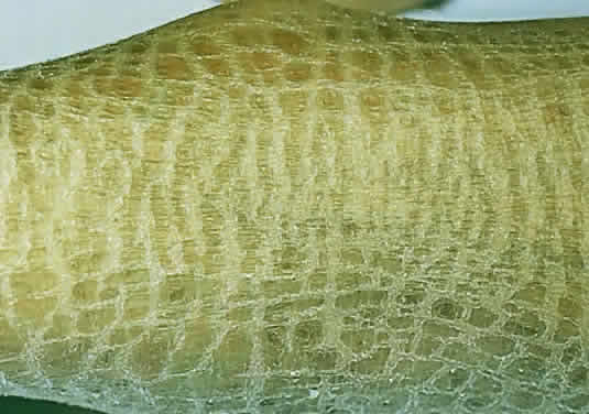
on diagnostic criteria, Hamburg 1992. J Inherit Metab Dis 16:241, 1993

385. Garborg I, Torvik A, Hals J et al: Congenital neuronal ceroid lipofuscinosis A case report. Acta Pathol Microbiol Immunol 95:119, 1987

386. Boustany R-M, Alroy J, Kolodny E: Clinical classification of neuronal ceroid-lipofuscinosis subtypes. Am J Med Genet 5:47, 1988

387. Goebel H: Neuronal ceroid lipofuscinoses: The current status. Brain Dev 14:203, 1992

388. Berkovic S, Carpenter S, Andermann F et al: Kufs' disease: A critical reappraisal. Brain 111:27, 1988

389. Constantinidis J, Wisniewski K, Wisniewski T: The adult and a new late adult forms of neuronal ceroid lipofuscinosis. Acta Neuropathol 83:461, 1992

390. Santavuori P: Neuronal ceroid lipofuscinoses in childhood. Brain Dev 10:80, 1988

391. Santavuori P, Vanhanen S, Sainio K et al: New aspects in the diagnosis of neuronal ceroid lipofuscinosis. Brain Dysfunction 4:211, 1991

392. Eiberg M, Gardiner R, Mohr J: Batten disease (Spielmeyer-Sjogren disease) and haptoglobin (HP): Indication of linkage and assignment to chromosome 16. Clin Genet 36:217, 1989

393. Callen D, Baker E, Lane S et al: Regional mapping of the Batten disease locus (CLN3) to human chromosome 16p12. Am J Hum Genet 49:1372, 1991

394. Jarvela I, Schleutker J, Haataja L et al: Infantile form of neuronal ceroid lipofuscinosis (CLN1) maps to the short arm of chromosome 1. Genomics 9:170, 1991

395. Vesa J, Hallsten E, Verkruyse L et al: Mutations in the palmitoyl protein thioesterase gene causing infantile neuronal ceroid lipofuscinosis. Nature 376:584, 1995

396. Williams R, Vesa J, Jarvela I et al: Genetic heterogeneity in neuronal ceroid lipofuscinosis (NCL): Evidence that the late-infantile subtype (Jansky-Bielschowsky disease; CLN2) is not an allelic form of the juvenile or infantile subtypes. Am J Hum Genet 53:931, 1993

397. Savukoski M, Kestila M, Williams R et al: Defined chromosomal assignment of CLN5 demonstrates that at least four genetic loci are involved in the pathogenesis of human ceroid lipofuscinoses. Am J Hum Genet 55:695, 1994

398. Boustany R, Kolodny E: Neurological progress. The neuronal ceroid-lipofuscinoses: A review. Rev Neurol (Paris) 145:105, 1989

399. Boustany R-M: Neurology of the neuronal ceroid lipofuscinoses: Late infantile and juvenile types. Am J Med Genet 42:533, 1992

400. Palmer D, Fearnley I, Walker J et al: Mitochondrial ATP synthase subunit c storage in the ceroid lipofuscinoses (Batten disease). Am J Med Genet 42:561, 1992

401. Kominami E, Ezaki J, Muno D et al: Specific storage of subunit c of mitochondrial ATP synthase in lysosomes of neuronal ceroid lipofuscinosis (Batten's disease). J Biochem 111:278, 1992

402. Tyynela J, Palmer D, Baumann M et al: Storage of saposins A and D in infantile neuronal ceroid lipofuscinosis. FEBS Lett 330:8, 1993

403. Santavuori P, Haltia M, Rapola J et al: Infantile type of so-called neuronal ceroid lipofuscinosis: I. A clinical study of 15 patients. J Neurol Sci 18:257, 1973

404. Goebel H, Zeman W, Damaske E: An ultrastructural study of the retina in the Jansky-Bielschowsky type of neuronal ceroid lipofucsinosis. Am J Ophthalmol 83:70, 1977

405. Schochet S Jr, Font R, Morris H: Jansky-Bielschowsky form of neuronal ceroid lipofuscinosis: Ocular pathology of-the Batten-Vogt syndrome. Arch Ophthalmol 98:1083,-1980

406. Copenhaver R, Goodman G: The electroretinogram in infantile, late infantile and juvenile amaurotic family idiocy. Arch Ophthalmol 63:203, 1960

407. Vogt H: Uber familiare omurotische Idiote und verwandte Krankenheitsbilder. Monatschrr Psychiatr Neurol 18:161, 1905

408. Spielmeyer W: Uber familiore amurotische Idiotie. Neurol Cbl 24:620, 1905

409. Spalton D, Taylor D, Sanders M: Juvenile Batten's disease: An ophthalmological assessment of 26 patients. Br J Ophthalmol 64:726, 1980

410. Traboulsi E, Green W, Luckenbach M et al: Neuronal ceroid lipofuscinosis: Ocular histopathologic and electron microscopic studies in the late infantile, juvenile, and adult forms. Graefes Arch Clin Exp Ophthalmol 225:391, 1987

411. Goebel H, Fix J, Zeman W: The fine structure of the retina in neuronal ceroid lipofuscinosis. Am J Ophthalmol 77:-25, 1974

412. Pardo C, Rabin B, Palmer D et al: Accumulation of the adenosine triphosphate synthase subunit c in the mnd mutant mouse: A model for neuronal ceroid lipofuscinosis. Am J Pathol 144:829, 1994

413. Raininko R, Santavuori P, Heiskala H et al: CT findings in neuronal ceroid lipofuscinosis. Neuropediatrics 21:95, 1990

414. Libert J: Diagnosis of lysosomal storage diseases by the ultrastructural study of conjunctival biopsies. Pathol Ann 15:37, 1980

415. Brod R, Packer A, Van Dyk H: Diagnosis of neuronal ceroid lipofuscinosis by ultrastructural examination of peripheral blood lymphocytes. Arch Ophthalmol; 105:1388, 1987

416. Conradi N, Uvebrant P, Hokegard K et al: First-trimester diagnosis of juvenile neuronal ceroid lipofuscinosis by demonstration of fingerprint inclusions in chorionic villi. Prenat Diagn 9:283, 1989

417. Rapola J, Salonen R, Ammala P et al: Prenatal diagnosis-of the infantile type of neuronal ceroid lipofuscinosis by electron microscopic investigations of human chorionic villi. Prenat Diagn 10:553, 1990

418. Jarvela I, Rapola J, Peltonen L et al: DNA-based prenatal diagnosis of the infantile form of neuronal ceroid lipofuscinosis (INCL; CLN1). Prenat Diagn 11:323, 1991

419. Uvebrant P, Bjork E, Conradi N et al: Successful DNA-based prenatal exclusion of juvenile neuronal ceroid lipofuscinosis. Prenat Diagn 13:651, 1993

420. Hellsten E, Vesa J, Jarvela I et al: Refined assignment of the infantile neuronal ceroid lipofuscinosis (INCL) locus at 1p32 and the current status of prenatal and carrier diagnosis. J Inherit Metab Dis 16:335, 1993

421. Gibbs K, Walshe J: Biliary excretion of copper in Wilson's disease. Lancet 2:538, 1980

422. Petrukhin K, Lutsenko S, Chernov I et al: Characterization of the Wilson disease gene encoding a P-type copper transporting ATPase: Genomic organization, alternative splicing and structure function predictions. Hum Mol Genet 3:-1647, 1994

423. Chowrimootoe G, Ahmed H, Seymour C: New insights-into the pathogenesis of copper toxicosis in Wilson's dis—ease: Evidence for copper incorporation and defective canalicular transport of caeruloplasmin. Biochem J 315:851, 1996

424. Sass-Kortsak A: Wilson's disease: A treatable cause of liver disease in children. Pediatr Clin North Am 22:963, 1975

425. Strickland G, Leu M: Wilson's disease: Clinical and laboratory manifestations in 40 patients. Medicine 54:113, 1975

426. Scheinberg I, Sternlieb I: Wilson's Disease. Philadelphia: WB Saunders, 1984

427. Lowe C, Terrey M, MacLachan E: Organic aciduria, decreased renal ammonia production, hydrophthalmos and mental retardation: A clinical entity. Am J Dis Child 83:-164, 1952

428. Zhang X, Jefferson A, Auethavekiat V et al: The protein deficient in Lowe syndrome is a phosphatidylinositol-4,5-biphosphate 5-phosphatase. Proc Natl Acad Sci USA 92:4853, 1995

429. Menkes J, Alter M, Steigleder G et al: A sex-linked recessive disorder with retardation of growth, peculiar hair-and focal and cerebellar degeneration. Pediatrics 29:764,-1962

430. Grover W, Johnson W, Henkin R: Clinical and biochemical aspects of trichopoliodystrophy. Ann Neurol 5:65, 1979

431. Horn N. Menkes' X-linked disease: Prenatal diagnosis and carrier detection. J Inherit Metab Dis 6:59, 1983

432. Billings D, Degnan M: Kinky hair syndrome: A new case and review. Am J Dis Child 121:447, 1971

433. Singh S, Bresnan M: Menkes kinky hair syndrome (trichopoliodystrophy): Low copper level in the blood, hair and urine. Am J Dis Child 125:572, 1973

434. Danks D, Campbell P, Stevens B et al: Menke's kinky hair syndrome: An inherited defect in copper absorption with widespread effects. Pediatrics 50:188, 1972

435. Van Bogaert L, Bertrand I: Sur une idiotie familiale avec degenerescence spongieuse de nevraxe. Acta Neurol Belg 49:572, 1949

436. Kaul R, Gao G, Balamurugan K et al: Cloning of the human aspartoacylase, DNA and a common missence mutation in Canavan disease. Nat Genet 5:118, 1993

437. Kaul R, Balamurugan K, Gao G et al: Canavan disease: genomic organization and localization of human ASPA to 17p13-ter and conservation of the ASPA gene during evolution. Genomics 21:364, 1994

438. Matalon R, Michals K, Sebesta D et al: Aspartoacylase deficiency and N-acetylaspartic aciduria in patients with Canavans disease. Am J Med Genet 29:463, 1988

439. Divry P, Mathieu M: Aspartoacylase deficiency and N-acetylaspartic aciduria in patients with Canavan disease. Am J Med Genet 32:550, 1989

440. Ungar M, Goodman R: Spongy degeneration of the brain in Israel. A retrospective study. Clin Genet 23:23, 1983

441. Kelley R, Stamas J: Quantification of N-acetyl-L-aspartic acid in urine by isotope dilution gas chromatography-mass spectrometry. J Inherit Metab Dis 15:97, 1992

442. Borash V, Flhor D, Morag B et al: A radiometric assay for aspartoacylase activity in human fibroblasts: Application for the diagnosis of Canavan's disease. Clin Chim Acta 201:175, 1991

Back to Top