Chapter 21
Endocrine Disease and the Eye
H. C. CHEN and EVA M. KOHNER
Main Menu   Table Of Contents

Search

DISORDERS OF THE THYROID GLAND
DISORDERS OF THE PITUITARY GLAND
DISORDERS OF THE ADRENAL GLAND
DISORDERS OF THE PARATHYROID GLANDS
MULTIPLE ENDOCRINE NEOPLASIA
REFERENCES

DISORDERS OF THE THYROID GLAND
The thyroid gland is situated anterior to and on either side of the upper part of the trachea and the thyroid cartilage. Its principal function is the secretion of thyroxine (T4) and triiodothyronine (T3). Both hormones increase cellular metabolism and have widespread physiologic action, including enhancing the adrenergic state; of the two, T3 possesses a greater level of activity.

The synthesis of thyroid hormones is dependent on the availability of dietary iodine, which is incorporated by the thyroid gland into individual tyrosyl residues of a complex molecule called thyroglobulin. These individual residues merge to form T3 and T4, which are cleaved from the parent thyroglobulin molecule for release into the blood stream. Thyroid hormones are transported in the blood stream bound principally to thyroxine-binding globulin. Only a very small percentage (less than 0.5%) circulate “free” or unbound, and it is this fraction that is responsible for the physiologic action.

The function of the thyroid gland is controlled by the anterior pituitary gland via the secretion of thyroid-stimulating hormone (TSH), which is itself influenced by the release of thyrotropin-releasing hormone by the hypothalamus; TSH release by the anterior pituitary is partly controlled by the thyroid hormones through a negative feedback mechanism.

HYPERTHYROIDISM

Hyperthyroidism or thyrotoxicosis is characterized by the overproduction of thyroid hormones. A small proportion of patients have only elevated T3 levels, but the majority possess high levels of both T3 and T4. Thyrotoxicosis may be classified broadly into two groups: (1) where the thyroid gland is either diffusely hypertrophic and hyperplastic (Graves' disease); or (2) where single or multiple hyperactive nodules exist in the gland.

Graves' disease is a condition that predominantly affects females and, although it may occur at any age, has a peak incidence in the third and fourth decades. In almost all patients with this autoimmune disease, immunoglobulins are directed against the TSH receptors on the thyroid cellular membrane. As with other autoimmune conditions, there is a strong familial tendency for Graves' disease: it has been associated with the HLA antigens B8 and DR3 in whites and with Bw35 in Asians.

It usually presents insidiously, although rapid and dramatic presentations are not uncommon; the exact precipitating mechanism is unknown. Systemic symptoms include weight loss, increased sweating and heat intolerance, palpitation, fatigue, and diarrhea. Examination of a patient with Graves' disease may reveal a goiter, a fine tremor, palmar erythema, finger clubbing, vitiligo, alopecia, pretibial myxedema, and a multitude of cardiovascular signs (e.g., tachycardia, atrial fibrillation, bounding peripheral pulses).

Ocular Involvement

Ocular involvement is an almost integral part of the clinical presentation of Graves' disease; however, the systemic and eye manifestations are generally considered to run independent courses. Although most of the systemic features of Graves' disease can be attributed to the increased activity of thyroid hormones, the ocular involvement is less obviously linked to this. Graves' ophthalmopathy, preferably termed thyroid-associated ophthalmopathy (TAO), may occur in the absence of overt systemic signs of hyperthyroidism (approximately 10% of cases; more subtle analysis often reveals evidence of systemic thyroid disease in most of these cases), or even after the systemic disease has been treated adequately. TAO is the most common cause of proptosis in adults; the term exophthalmos is used exclusively to describe the proptosis of TAO and will be used henceforth in this section.

Compared with Graves' disease, hyperthyroidism due to “toxic” thyroid nodules is less common and generally occurs in older patients. Ocular involvement is not a common feature of this condition.

Presentation

Patients with TAO may present with a wide range of symptoms. Both eyes are usually affected, often to differing degrees, or the involvement may be unilateral. Although the condition may be selflimiting, it often has a tendency to produce episodes of acute activity lasting several months interspersed by relative quiescence, but the progression in severity from any one acute episode is not likely to regress as a result of fibrotic changes. The natural tendency of the disease process is to eventually “burn out” within 2 to 3 years.

Lid puffiness and irritable, watery eyes are common symptoms; complaints include a feeling of “grittiness” in the eyes and profuse tearing despite the absence of apparent irritants. Affected eyes may feel gritty for several reasons: (1) lid retraction and exophthalmos may cause problems with corneal lubrication, and superior limbic keratitis is a well-recognized association; (2) thickened conjunctival folds may obstruct the drainage of tears into the lower lid punctum, evidenced by a fullness of the conjunctiva at the medial canthus; and (3) conjunctival hyperemia commonly accompanies inflammation of the extraocular muscles. The injection is usually most marked over the insertions of the extraocular muscles (Fig. 1).

Fig. 1. Patient with active thyroid-associated ophthalmopathy. Features exhibited include conjunctival hyperemia, periorbital edema, upper lid retraction, and exophthalmos (Courtesy of Mr. Peter Fells, Moorfields Eye Hospital, London.)

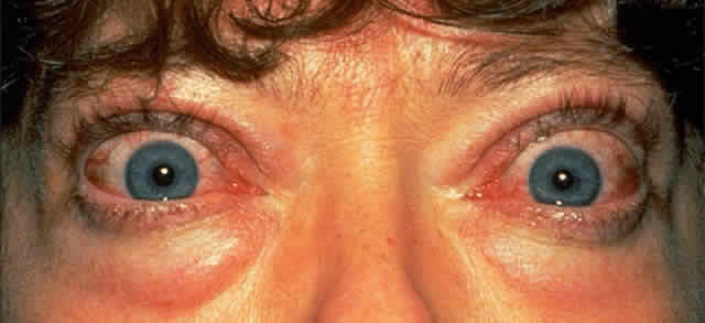
Cosmetic problems associated with protruding eyes are also common; this protrusion is usually asymmetric and is due to a combination of upper lid retraction and exophthalmos. Lid retraction is among the earliest and most common signs causing exposure of the superior sclera and widening of the palpebral fissure. It is one feature of TAO that can be partly attributed to the systemic effects of hyperthyroidism; increased stimulation of the smooth muscle retractors in both the upper and lower lids can result from heightened adrenergic activity. Upper lid retraction also results from a tightened inferior rectus due to inflammation and fibrosis, and this tightening leads to compensatory overactivity of the superior rectus/levator palpebrae complex. Similar inflammatory and fibrotic involvement of the levator palpebrae muscle will add further to upper lid retraction.1 This wide-eyed staring appearance is often accentuated by the presence of exophthalmos (see Fig. 1).

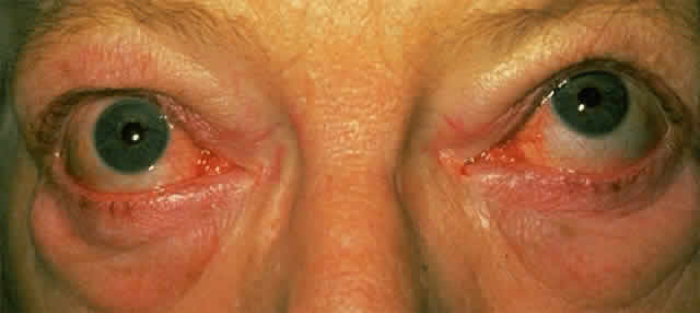
When mild, the signs and symptoms of exophthalmos may be limited to a feeling of grittiness, conjunctival injection, and chemosis; however, severe corneal exposure, which is often complicated by a poor Bell's phenomenon because of restrictive myopathy, can lead to pannus formation and ulceration, corneal scarring and infection, and visual impairment. Because exophthalmos is secondary to increased retro-orbital mass, it can be associated with optic nerve compression, although significant nerve compression may be present in its absence.

The principal visual symptoms are diplopia and reduced visual acuity. According to Fells,2 patients with thyroid-associated diplopia see vertically separated images upon waking in the morning, which improve within minutes but recur later during periods of fatigue or alcohol intoxication. Defective ocular motility in TAO is due to inflammatory engorgement of the extraocular muscles: the inferior and medial recti more commonly and the lateral and superior recti less commonly. The restriction in function may initially be reversible, but is eventually permanent due to cicatricial, fibrotic elements (Fig. 2). Since the inferior rectus muscle is the most commonly affected, upgaze can often lead to elevated intraocular pressure.

Fig. 2. Patient in the quiescent cicatricial phase of thyroid-associated ophthalmopathy. There is limited upgaze in the right eye due to fibrosis of the inferior rectus muscle. (Courtesy of Mr. Peter Fells, Moorfields Eye Hospital, London.)

Optic nerve compression (compressive neuropathy) is one of the serious complications of TAO and leads to reduced visual acuity. Its prevalence has been reported at approximately 10% in a prospective study of 101 patients attending a combined thyroid-eye clinic during a 5-year period3; this figure may be artificially high because of the specialized nature of this clinic. It is more likely that less than 5% of patients with Graves' disease will develop sight-threatening compressive neuropathy. Optic nerve involvement is generally accepted to be due to compression at the orbital apex by enlarged extraocular muscles.4 These patients are likely to be older and male; they commonly exhibit limitations of extraocular movements with significant vertical deviation.5,6 Other causes of reduced visual acuity are corneal exposure and desiccation, with their attendant complications, and chorioretinal striae involving the macula, due to an increased retro-orbital mass.

In an attempt to clarify the nomenclature pertaining to thyroid-related eye disease, Werner classified the eye changes into seven categories, based on an original classification by the American Thyroid Association7; Werner's classification is presented in Table 1. Although useful as a means of classifying the severity of involvement, its usage is limited because it lacks a means of measuring the rate of progression or treatment-induced regression of clinical activity. The signs of TAO can be subdivided into those pertaining to the lids, conjunctiva and cornea, globe, extraocular muscles, and fundus. These signs are summarized in Table 2.


TABLE 1. Werner's Classification of Thyroid-Associated Ophthalmopathy*


ClassDefinition
0No signs or symptoms
1Only signs, no symptoms (signs limited to upper lid retraction ± lid lag and proptosis)
 Proptosis is graded into:
   0: Absent (20 mm or less)
   a: Minimal (21–23 mm)
   b: Moderate (24–27 mm)
   c: Marked (28 mm or greater)
2Soft tissue involvement
  i: Symptoms of excessive lacrimation, “gritty” sensation, retrobulbar discomfort, and photophobia
  ii: Signs are subdivided into the following grades:
   0: Absent
   a: Minimal (edema of conjunctiva and lids, conjunctival injection, orbital fat extrusion, palpable lacrimal glands, swollen extraocular muscles)
   b: Moderate (minimal signs plus chemosis, lagophthalmos, eyelid “fullness”)
   c: Marked
3Proptosis (subdivided as in class 1)
4Extraocular muscle involvement
   0: Absent
   a: Minimal (limitation of motion, evident at extremes of gaze)
   b: Moderate (evident restriction of motion without fixation of position)
   c: Marked (fixation of position of a globe or globes)
5Corneal involvement
   0: Absent
   a: Minimal (stippling of cornea)
   b: Moderate (ulceration)
   c: Marked (clouding, necrosis, perforation)
6Sight loss (optic nerve compression)
   0: Absent
   a: Minimal (disc pallor or choking, or visual field defect; vision 20/20–20/60)
   b: Moderate (minimal signs, but vision 20/70–20/200)
   c: Marked (vision less than 20/200)

* As indicated, each class is further subdivided into absent, mild, moderate, or marked. The first two classes (0 and 1) represent non-sight-threatening disease, the latter five classes (2 to 5) represent more severe infiltrative disease, increasing numbers signifying more advanced involvement. The first letters of each definition constitute the acronym NO SPECS, which is the name by which this classification is commonly referred.7
(Adapted from Werner SC: Classification of the eye changes of Graves' disease. Am J Ophthalmol 68:646, 1969.)


TABLE 2. Signs of Thyrotoxicosis

  Lids
  Puffy lids (Enroth's sign)
  Lid retraction (Dalrymple's sign)
  Lid lag:

  1. Delay of upper lid in following globe movement in downward gaze (Graefe's sign)
  2. Jerky downward movement of the eyelid (Boston's sign)
  3. Delay of lower lid in following globe movement in upward gaze (Griffith's sign)
  4. Globe lagging behind upper lid on upward gaze (Mean's sign)

  Tremor of closed eyelids (Rosenbach's sign)
  Reduced blinking (Stellwag's sign)
  Increased pigmentation of skin of the eyelids (Jellinek's sign)
  Conjunctiva and Cornea
  Injected, chemotic conjunctiva
  Superior limbic keratoconjunctivitis
  Signs of corneal exposure ± pannus formation and ulceration
  Globe
  Exophthalmos
  Ocular bruit
  Extraocular Muscles
  Apparent overactivity of extraocular muscles, most commonly:
  1. Inferior rectus with weakness of upgaze
  2. Medial rectus with weakness of convergence

  Elevated intraocular pressure on upgaze
  Fundus
  Congested retinal veins
  Swollen optic disc
  Chorioretinal striae
  Pale, atrophic optic disc


Pathogenesis

Present understanding of the pathogenesis of TAO remains unclear; however, as with the thyroid component, autoimmune mechanisms have been implicated. The association between the ocular disorder and a hyperactive thyroid gland may be the result of a linkage between the two conditions and a primary autoimmune disorder manifested through a variety of circulating autoantibodies acting on two different end-organs sharing a hypothetical autoantigen. Currently it is unclear what this autoantigen is, but several candidates have been proposed, including a 64-kilodalton (kd) protein found in thyroid and eye-muscle plasma membranes8,9 and the TSHreceptor protein.10,11 Antibodies directed against a 64-kd protein expressed by extraocular muscle has been reported to be present in 33% of patients with TAO and in 75% of patients with severe eye disease.12 There may also be other antigens expressed in the orbit similar to both thyroglobin and antigens expressed on thyroid microsomal cells, since antibodies directed against them have been identified in the orbit.13

The stimulatory strength of these respective antibodies vary among patients. In cases predominantly involving the eye, the antibodies directed against the thyroid may be weak. In cases where these antibodies prevent the normal binding of TSH, there may be an association between hypothyroidism and TAO. It has also been suggested that a common autoantigen exists (e.g., the 64-kd antigen) that is responsible for the milder eye disease seen in the majority of patients with Graves' disease; in patients with severe disease, other eye-muscle-specific antigens have been proposed.14 It has been demonstrated that IgG from patients with TAO stimulated the growth of extraocular myoblasts, compared with sera from controls and patients with Graves' disease who had no ocular involvement. The effect was relatively specific to these cells, whereas the effect on skeletal myoblasts was less marked.15

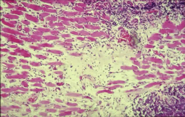
The inflammatory infiltration of the extraocular muscles consists principally of activated T-cells together with smaller numbers of B-cells, macrophages, and mast cells.16 The infiltration is mainly interstitial and is accompanied by increased fibroblastic activity induced by cytokines derived from the immune cells, leading to deposition of glycosaminoglycans and collagen as well as edema. The eventual outcome is fat cell infiltration and fibrosis (Fig. 3). The muscle fibers appear normal, with the exception of subsarcolemmal deposits of lipid and glycogen, and there is an absence of muscle-cell destruction.

Fig. 3. Histologic section of an extraocular muscle in thyroid-associated ophthalmopathy. There is diffuse lymphocytic infiltration accompanied by fibrotic elements (light pink) affecting muscle fibers (dark pink). (Courtesy of Mr. Peter Fells, Moorfields Eye Hospital, London.)

It is unclear which cell type expresses the offending antigen, but current understanding favors the fibroblast. According to Weetman,17 the histologic picture reveals a pathologic process directed principally against the retrobulbar fibroblast, rather than muscle cells, but this may be just a manifestation of the presence of large numbers of immune cells attracted there by a nonfibroblastic source. Antibodies against the TSH-receptor have been shown to stimulate collagen synthesis by fibroblasts18; retrobulbar fibroblasts have also been demonstrated to possess TSH-receptor-encoding RNA.19 However, an immune process directed against muscle cells is suggested by the finding of higher levels of eye-muscle-binding antibody in patients with TAO, whereas levels of antifibroblast antibody were unremarkable.20

Also unclear is why the extraocular muscles are selectively involved in TAO while other skeletal muscles in the body are spared. Schmidt and associates21 reported that hyperthyroidism alters the immunocompetent cell population in extraocular muscles, but not skeletal muscles. Another possible explanation is that extraocular muscles also possess more spindles and connective tissue and have a greater blood supply than skeletal muscles.

Most of the infiltrative pathology seen in TAO traditionally has been attributed to the retro-orbital deposition of glycosaminoglyans, with associated edema and inflammatory cell infiltration of the muscles. More recently, Hudson and colleagues22 proposed that, in some cases, the exophthalmos may be due to passive orbital venous congestion from a partial obstruction of the superior ophthalmic vein. They based this view on the observation that exophthalmos is sometimes present with little enlargement of the extraocular muscles, with the exception of the superior rectus muscle together with congestion of the superior ophthalmic vein with which it is closely linked. The obstruction could be purely compressive, or there may be an element of contiguous inflammation from the adjacent muscle.

Orbital fat content is not thought to play a significant role in compressive optic neuropathy. Feldon and co-workers23 demonstrated that patients with optic nerve compression had greater extraocular muscle volumes than those without optic nerve involvement, whereas both groups had similar fat content; orbital fat volume was found to decrease with increasing muscle size.

Diagnosis

Diagnosis of TAO is based on clinical findings, sometimes in the absence of hyperthyroidism. Symptoms and signs indicating active inflammation (e.g., pain, conjunctival injection, edema) are particularly important because they influence the management strategy.24 The degree of exophthalmos should be assessed with an exophthalmometer (a protrusion of 20 mm or more is suspicious), and the intraocular pressure should be measured with the patient in the primary position and with attempted upgaze. Photography is a useful method of recording clinical status.

Clinical investigations may reveal elevated plasma T3 and T4 levels (the free thyroxine index can be determined after correction for plasma protein binding) and in most cases decreased plasma TSH. Antibodies that may be detected include those directed against TSH-receptor, thyroglobulin, and thyroid membranes.

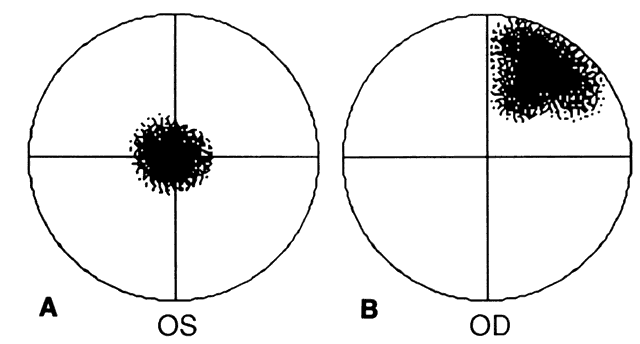
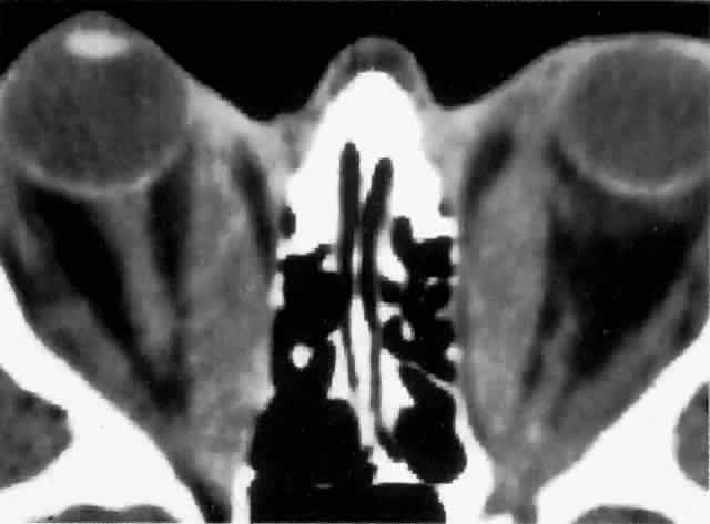
Computed tomography (CT), most revealing in the coronal plane, and ultrasonography are useful methods for assessing extraocular muscle thickness (Fig. 4). Magnetic resonance imaging (MRI) has been advocated as a means of differentiating between extraocular muscles that are actively inflamed and those that are fibrosed25–27; both muscular problems can restrict ocular movement, but the former is amenable to immunosuppressive intervention. The T2 relaxation time of MRI is directly proportional to the water content of the tissue scanned: since inflamed tissues are edematous, they should have longer T2 times. MRI is also thought to be better than CT in providing views of the orbital apex and may therefore be better for evaluating optic nerve compression.2

Fig. 4. CT scan through the orbits of a patient with thyroid-associated ophthalmopathy, showing bilateral exophthalmos with considerably thickened medial rectus muscles. (Courtesy of Mr. Peter Fells, Moorfields Eye Hospital, London.)

Extraocular muscle volume is inversely proportional to ocular motility, especially for horizontal movements. Therefore, the peak velocity of saccadic eye movement, measured with reflective infrared oculography, has been proposed as another means of assessing clinical severity.28–30 The degree of optic nerve compression has been found to increase as peak velocity decreases, particularly with larger angles of eye movement.29

Patients with optic nerve compression present with reduced visual acuity, which may be clinically substantiated by the presence of an afferent pupillary defect and visual field loss. More sophisticated means of detecting early compressive pathology include the pattern electroretinogram, cortical visual evoked potentials, and color contrast sensitivity along the tritan axis.31 Depending on the degree of severity, fundal examination may reveal varying degrees of venous congestion, swollen optic discs, and chorioretinal striae (usually horizontal). It is important to note that 40% to 50% of patients with optic nerve compression may have a normal fundal appearance.5,32

Prognosis and Risk Factors

Unlike Graves' disease, which is far more prevalent in females, TAO is much more evenly distributed between the sexes; in fact, when TAO is present in males, there is a greater likelihood of more severe involvement.3 TAO is a part of Graves disease and therefore cannot be differentiated from it reliably. Older patients are more likely to develop more severe ocular disease, possibly because of age-dependent alterations in the elasticity of the extraocular muscles, rendering them stiffer and more likely to impinge on the optic nerve rather than molding around it.29 Kendler and associates,33 in a series of 557 cases of Graves' disease, reported that patients older than 50 years have an increased incidence of ocular motility impairment (32% versus 12%) and more severe reduction in visual acuity.

Smoking is strongly associated with an increased risk of TAO development as well as with more severe involvement34–36; this effect may be related partly to a smoking-induced increase in thyroglobulin release from the thyroid gland and to alterations in immunoregulatory cell function.

Various forms of treatment for hyperthyroidism, including radioactive iodine37,38 and surgery,39 have been found to increase the severity of TAO. It has been suggested that this may be due to the increased release of antigenic thyroidal elements into the blood stream. The presence of pretibial myxedema is a poor prognostic sign and usually signifies the presence of significant ophthalmopathy and a prolonged clinical course.

Management

It is unclear whether treatment of the underlying thyrotoxic state produces any reduction in the ocular symptoms apart from lid retraction, which if it is secondary to increased adrenergic activity, may be expected to improve. If the cornea is exposed, it is important to prescribe artificial tears as a means of corneal lubrication. To help reduce the degree of periorbital edema accumulation overnight, the patient should be instructed to sleep on several pillows or to have the mattress at the head end of the bed elevated.

For more severely affected eyes, immunosuppressive therapy with glucocorticoids and retrobulbar irradiation has been reported to benefit approximately 60% of patients with TAO, particularly in improving visual acuity and cosmesis; this treatment is less effective in combating exophthalmos and movement dysfunction.40 Immunosuppressive agents have the greatest effect against active inflammatory disease of short duration, rather than changes that are principally cicatricial. Plasmapheresis has also been used to reduce the degree of immune activity, although it usually has only a transitory effect.

Steroids are the most widely used immunosuppressive drugs; to be effective, high doses of prednisolone, 80 to 100 mg daily, are usually required and relatively high doses are often needed during a course of several weeks to maintain the effect. The addition of a second drug, such as cyclosporin A or azathioprine, may allow for lower doses of prednisolone. Based on the fact that methylprednisolone has been used with good effect in other autoimmune conditions, Kendall-Taylor and colleagues41 described the successful use of a large-dose IV bolus of methylprednisolone, carefully monitoring the side effects, particularly those involving the cardiovascular system. Their protocol used a dose of 0.5 g methylprednisolone in 200 mL isotonic saline infused in a 30-minute period, with a second 0.5 g given 48 hours later. A maintenance dosage of oral prednisolone, 40 mg daily, was then commenced; within a four-week period, it is gradually reduced to 10 mg. Subsequent reduction in dosage depended on clinical status. The principal advantages of this technique over entirely oral prednisolone appears to be the rapid improvement in visual acuity (within 48 hours) and a relatively lower incidence of side effects.

Low-dose external-beam megavoltage radiotherapy has been employed successfully in treating sight-threatening TAO; the improvement in radiotherapy techniques, principally the use of tightly collimated beams with small treatment portals through a lateral port, means that the incidence of long-term side effects such as cataract formation and radiation retinopathy is low.42 A total dose of 20 Gy delivered in 12 fractions in a period of 15 days is a recommended treatment regimen.43

A prospective study by Prummel and co-workers reported similar efficacy between steroid therapy and radiotherapy in patients with moderately severe TAO, but without optic nerve involvement (approximately 50% of patients responded in each group); of patients treated with steroids, a higher proportion had side effects compared with those treated with radiotherapy.44 Prednisone (a 3-month oral course) was found to be more effective in reducing soft tissue edema, whereas radiotherapy (2 Gy daily in 10 fractions in a period of 2 weeks) was considered better at improving motility. Because of the lack of patients with compressive optic neuropathy, the primary use of radiotherapy in these patients currently cannot be advocated because of its delayed effect.45

If medical decompression is not successful (i.e., vision remains poor, continues to deteriorate or corneal exposure remains severe), then surgical decompression is the next treatment option. Decompression allows some of the congested orbital contents to prolapse into the surrounding sinuses. With improvements in surgical technique, surgical decompression is also frequently performed in patients with lesser degrees of involvement to improve cosmesis.46 Several approaches toward this end have been described, ranging from the minimum requirement of removing the medial wall and medial half of the orbital floor, to four-wall decompression; although the latter achieves the greatest degree of retrodisplacement, it is also associated with the highest complication rate and therefore is not commonly performed.

Orbital decompression in cases of compressive neuropathy has been reported to have high success rates, approximately 90% of patients experiencing an improvement in vision or maintaining visual stability; no major complications have been reported after transantral decompression.47,48 From the results of a postal survey of ophthalmic surgeons, McCord49 reported that 41% of orbital decompressions were performed to reduce corneal exposure from severe exophthalmos, 39% to alleviate optic nerve compression, and 20% to improve the cosmetic impairment caused by milder forms of exophthalmos. The series also described antralethmoidal and three-wall decompressions (medial, floor, and lateral walls) as producing an average of 4 to 6 mm of retrodisplacement of the globe; the antral-ethmoidal approach was the most commonly applied (75% of the surgeons surveyed). Decompression may be associated with worsening extraocular muscle imbalance postoperatively. The translid approach is thought to produce fewer problems than the transantral approach, although the latter is more efficacious in relieving nerve compression because it allows decompression up to the orbital apex. Orbital decompression for lesser degrees of exophthalmos with removal of orbital fat without bone also has been described, and a retrodisplacement of 2.2 to 5.9 mm has been achieved.50,51

Up to 15% of patients with TAO may suffer from diplopia.52 Regular orthoptic assessment is needed to manage this symptom satisfactorily. Diplopia may be relieved initially with the application of temporary prisms (e.g., Fresnel prisms), the adoption of a compensatory head posture, or in severe cases, occlusion. After a period of stability (Fells2 recommends a period of 6 months), extraocular muscle surgery, or in milder cases, the incorporation of prisms into the spectacle prescription, may be contemplated. The most common form of extraocular muscle surgery is the recession of tight, fibrous inferior rectus muscles with the aid of adjustable sutures to optimize positioning.

The use of botulinum toxin in patients with TAO suffering from diplopia has been advocated by some.53,54 Lyons and associates53 injected 62.5 picograms of botulinum neurotoxin A in 0.1 mL normal saline into the affected muscle under electromyographic monitoring in a series of 38 patients with an average hypotropia of 23 prism diopters and esotropia of 28 prism diopters; 75% of the patients achieved a mean reduction of 14 prism diopters in the angle of deviation. The effect was undetectable after 2 months in the majority of patients, most of whom eventually required strabismus surgery. Six patients did obtain long-term stability (at least 12 months); however, of these patients three had previously undergone strabismus surgery. Lyons and colleagues advocated this form of treatment for two reasons:

  1. With conventional treatment, patients with diplopia must wait for the condition to stabilize before surgery can be contemplated, which may mean that they will have a prolonged period of visual disturbance. There is one caveat, however: botulinum toxin will interfere with orthoptic assessments during this period.
  2. There is a possibility that the improvement achieved with botulinum toxin will be permanent.

Lid retraction may be improved by orbital decompression, especially of the lower lid; however, the backward and downward movement of the globe following decompression may accentuate upper lid retraction. Repositioning (recession) of the upper lid retractors may have to be performed as an adjunct. To improve cosmesis in patients who do not have significant exophthalmos, primary recession of the lid retractors can be performed without decompression. Any required lid procedures, however, should be undertaken after decompression (which may worsen extraocular muscle imbalance) and strabismus surgery. Such techniques include disinsertion of Müller's muscle, with or without levator aponeurosis incision or recession for upper lid retraction. Botulinum toxin has also been used to reduce lid retraction, producing an improvement lasting up to 32 weeks.55 Spacers may be inserted between the lower lid retractors and the tarsal plate to improve lower lid retraction; sclera and hard palate mucosal grafts have been employed to this end.

HYPOTHYROIDISM

Most commonly, hypothyroidism occurs secondary to idiopathic atrophy of the thyroid gland; it may also result from treatment for hyperthyroidism (particularly after radioactive iodine therapy), iodine deficiency, and Hashimoto's thyroiditis. Hypothyroidism usually has an insidious onset: patients present with complaints of lethargy, weight gain, dry and thickened skin, coarse hair, anorexia, constipation, thickening of the voice, and psychiatric symptoms.

Ophthalmologic features of the condition include periorbital swelling, which is part of the generalized nonpitting skin edema of myxedema and the characteristic loss of the outer third of the eyebrows. A more unusual ophthalmic feature is open-angle glaucoma, which has been reported to be associated with the deposition of a mucopolysaccharide within the trabecular meshwork.56

Back to Top
DISORDERS OF THE PITUITARY GLAND
The pituitary gland, also known as the hypophysis, is often known as the “orchestrator” of endocrine function because it controls the function of several other glands. It is divided anatomically, embryologically, and functionally into two principal parts, the anterior and posterior lobes; there is also a poorly developed intermediate region between the two. The posterior lobe is attached to the hypothalamus by the pituitary stalk, which embraces the anterior aspect of the third ventricle. The six hormones secreted by the anterior lobe are thyroid-stimulating hormone (TSH), adrenocorticotropic hormone (ACTH), follicle-stimulating hormone, luteinizing hormone, growth hormone, and prolactin. The two principal products of the posterior lobe are vasopressin (antidiuretic hormone) and oxytocin.

The pituitary gland is situated in a bony cavity within the sphenoid bone called the sella turcica (pituitary fossa). This cavity is roofed over by a covering of dura mater called the diaphragma sellae which is attached to bony protrusions, the anterior and posterior clinoids situated in the four corners of the sella turcica. The diaphragma sellae is pierced by the pituitary stalk. The pituitary fossa is intimately related to various structures. Lying superior to the gland and anterior to the pituitary stalk is the optic chiasma, with the optic nerves and tracts entering and leaving it, respectively; it is principally this anatomic arrangement that takes pituitary disorders into the realm of ophthalmology (see next section; Fig. 5). On either side of the pituitary fossa lie the cavernous sinuses. Their contents consist of the third, fourth, and sixth cranial nerves, as well as the ophthalmic and maxillary divisions of the fifth nerve; the internal carotid artery also passes through the sinus. The sphenoid sinus lies anteriorly and inferiorly.

Fig. 5. The pituitary gland and surrounding structures. AC = anterior clinoid process; AL = anterior lobe; OC = optic chiasm; PC = posterior clinoid process; PL = posterior lobe; PS = pituitary stalk; SL = sella turcica; 3V = third ventricle.

PRESENTATION

The symptomatology of a dysfunctional pituitary gland may be related to pressure exerted by an enlarged gland on neighboring structures or to overproduction or underproduction of its hormones. Of interest to the ophthalmologist are patients with pituitary tumors who may present with visual symptoms arising from pressure effects exerted on the optic nerves, chiasma, or tracts, with the combination of optic nerve and chiasmatic symptoms being the most common. Additionally, extension of the tumor into the cavernous sinus can lead to paresis of the third, fourth, or sixth optic nerves, causing disorders of extraocular movement; the extension of a pituitary tumor into the cavernous sinuses suggests an aggressive, rapidly enlarging tumor.

Upward extension of a pituitary tumor can impinge on the optic chiasm, which is located in the suprasellar arachnoidal cistern, approximately 1 cm above the diaphragma sellae, with the optic nerve inclining up to 45° from the optic canal to approach it. For direct compression of the chiasm to occur, the pituitary tumor must be extrasellar and of considerable size, although smaller tumors may impinge on an optic nerve situated closer to the pituitary fossa (Fig. 6). Within the chiasm, the inferior nasal fibers cross anteriorly (and are therefore situated more inferiorly, since the chiasm is tilted with its anterior aspect sited relatively lower). Before crossing over to the opposite optic tract, the inferior nasal fibers first enter the posterior aspect of the contralateral optic nerve, forming the anterior knee of von Willebrand; the superior nasal fibers do the opposite, crossing the chiasm posteriorly, having first entered the anterior aspect of the ipsilateral optic tract for a short distance (Fig. 7).

Fig. 6. Schematic representation of the angled orientation of the optic chiasm along the anteroposterior axis, with its anterior aspect located more inferiorly and therefore closer to the pituitary fossa. The optic nerves are located further inferiorly as they “climb upward” from the optic canal to reach the chiasm.

Fig. 7. The crossing of nasal optic nerve fibers within the chiasm.

The position of the optic chiasm relative to the pituitary fossa is variable, the most common relationship being for the chiasm to overlie the fossa directly. The prefixed and postfixed positions, with the chiasm overlying the posterior and anterior chiasmal notch, respectively, are far less common, each accounting for approximately 10% of cases (Fig. 8).

Fig. 8. Variations in the relationship of the optic chiasm relative to the pituitary fossa.

An upwardly growing pituitary tumor is most likely to impinge on the anterior notch of the chiasm because this is its lowest lying aspect. The inferior nasal fibers of each optic nerve crossing in this part of the chiasm to the opposite optic tract carry visual information of the respective upper temporal field of each eye. Therefore, chiasmatic compression from a pituitary tumor classically begins with an upper quadrantic bitemporal hemianopia. The onset of visual field loss is usually insidious and asymmetric; the field loss gradually increases to affect the lower temporal field. Visual acuity in the eye with the greater field defect may be reduced as the ipsilateral optic nerve is compressed simultaneously.

Transitory worsening of visual acuity that shows responsiveness to steroids, thus mimicking optic neuritis, has been reported. Steroid-induced improvement of visual acuity in patients with tumor compression of the optic nerve is usually short lived57; however, prolonged improvement may delay the diagnosis of a tumor.58

If there is a postfixed relationship between the optic chiasm and the pituitary fossa, the patient may present with reduced visual acuity, also associated with a contralateral junctional scotoma (if the anterior knee of von Willebrand is affected) (Fig. 9). A prefixed relationship may lead to optic tract compression, resulting in an incongruous homonymous hemianopia; however, because the tracts are located at a higher level than the chiasm (i.e., further away from the diaphragma sella), the pituitary tumor must be relatively extensive for this to occur.

Fig. 9. Compression of the left optic nerve at its junction with the optic chiasm produces the following: a left (OS) central scotoma (A) and a right (OD) junctional scotoma (B). The junctional scotoma results from compression of the crossing nasal fibers of the right eye that enter the proximal end of the left optic nerve (anterior knee of von Willebrand).

Occasionally, the field defects may be difficult to distinguish from those seen in patients with glaucoma (i.e., arcuate losses emanating from the blind spot, which may not obey the vertical meridian). Extensive terminal visual field loss, where only a small island of the nasal field remains, is impossible to localize to the chiasmal region.

The presence of bitemporal hemianopia can give rise to a collection of ill-defined perceptual problems because of the phenomenon of hemifield slide. Patients complain of double vision and symptoms associated with lack of binocularity (e.g., poor depth perception). Although binocular testing reveals an almost full visual field, this is composed of the nasal field of each eye with loss of cortical fusion of visual information from corresponding retinal points in each eye. This “uncouples” the two hemispheres, and therefore ocular alignment suffers as a result.

In addition to having visual symptoms, patients with pituitary tumors can present with overproduction of pituitary hormones, leading to a variety of symptoms: amenorrhea and hirsutism in women; impotence, gynecomastia, and loss of libido in men; and galactorrhea, infertility (prolactin excess), acromegaly or gigantism (growth hormone excess), and Cushing's disease (ACTH excess) in both sexes. TSH- and gonadotropin-secreting adenomas are usually due to reactive pituitary hyperplasia caused by prolonged hypofunction of the thyroid gland and gonads, respectively, although primary secreting pituitary tumors can occur.

The prolactin-secreting tumor (prolactinomas) is the most common type of pituitary tumor. Prolactinomas are more common in women, but when they occur in men they are more likely to extend beyond the sella. Wilson59 reported a series of surgically removed pituitary tumors where approximately 25% were nonsecretory; of the secretory tumors, prolactinomas were most common, followed by tumors that secreted growth hormone and ACTH. The tumors most likely to have suprasellar extension are the prolactin-secreting and the nonsecretory types. Pituitary adenomas commonly present after the fourth decade of life; adenomatous hyperplasia is very common, although the majority of cases are symptomless. The majority of these tumors are benign and produce symptoms by virtue of their size and hormonal disturbance.

The growth of the tumor may lead to compressive atrophy of the rest of the gland with underproduction of the other hormones. The most common cause of hypopituitarism in adults is prolactinoma; in children, it is craniopharyngioma. The latter is not a pituitary tumor, but arises from remnants of Rathke's pouch, from which the anterior pituitary lobe is derived embryologically.

In addition to the symptoms elucidated above, local extension of the tumor can also lead to headaches due to pressure on the surrounding dura or blood vessels.

DIAGNOSIS

Radiography employing a lateral view with accurately superimposed anterior clinoid processes allows for the measurement of the vertical and anteroposterior parameters of the sella turcica; however, these measurements can be highly variable in healthy persons, and therefore they often are not helpful. A coned lateral view and an occipitofrontal view may allow abnormalities of the pituitary fossal floor to be identified, appearing as a double floor due to asymmetric enlargement of the gland. This finding may, however, be due either to a normal variant or to poor radiographic technique.

Currently, the most widely used imaging technique is CT; it should be able to produce thin coronal cuts through the region of the gland with contrast. With the advent of MRI, another useful tool has been added to the armamentarium. Because it is better at imaging soft tissues and does not employ radiation, it has certain advantages over CT.

The principal serologic investigation is the measurement of prolactin levels (levels greater than 200 ng/mL are commonly considered abnormal); assays of growth hormone and other pituitary hormones may also be conducted as indicated clinically.

TREATMENT

Bromocriptine, a dopamine agonist, is useful in shrinking the size of prolactinomas, especially small ones60; however, its success in shrinking large tumors, with improvement in visual function, has also been reported.61 The principal advantages of bromocriptine are that it is well tolerated, normal pituitary function is preserved, and it lacks the invasiveness of surgery. The disadvantage is that cessation of treatment is likely to be accompanied by tumor enlargement.

Because bromocriptine treatment is prolonged and there is a lower success rate in its use for the treatment of large tumors, Wilson59 recommended preshrinking tumors larger than 2 cm with bromocriptine, followed by transsphenoidal resection. Surgical resection can be supplemented with radiotherapy as indicated. The visual improvement following treatment may be quite dramatic, the greatest degree of improvement occurring within the first few months. Serial visual field examinations should be conducted frequently in the early postoperative period and gradually tapered to annual examinations. The risk of recurrence is directly proportional to the tumor size.62

Back to Top
DISORDERS OF THE ADRENAL GLAND
The adrenal glands are situated above the superior poles of the kidneys. They are anatomically and functionally divided into two separate parts: (1) the inner cortex, which secretes three categories of steroid compounds—glucocorticoids (e.g., cortisol, corticosterone), mineralocorticoids (e.g., aldosterone), and sex steroids (e.g., androgen, estrogen, progesterone); and (2) the outer medulla, which secretes adrenaline and noradrenaline.

DISORDERS OF THE ADRENAL CORTEX

Cushing's Syndrome

Excessive production of adrenocortical products lead to the characteristic features of Cushing's syndrome: rounded plethoric appearance (“moon face”), muscle wasting, redistribution of body fat giving rise to central obesity and a “buffalo hump,” easy bruising, and purple skin striae. Other features include osteoporosis, impaired glucose tolerance, hypertension, hirsutism, and acne. Cushing's initial description of the condition was adrenal hypersecretion secondary to overproduction of ACTH by a pituitary adenoma (Cushing's disease); however, this is not a common cause of Cushing's syndrome, which is usually secondary to a malfunction of the hypothalamic-pituitary-adrenal feedback axis. Cushing's syndrome may also result from iatrogenic causes, such as high doses of steroids prescribed for their anti-inflammatory property or adenomas or carcinomas of the adrenal cortex.

Ocular involvement in Cushing's syndrome is poorly defined. Prolonged administration of steroids is associated with cataractogenesis (posterior subcapsular type), although this is not a feature of endogenous steroid overproduction. Bouzas and colleagues63 suggested that this difference may be due to exposure to a natural glucocorticoid in the latter condition and to pharmacokinetic differences between the two types of glucocorticoids, cortisol and the principal exogenous steroid used, prednisolone. Topical ocular application of glucocorticoids is a well-recognized cause of elevated intraocular pressure in susceptible persons; however, elevated serum cortisol levels have been reported to be associated with only slight elevation of intraocular pressure, with no great risk of glaucomatous optic nerve damage.64 The features of hypertensive retinopathy and proptosis have also been described with both endogenous and iatrogenic Cushing's syndrome.65,66

Addison's Disease

Addison's disease is caused by insufficiency of the adrenal cortex; it is an uncommon condition that is usually secondary to an autoimmune adrenalitis. Other causes include tuberculosis, amyloidosis, infarction, and ACTH deficiency due to steroid therapy or Sheehan's syndrome.

The symptomatology includes weight loss associated with abdominal pain, vomiting, diarrhea, fever, myalgia, and patients may present with an addisonian crisis (i.e., shock, hypotension, and hyponatremia), which is sometimes provoked by an intercurrent disease or infection. Addisonian skin pigmentation, which is indirectly related to increased secretion of ACTH by the pituitary gland (the precursor molecule of ACTH also produces sequences that cause increased pigmentation), may be seen in areas of pressure and in skin creases. Of interest to the ophthalmologist is the possible presence of pigmentation involving the eyelids and conjunctiva. Papilledema caused by increased intracranial pressure has also been described in association with Addison's disease.67

DISORDERS OF THE ADRENAL MEDULLA

Neuroblastoma

Neuroblastomas arise from primitive neuroectodermal elements from which the adrenal medulla is derived. These tumors commonly arise from the adrenal medulla and are among the most common of childhood malignancies, usually developing before the age of 6 years; one third of patients present before 1 year of age.

Patients may present with an abdominal mass and, because most of these tumors secrete catecholamines, symptoms related to excess levels may be present: diarrhea, vomiting, weight loss, pallor, and hypertension. Because these tumors tend to metastasize early, the site of metastases may quite often be the presenting feature.

High orbital metastatic rates of 20% to 38% have been reported68,69; in a series of children with systemic malignancies and orbital metastases, 35% were from neuroblastomas.70 Patients with orbital metastatic disease usually present with proptosis, and this is commonly associated with subconjunctival hemorrhage and ecchymosis of the eyelids. The latter is due to the highly vascular nature of the tumor and its predisposition to bleeding. Metastatic disease involving both orbits is common. Intraocular metastasis is rare, and intraocular neuroblastomas are probably just as likely to be primary tumors of the eye.71

A less common mode of presentation is with Horner's syndrome, usually associated with heterochromia iridis (lighter iris on the involved side); these patients may have a neuroblastoma arising from the thoracic or cervical sympathetic chain. These patients have a better survival rate than those presenting with exophthalmos.

The rare syndrome of infantile myoclonic encephalopathy or opsomyoclonus (dancing-eye syndrome) is associated with an occult neuroblastoma in 20% to 50% of cases (those unrelated to neuroblastoma may have a viral pathology) and is the initial presentation of neuroblastoma in 1% to 3% of children.72,73 These children present with opsoclonus, myoclonus, and ataxia; these neurologic manifestations have been proposed to have an immunologic basis and may be controlled by treatment with steroids or ACTH. Urinary catecholamines in these children may be within normal limits, and careful screening for a small occult neuroblastoma is mandatory. Treatment of the underlying tumor (e.g., surgery, radiotherapy, chemotherapy) results in resolution of the neurologic symptoms, usually with no long-term neurologic sequelae.

Imaging techniques useful in screening for occult and metastatic neuroblastoma include chest radiography, abdominal CT, and scanning with radiolabeled Metaiodobenzylguanidine (transported into and stored by chromaffin cells). Radiographic examination of the orbit may reveal localized areas of reduced bone density from orbital metastasis, although this appearance may not be present for several months after the first clinical signs. Elevated urinary excretion of catecholamines, particularly of vanillylmandelic acid and its products, is present in the majority of cases (reported in one series to be more than 85%, with the excretionary rate correlated to the stage of the tumor and therefore to prognosis).74

Pheochromocytoma

This rare catecholamine-secreting tumor originates in chromaffin cells of neuroectodermal origin and is found most commonly in the adrenal medulla. The disease has a familial tendency, and when inherited, the condition is likely to be bilateral. Approximately 10% of these tumors are malignant, with a higher prevalence in familial cases. The principal catecholamine secreted is noradrenaline; a high secretion of dopamine suggests malignancy.

Between 0.5% and 1% of patients with severe hypertension have an underlying pheochromocytoma.75 These patients, usually aged 20 to 50 years, present with complaints of paroxysmal attacks of headache, sweating, palpitations, anxiety, tachycardia, tremors, chest pain, and flushing or pallor; the attacks usually last less than 1 hour. Hypertension is the most characteristic feature, which may also be paroxysmal, returning to normal between attacks; however, sustained hypertension is also common. These paroxysms may be precipitated by exercise, anxiety, laughter, or a concurrent event, such as pregnancy, trauma, and anesthesia.

The major ophthalmic feature of the condition is hypertensive retinopathy with flame-shaped hemorrhages, cotton-wool spots, narrowed arteries, and swollen optic discs. Pheochromocytoma is also associated with several of the phakomatoses, particularly neurofibromatosis: 5% of patients with pheochromocytoma also suffer from neurofibromatosis, whereas 1% of patients with neurofibromatosis also have an associated pheochromocytoma. Less commonly, there is also an association with Sturge-Weber disease, von Hippel-Lindau disease, and tuberous sclerosis.

Diagnosis is based on elevated serum or urinary catecholamine levels. One negative assay, however, does not exclude the condition because of its episodic nature.

Back to Top
DISORDERS OF THE PARATHYROID GLANDS
There are usually four parathyroid glands, located respectively near the four poles of the thyroid gland. The major hormone secreted is the parathyroid hormone, and its secretion is prompted by a decrease in plasma ionized calcium, although this control mechanism is modified by several other factors, including levels of vitamins A and D, growth hormone, and somatostatin. Parathyroid hormone acts on bones, kidneys, and intestines and produces an increase in plasma calcium and a decrease in plasma phosphate.

HYPERPARATHYROIDISM

Hyperparathyroidism may be subdivided into primary, secondary, tertiary, and pseudohyperparathyroidism. With the exception of secondary causes, the other three conditions are associated with increased plasma calcium. Primary hyperparathyroidism is due to hyperfunction of the parathyroid glands and is usually secondary to hyperplasia or a single adenoma of the parathyroid glands; carcinoma is rare. It is most common between the ages of 30 to 50 years and is twice as common in women. Tertiary hyperparathyroidism is a consequence of prolonged secondary hyperparathyroidism (due to hypocalcemia), when the “control settings” for the glands have been reset and they take on an autonomous role. Pseudohyperparathyroidism results from the production of parathyroid hormone by extraglandular tissue, as happens in some malignancies (e.g., bronchial carcinoma).

The clinical manifestations of hypercalcemia are polyuria and polydipsia, nausea and vomiting, fatigue and hypotonicity of muscles and ligaments, behavioral disorders, and constipation. Chronicity leads to metastatic calcification of soft tissues, skeletal decalcification with deformation and pathologic fractures, and an increased incidence of renal calculi. A large proportion of patients with hypercalcemia are, however, symptomless.

Hypercalcemia can lead to calcification of the conjunctiva, which produces white or transparent, crystal-like particles; calcified nodules of the eyelids may also develop. Corneal involvement produces the appearance described as band keratopathy. The area of corneal involvement is related to the palpebral fissure. The band begins near the limbus, albeit separated from it by a sharply demarcated clear zone, and tapers as it advances toward the central cornea. It is produced by the deposition of calcium in Bowman's layer, the superficial stroma, and the deep epithelium; calcium deposition in the interpalpebral zone has been proposed to be secondary to the evaporation of tears, which causes precipitation of calcium. A band keratopathy produces symptoms through an irregular epithelium, epithelial erosion, and through advancement of the band to cover the central cornea causing reduced acuity. The band can be removed by surgical debridement of the overlying epithelium, followed by the application of the chelating agent ethylenediaminetetraacetic acid (EDTA); more recently, band keratopathies have been successfully removed with the excimer laser.76 Calcium deposition in the sclera has also been described.77

Radiographic evidence of bone disease is rare in primary hyperparathyroidism. If present, bone disease manifests as subperiosteal erosion (more commonly in the bones of the hands), diffuse osteoporosis or osteosclerosis (more commonly seen in patients with secondary hyperparathyroidism), and cystic lesions (brown tumors).

HYPOPARATHYROIDISM

This condition is usually the result of the accidental removal of the parathyroid glands during thyroidectomy, although it may be idiopathic in origin. The lack of parathyroid hormone produces a clinical state of hypocalcemia and hyperphosphatemia, which produce hyperexcitability of muscles (tetany). Tetany is characterized by muscle cramps, carpopedal spasms, laryngeal stridor, and convulsions. It can be induced clinically by tapping a finger over the facial nerve, which produces twitching of the facial muscles (Chvostek's sign) and by creating forearm ischemia with a sphygmomanometer cuff inflated for 3 minutes, which leads to carpal spasm (Trousseau's sign). More chronic manifestations of hypocalcemia include skin changes, loss of pubic and axillary hair, brittle nails, hypoplastic teeth, and personality changes; calcification of the basal ganglia may be observed in chronic cases of idiopathic hypocalcemia.

The principal ocular response to hypocalcemia is cataractogenesis, usually observed when serum calcium is at a level that induces tetany. The lens develops multiple, small, white or multicolored punctate opacities usually located in the subcapsular region, which with progression, involves the lenticular cortex. Eventually, more widespread changes develop with vacuolation and diffuse opacities. Treatment with vitamin D and calcium supplements can halt the progression of cataract formation. A less common manifestation is papilledema due to increased intracranial pressure. Children with idiopathic hypocalcemia tend to have a form of chronic keratoconjunctivitis.

Pseudohypoparathyroidism, a rare genetic disorder in which tissues are resistant to the effect of parathyroid hormone, can also give rise to the ocular features already mentioned. These patients have a characteristic short stature with a stocky build and short metatarsals, metacarpals, and phalanges. In pseudo-pseudohypoparathyroidism, the features of pseudohypoparathyroidism are present, but there is no serologic evidence of dysfunctional calcium metabolism.

Back to Top
MULTIPLE ENDOCRINE NEOPLASIA
Patients with multiple endocrine neoplasia (MEN) syndrome have tumors of two or more endocrine glands. These tumors are classified according to which glands are involved. MEN type IIb (also classified as MEN type III) consists of medullary thyroid carcinoma and pheochromocytoma together with mucosal neuromas, intestinal ganglioneuromatosis, and a marfanoid habitus. It is usually inherited in an autosomal-dominant pattern.

Patients with MEN type IIb have prominent corneal nerves, and some may also have thickened conjunctival nerves. Other ocular features include conjunctival neuromas, thickened eyelids, and prominent eyebrows.

Back to Top
REFERENCES

1. Ohnishi T, Noguchi S, Murakami N et al: Levator palpebrae superioris muscle: MR evaluation of enlargement as a cause of upper eyelid retraction in Graves' disease. Radiology 188:115, 1993

2. Fells P: Thyroid-associated eye disease: Clinical management. Lancet 338:29, 1991

3. Perros P, Crombie AL, Matthews JN, Kendall-Taylor P: Age and gender influence the severity of thyroid-associated ophthalmopathy: a study of 101 patients attending a combined thyroid-eye clinic. Clin Endocrinol (Oxf) 38:367, 1993

4. Day RM, Carroll FD: Optic nerve involvement associated with thyroid dysfunction. Arch Ophthalmol 67:289, 1962

5. Neigel JM, Rootman J, Belkin RI et al: Dysthyroid optic neuropathy: the crowded orbital apex syndrome. Ophthalmology 95:1515, 1988

6. Trobe JD, Glaser JS, Laflamme P: Dysthyroid optic neuropathy: clinical profile and rationale for management. Arch Ophthalmol 96:1199, 1978

7. Werner SC: Classification of the eye changes of Graves' disease. Am J Ophthalmol 68:646, 1969

8. Miller A, Arthurs B, Boucher A et al: Significance of antibodies reactive with a 64 kDa eye muscle membrane antigen in patients with thyroid autoimmunity. Thyroid 2:197, 1992

9. Boucher A, Bernard NF, Zhang ZG et al: Nature and significance of orbital autoantigens and their corresponding autoantibodies in thyroid-associated ophthalmopathy. Autoimmunity 13:89, 1992

10. Nishikawa M, Yoshimura M, Toyoda N et al: Correlation of orbital muscle changes evaluated by magnetic resonance imaging and thyroid-stimulating antibody in patients with Graves' ophthalmopathy. Acta Endocrinol 129:213, 1993

11. Molnar I, Balazs C: TSH binding site structures in human eye muscle fractions identified by using covalent-crosslinking. Biomed Pharmacother 46:121, 1992

12. Salvi M, Bernard N, Miller A et al: Prevalence of antibodies reactive with a 64 kDa eye muscle membrane antigen in thyroid-associated ophthalmopathy. Thyroid 1:207, 1991

13. Bahn RS, Heufelder AE: Pathogenesis of Graves' ophthalmopathy. N Engl J Med 329:1468, 1993

14. Wall JR, Bernard N, Boucher A et al: Pathogenesis of thyroid-associated ophthalmopathy: an autoimmune disorder of the eye muscle associated with Graves' hyperthyroidism and Hashimoto's thyroiditis. Clin Immunol Immunopathol 68:1, 1993

15. Perros P, Kendall-Taylor P: Biological activity of autoantibodies from patients with thyroid-associated ophthalmopathy: In vitro effects on porcine extraocular myoblasts. Q J Med 84:691, 1992

16. Weetman AP, Cohen S, Gatter KC et al: Immunohistochemical analysis of the retrobulbar tissues in Graves' ophthalmopathy. Clin Exp Immunol 75:222, 1989

17. Weetman AP: Thyroid-associated eye disease: pathophysiology. Lancet 338:25, 1991

18. Rotella CM, Zonefrati R, Toccafondi R et al: Ability of monoclonal antibodies to the thyrotropin receptor to increase collagen synthesis in human fibroblasts: an assay which appears to measure exophthalmogenic immunoglobulins in Graves' sera. J Clin Endocrinol Metab 62:357, 1986

19. Heufelder AE, Dutton CM, Sarkar G et al: Detection of TSH receptor RNA in cultured fibroblasts from patients with Graves' ophthalmopathy and pretibial dermopathy. Thyroid 3:297, 1993

20. Perros P, Kendall-Taylor P: Antibodies to orbital tissues in thyroid-associated ophthalmopathy. Acta Endocrinol 126:137, 1992

21. Schmidt ED, van der Gaag R, Mourits MP, Koornneef L: Site-dependent distribution of macrophages in normal human extraocular muscles. Invest Ophthalmol Vis Sci 34:2130, 1993

22. Hudson HL, Levin L, Feldon SE: Graves exophthalmos unrelated to extraocular muscle enlargement: Superior rectus muscle inflammation may induce venous obstruction. Ophthalmology 98:1495, 1991

23. Feldon SE, Lee CP, Muramatsu MS, Weiner JM: Quantitative computed tomography of Graves' ophthalmopathy: extraocular muscle and orbital fat in development of optic neuropathy. Arch Ophthalmol 103:213, 1985

24. Mourits MP, Koorneef L, Wiersinga WM et al: Clinical criteria for the assessment of disease activity in Graves' ophthalmopathy: a novel approach. Br J Ophthalmol 73:639, 1989

25. Laitt RD, Hoh B, Wakeley C et al: The value of the short tau inversion recovery sequence in magnetic resonance imaging of thyroid eye disease. Br J Radiology 67:244, 1994

26. Just M, Kahaly G, Higer H et al: Graves' ophthalmopathy: role of MR imaging in radiation therapy. Radiology 179:187, 1991

27. Hosten N, Sander B, Cordes M et al: Graves ophthalmopathy: MR imaging of the orbits. Radiology 172:759, 1989

28. Feldon SE, Muramatsu S, Weiner JM: Clinical classification of Graves' ophthalmopathy: identification of risk factors for optic neuropathy. Arch Ophthalmol 102:1469, 1984

29. Feldon SE, Levin L, Liu SK: Graves' ophthalmopathy: correlation of saccadic eye movements with age, presence of optic neuropathy, and extraocular muscle volume. Arch Ophthalmol 108:1568, 1990

30. Hallin ES, Feldon SE: Graves' ophthalmopathy: II. Correlation of clinical signs with measures derived from computed tomography. Br J Ophthalmol 72:678, 1988

31. Fells P: Management of dysthyroid eye disease. Br J Ophthalmol 75:245, 1991

32. Trobe JD: Optic nerve involvement in dysthyroidism. Ophthalmology 88:488, 1981

33. Kendler DL, Lippa J, Rootman J: The initial clinical characteristics of Graves' ophthalmopathy vary with age and sex. Arch Ophthalmol 111:197, 1993

34. Shine B, Fells P, Edwards OM, Weetman AP: Association between Graves' ophthalmopathy and smoking. Lancet 335:1261, 1990

35. Winsa B, Mandahl A, Karlsson FA: Graves' disease, endocrine ophthalmopathy and smoking. Acta Endocrinol 128: 156, 1993

36. Prummel MF, Wiersinga WM: Smoking and risk of Graves' disease. JAMA 269:479, 1993

37. Bartalena L, Marcocci C, Bogazzi F et al: Use of corticosteroids to prevent progression of Graves' ophthalmopathy after radioiodine therapy for hyperthyroidism. N Engl J Med 321:1349, 1989

38. Tallstedt L, Lundell G, Torring O et al: Occurrence of ophthalmopathy after treatment for Graves' hyperthyroidism: The Thyroid Study Group. N Engl J Med 326:1733, 1992

39. Jacobson DH, Gorman CA: Endocrine ophthalmopathy: current ideas concerning etiology, pathogenesis and treatment. Endocr Rev 5:200, 1984

40. Wiersinga WM: Immunosuppressive treatment of Graves' ophthalmopathy. Thyroid 2:229, 1992

41. Kendall-Taylor P, Crombie AL, Stephenson AM et al: Intravenous methylprednisolone in the treatment of Graves ophthalmopathy. Br Med J 297:1574, 1988

42. Donaldson SS, Bagshaw MA, Kriss JP: Supervoltage orbital radiotherapy for Graves' ophthalmopathy. J Clin Endocrinol Metab 37:276, 1973

43. Harnett AN, Doughty D, Hirst A, Plowman PN: Radiotherapy in benign orbital disease: II. Ophthalmic Graves' disease and orbital histiocytosis X. Br J Ophthalmol 72:289, 1988

44. Prummel MF, Mourits MP, Blank L et al: Randomized double-blind trial of prednisone versus radiotherapy in Graves' ophthalmopathy. Lancet 342:949, 1993

45. Larkin R: Treatment of Graves' ophthalmopathy. Lancet 342:941, 1993

46. Lyons CJ, Rootman J: Orbital decompression for disfiguring exophthalmos in thyroid orbitopathy. Ophthalmology 101:223, 1994

47. Garrity JA, Fatourechi V, Bergstralh EJ et al: Results of transantral orbital decompression in 428 patients with severe Graves' ophthalmopathy. Am J Ophthalmol 116: 533, 1993

48. Girod DA, Orcutt JC, Cummings CW: Orbital decompression for preservation of vision in Graves' ophthalmopathy. Arch Otolaryngol Head Neck Surg 119:229, 1993

49. McCord CD: Current trends in orbital decompression. Ophthalmology 92:21, 1985

50. Trokel S, Kazim M, Moore S: Orbital fat removal: decompression for Graves' orbitopathy. Ophthalmology 100: 674, 1993

51. Stark B, Olivari N: Treatment of exophthalmos by orbital fat removal. Clin Plast Surg 20:285, 1993

52. Scott WE, Thalacker JA: Diagnosis and treatment of thyroid myopathy. Ophthalmology 88:493, 1981

53. Lyons CJ, Vickers SF, Lee JP: Botulinum toxin therapy in dysthyroid strabismus. Eye 4:538, 1990

54. Elston JS, Lee JP, Powell CM et al: Treatment of strabismus in adults with botulinum toxin A. Br J Ophthalmol 69:718, 1985

55. Ebner R: Botulinum toxin type A in upper lid retraction of Graves' ophthalmopathy. J Clin Neuro Ophthalmol 13: 258, 1993

56. McLenachan J, Davies D: Glaucoma and the thyroid. Br J Ophthalmol 49:441, 1965

57. Knight CL, Hoyt WF, Wilson CB: Syndrome of incipient prechiasmal optic nerve compression: progress toward early diagnosis and surgical management. Arch Ophthalmol 87:1, 1972

58. Senelick RC, Van Dyk HJL: Chromophobe adenoma masquerading as corticosteroid-responsive optic neuritis. Am J Ophthalmol 78:485, 1974

59. Wilson CB: A decade of pituitary microsurgery: The Herbert Olivecrona lecture. J Neurosurg 64:814, 1984

60. Spark RF, Baker R, Bienfang DC, Bergland R: Bromocriptine reduces pituitary tumor size and hypersecretion: requiem for pituitary surgery? JAMA 247:311, 1982

61. Wass JA, Williams J, Charlesworth M et al: Bromocriptine in management of large pituitary tumours. Br Med J 284:1908, 1982

62. Salmi J, Grahne B, Valtonen S, Pelkonen R: Recurrence of chromophobe pituitary adenomas after operation and postoperative radiotherapy. Acta Neurol Scand 66:681, 1982

63. Bouzas EA, Mastorakos G, Friedman TC et al: Posterior subcapsular cataract in endogenous Cushing syndrome: an uncommon manifestation. Invest Ophthalmol Vis Sci 34: 3497, 1993

64. Jonas JB, Huschle O, Koniszewski G et al: Intraocular pressure in patients with Cushing's disease. Graefes Arch Clin Exp Ophthalmol 228:407, 1990

65. Morgan DC, Mason AS: Exophthalmos in Cushing's syndrome. Br Med J 2:481, 1958

66. Slansky HH, Kolbert G, Gartner S: Exophthalmos induced by steroids. Arch Ophthalmol 77:578, 1967

67. Walsh FB: Papilledema associated with increased intracranial pressure in Addison's disease. Arch Ophthalmol 47:86, 1952

68. Albert DM, Rubenstein RA, Scheie HG: Tumor metastasis to the eye: II. Clinical study in infants and children. Am J Ophthalmol 63:727, 1967

69. Musarella MA, Chan HS, DeBoer G, Gallie BL: Ocular involvement in neuroblastoma: prognostic implications. Ophthalmology 91:936, 1984

70. Jain IS, Dinesh K, Mohan K: Ocular and orbital metastasis from systemic malignancies. Indian J Ophthalmol 35:437, 1987

71. Cibis GW, Freeman AI, Pang V et al: Bilateral choroidal neonatal neuroblastoma. Am J Ophthalmol 109:445, 1990

72. Mitchell WG, Snodgrass SR: Opsoclonus-ataxia due to childhood neural crest tumors: a chronic neurologic syndrome. J Child Neurol 5:153, 1990

73. Parisi MT, Hattner RS, Matthay KK et al: Optimized diagnostic strategy for neuroblastoma in opsoclonusmyoclonus. J Nucl Med 34:1922, 1993

74. Carlsen NL, Schroeder H, Christensen IJ et al: Signs, symptoms, metastatic spread and metabolic behaviour of neuroblastomas treated in Denmark during the period 1943. Anticancer Res 7:465, 1987

75. Ledingham JGG: Secondary hypertension. In Weatherall DJ, Ledingham JGG, Warrell DA (eds): Oxford Textbook of Medicine, p 1382 Oxford, Oxford University Press, 1987

76. O'Brart DP, Gartry DS, Lohmann CP et al: Treatment of band keratopathy: surgical techniques and long term follow up. Br J Ophthalmol 77:702, 1993

77. Patrinely JR, Green WR, Connor JM: Bilateral posterior scleral ossification. Am J Ophthalmol 94:351, 1982

Back to Top