Chapter 30
Ocular Manifestations of Gastrointestinal Diseases
ANIL K. V. ARALIKATTI, LOUISE M. DOWNEY and DAMIAN O'NEILL
Main Menu   Table Of Contents

Search

EYELID MANIFESTATIONS
ORBITAL MANIFESTATIONS
CONJUNCTIVAL MANIFESTATIONS
CORNEAL MANIFESTATIONS
LENS MANIFESTATIONS
RETINAL MANIFESTATIONS
UVEAL, SCLERAL, AND VASCULAR MANIFESTATIONS
NEURO-OPHTHALMOLOGIC MANIFESTATIONS
VIRAL HEPATITIS AND CORNEAL TRANSPLANTATION
REFERENCES

Ocular changes are associated with many developmental and metabolic conditions affecting the liver, pancreas, and intestine. These ocular features have diagnostic, therapeutic, and prognostic implications. In this chapter, we deal topographically with the ocular findings in a range of gastrointestinal diseases.
Back to Top
EYELID MANIFESTATIONS
Xanthelasma (Fig. 1) is a feature of hyperlipidemia and prolonged cholestasis. It is particularly evident in patients with primary biliary cirrhosis.1

Fig. 1. Xanthelasma.

Primary biliary cirrhosis is a chronic cholestatic liver disease of presumed autoimmune etiology. Antimitochondrial antibody is usually present at a titer of greater than or equal to 1:40. No drug treatment has been shown to have a significant effect in primary biliary cirrhosis, although many of the complications of the disease are amenable to treatment. Liver transplantation has been shown to prolong survival and is the only effective treatment for end-stage primary biliary cirrhosis.2

Back to Top
ORBITAL MANIFESTATIONS
Frontal bossing with a shallow nasal bridge is a feature of many of the lysosomal storage diseases (mucopolysaccharidoses and mucolipidoses), while hypertelorism is sometimes seen in patients with generalized gangliosidosis.3 Congenital intrahepatic cholestasis is frequently accompanied by hypertelorism.4
Back to Top
CONJUNCTIVAL MANIFESTATIONS

JAUNDICE

Hyperbilirubinemia causes a yellow discoloration of tissues containing elastin, such as skin and mucosa appreciable when bilirubin concentrations exceed 34 μmol/L (2 mg/100 dL).

Leptospirosis is a zoonosis acquired when humans are exposed to the infected urine of carrier mammals during activities such as agriculture and water-sports.5 It presents as conjunctival chemosis and subconjunctival hemorrhage accompanied by fever, myalgia, headache, vomiting, and aseptic meningitis, while severe illness is characterized by jaundice, renal failure, and hemorrhagic manifestations. Uveitis is a known late complication.6 This disease is potentially fatal. Treatment involves supportive management and administration of appropriate antibiotics. Systemic doxycycline is recommended for prophylaxis and mild disease, whereas penicillin G and ampicillin are indicated for severe disease.7,8

KERATOCONJUNCTIVITIS SICCA

Keratoconjunctivitis sicca (KCS) affects many patients with autoimmune diseases and is particularly associated with primary biliary cirrhosis. About 25% of patients with primary biliary cirrhosis have KCS.9,10

CONJUNCTIVAL ANOMALIES IN LYSOSOMAL STORAGE DISEASES

Minor tear and conjunctival anomalies are common in lysosomal storage diseases. Bulbar conjunctival microaneurysms are frequently seen in the mucolipidoses, and pingueculae are a feature of adult Gaucher's disease.11,12 Conjunctival cytologic studies are useful in the diagnosis of mucolipidosis type IV.13

Back to Top
CORNEAL MANIFESTATIONS
Asymptomatic peripheral corneal opacification is found in several chronic hepatobiliary disorders, while progressive central corneal opacification is a feature of many of the mucolipidoses and mucopolysaccharidoses.

THE CORNEA IN WILSON'S DISEASE

Wilson's disease is an autosomal-recessive disorder in which copper pathologically accumulates in the liver, central nervous system, and other tissues.

Liver damage typically presents in a child as cirrhosis, jaundice, and signs of portal hypertension, which includes ascites, splenomegaly, and esophageal varices. Central nervous system damage chiefly affects the basal ganglia, causing incoordination, spasticity, rigidity, tremor, or dysarthria. Psychiatric disturbances are common.14

The distinctive ocular sign of Wilson's disease is the Kayser-Fleischer ring (Fig. 2). This peripheral corneal copper deposition is at the level of Descemet's membrane and may appear green, brown, or red. It is not pathognomonic of Wilson's disease because similar rings may occasionally be seen in other hepatic conditions.15 The Kayser-Fleischer ring is usually found in patients with neurologic manifestations of Wilson's disease.16 Lenticular copper deposition may lead to pathognomonic sunflower cataract formation (Fig. 3).17

Fig. 2. Kayser-Fleischer ring in Wilson's disease. (Courtesy of P. McDonnell.)

Fig. 3. Sunflower cataract in Wilson's disease.

The treatment of Wilson's disease includes lifelong administration of chelating agents (d-penicillamine, trientine) or zinc. Kayser-Fleischer rings regress after systemic therapy, but this does not correlate with clinical improvement.18,19 Advanced liver disease and treatment-resistant cases require liver transplantation.16,20

The gene for Wilson's disease, ATP7B, is located on chromosome 13. Genetic mutation analysis is a reliable tool for screening first-degree relatives of an index case with known mutation.20,21 Hence, it is possible to identify and treat other affected siblings at an early age before they develop overt biochemical changes with irreversible hepatic or neurologic damage.

PERIPHERAL CORNEAL RINGS IN NON-WILSONIAN CHRONIC LIVER DISEASE

Pigmented peripheral corneal rings resembling Kayser-Fleischer rings have been reported in patients with primary biliary cirrhosis22 and other non-Wilsonian liver diseases. 15,23

PERIPHERAL CORNEAL CHANGES IN ALAGILLE SYNDROME

Alagille syndrome is an autosomal-dominant disorder characterized by paucity of interlobular bile ducts. There are five major clinical features: chronic cholestasis, characteristic facies, cardiovascular abnormalities, vertebral-arch defects, and posterior embryotoxon.24 It is caused by mutations in the human Jagged 1 gene on chromosome 20p12.25 The distinctive facial appearance (prominent forehead, deep-set eyes, hypertelorism, straight nose, and small pointed chin) is an important clinical sign in the diagnosis of Alagille syndrome.4

Posterior embryotoxon occurs in more than 80% of patients.24,26 It is, however, found in 10% of the general population24; therefore, it is possible that posterior embryotoxon may be a coincidental finding in a jaundiced newborn rather than a manifestation of Alagille syndrome. It is important not to make an erroneous diagnosis of Alagille syndrome and or overlook treatable extrahepatic biliary atresia.

Other common ocular abnormalities of Alagille syndrome include microcornea, iris abnormalities, diffuse fundus hypopigmentation, retinal pigment granularity, and optic disc anomalies.27 Optic disc drusen is very common, and its presence is a useful tool in the diagnosis of the condition.28 End-stage liver failure caused by Alagille syndrome requires liver transplantation.29

CORNEAL ARCUS

Corneal arcus (Fig. 4) is the most common metabolic corneal deposit. Arcus is caused by extracellular lipid deposition and presents as a yellow-white band in the peripheral cornea that is separated from the limbus by a narrow clear zone.

Fig. 4. Corneal arcus.

Arcus does not interfere with vision. The prevalence of arcus increases with age.30 Arcus is also associated with hyperlipidemia, cardiovascular disease, and coronary heart disease.31,32

Many primary hyperlipidemias are due to abnormalities of hepatic lipid metabolism. Premature corneal arcus is frequently seen in association with familial hypercholesterolemia, and has also been reported in type III, type IV, and type V hyperlipoproteinemia.33,34 Winder35 has surmised that the prevalence of corneal arcus in familial hypercholesterolemia is related to the age of the patient and the duration of hyperlipidemia rather than to the severity or type of hyperlipidemia.

Familial hypercholesterolemia is an autosomal-dominant defect of low-density lipoprotein receptors caused by a mutation in chromosome 19.36 The clinical criteria for the diagnosis of this condition include elevated cholesterol levels, family history of hypercholesterolemia, premature cardiovascular disease, presence of tendon xanthomas, and corneal arcus before 45 years of age.36 In homozygotes, corneal arcus is present in childhood, while, in heterozygotes, the arcus usually develops after 30 years of age.37

Corneal arcus is a prominent and early finding in type III hyperlipoproteinemia (familial dysbetalipoproteinemia or broad beta disease). Affected patients have xanthomas (particularly of the palmar creases: “xanthoma striatum”) and premature atherosclerosis. This condition is caused by a mutation of the apo E gene, which leads to impaired clearance of remnant lipoproteins by the liver.38

Lecithin cholesterol acyl transferase (LCAT) deficiency is a rare autosomal-recessive metabolic abnormality localized to chromosome 16q22.39 Very high concentrations of unesterified cholesterol are found because of the almost total absence of lysolecithin and absent or reduced LCAT function. Premature corneal arcus occurs before the other systemic features, which include atherosclerosis, proteinuria, renal failure, and hemolytic anemia.40

Fish-eye disease is a defect of lipid metabolism believed to be related to LCAT deficiency.41 The peculiar ocular finding is of profound diffuse corneal stromal lipid infiltration and opacification reckoned to be similar to the corneal opacification found in boiled fish.42 The clinical abnormality is confined to the cornea in this condition, and progressive disabling opacification is apparent from the second decade onward.

Tangier disease is a rare autosomal-recessive disorder associated with deficiency of high-density lipoproteins. It is caused by mutations in the gene encoding ATP-binding cassette transporter 1.43 Cholesterol esters accumulate in lymph nodes, liver, spleen, intestinal tract, and peripheral nerves. There is visible enlargement of the tonsils, which take a characteristic orange-yellow color. The most common ocular abnormality is corneal opacification, usually only apparent on slit lamp examination.44 Strabismus, ectropion, and retinal pigment epithelial abnormalities have also been reported in this condition.37

The association of corneal arcus with coronary heart disease and cardiovascular disease in the Lipid Research Clinics mortality follow-up study yielded definitive advice on which patients with corneal arcus should be investigated for hyperlipidemia. This study followed more than 10,000 patients and concluded that all patients younger than 50 years of age with corneal arcus should have a detailed evaluation of their coronary vascular disease risk factors, including serum lipid measurements.32

CENTRAL CORNEAL OPACIFICATION AND HEPATOMEGALY: MUCOLIPIDOSES AND MUCOPOLYSACCHARIDOSES

Mucopolysaccharidoses and mucolipidoses are inherited lysosomal enzyme deficiencies. The enzyme deficiency leads to the accumulation of the mucopolysaccharides (glycosaminoglycans) or mucolipids, which the deficient enzyme would normally metabolize. The enzyme deficiencies in these diseases occur in bone marrow stem cells, and thus these disorders fall into the relatively small number of inborn (errors of metabolism) that are amenable to correction by allogenic bone marrow transplantation.45,46 They are all autosomally recessive with the exception of mucopolysaccharidosis type II (Hunter syndrome), which is X-linked recessive.

The resultant accumulation of intracellular material causes hepatosplenomegaly, mental retardation, and skin, skeletal, and ocular changes with varying prevalence, depending on the subtype of disease involved and the age at which the patient is examined. There are 7 types of mucopolysaccharidoses with a broad continuum of severity and range of manifestations.47

Increased urinary excretion of glycosaminoglycan is a feature of the mucopolysaccharidoses but is not a feature of the mucolipidoses. Further subclassification depends on the patient's phenotype and family pedigree and identification of the deficient enzyme from cultured skin fibroblasts, cultured amniotic cells, peripheral blood leukocytes, or conjunctival biopsy (although the conjunctiva is usually clinically normal in these groups of conditions).48

The prevalence of clinically apparent corneal opacification depends on the type of disease, the age of the patient, the method of examination, and the experience of the examining physician.

Severe corneal clouding within the first few years of life is typical of mucopolysaccharidoses type I-H and type VI (Maroteaux-Lamy syndrome), while the apparent onset of corneal opacification may occur at any age from birth to age 20 in mucopolysaccharidosis type I-S (Scheie's syndrome).49–51

Corneal opacification is found much less frequently in other mucopolysaccharidoses. Approximately 10% of patients with mucopolysaccharidosis type IV (Morquio's syndrome) develop corneal opacities after age 10; corneal opacities are also seen in a small number of patients who have the milder phenotypes of mucopolysaccharidosis type II (Hunter syndrome) and, rarely, mucopolysaccharidosis type III (Sanfilippo's syndrome).48,50,52

Corneal opacification is present in almost all cases of mucolipidosis type IV in early childhood.53 Mild opacification, usually evident by age 10, is found in all cases of type III mucolipidosis.54 Mild corneal opacification is a feature of 40% of cases of type II mucolipidosis and less than 20% of type I mucolipidosis and generalized gangliosidosis.55–57

Episodic ocular pain is an important symptom in mucolipidosis type IV. It is caused by corneal epithelial cytoplasmic accumulation of abnormal material with subsequent corneal surface irregularities.58

Severe corneal opacities can be treated by penetrating keratoplasty, although the visual prognosis may be limited by several factors, including simultaneous retinopathy, glaucoma, and optic nerve disease, as well as the risk of cataract and recurrence of opacification in the corneal graft.48,59

The mucopolysaccharidoses are occasionally complicated by glaucoma. Both open-angle glaucoma and angle-closure glaucoma have been reported.60–62 There are also reports of glaucoma and ocular hypertension following bone marrow transplantation in patients with mucopolysaccharidoses.45,46

Successful early bone marrow transplantation for systemic mucopolysaccharidoses has been shown to improve systemic health and reduce corneal opacification in some patients.46 Recent trials of enzyme replacement therapy for mucopolysaccharidoses have been encouraging, and enzyme replacement therapy recently became commercially available for mucopolysaccharidosis type I.47,63

Back to Top
LENS MANIFESTATIONS
Prolonged systemic adrenocorticosteroid therapy (such as in the treatment of chronic inflammatory bowel disease, inflammatory eye disease, or after liver transplantation) may be complicated by secondary cataract formation. Liver disease with cataract formation is a feature of galactosemia, Zellweger (cerebrohepatorenal) syndrome, and cerebrotendinous xanthomatosis.

GALACTOSEMIA

Classical galactosemia is an autosomal-recessive defect of galactose metabolism due to a deficiency of the enzyme galactose-1-phosphate uridyl transferase. The resultant accumulation of galactose leads to hepatocellular damage (with cirrhosis and ascites), renal tubular acidosis (with aminoaciduria), mental retardation, and cataract formation. Severe cases present in infancy as diarrhea, vomiting, and hepatomegaly, followed by cataract formation after commencing milk feeding. Recently, coagulopathy-associated vitreous hemorrhage has been reported in cases with severe galactosemia.64 The diagnosis of galactosemia is confirmed by demonstrating deficient erythrocyte galactose-1-phosphate uridyl transferase enzyme activity.

Routine neonatal metabolic screening for galactosemia is undertaken in many countries. Early diagnosis and galactose-restricted diet reduces acute morbidity and mortality, and prevents cataract formation and progression.65,66 However, it does not prevent the long-term complications of the disease.65,67

The ophthalmologist plays an important role by diagnosing galactosemia, treating galactosemic cataracts, and detecting new lens opacities. The development of new or progressive lens opacities is a sensitive index of inadequate biochemical control.66,68

ZELLWEGER SYNDROME

Peroxisomes are subcellular organelles that perform a number of important anabolic and catabolic functions, including the degradation of very-long-chain fatty acids. Disorders of peroxisome biogenesis include Zellweger syndrome and neonatal adrenoleukodystrophy.69,70 Cataracts are a feature of Zellweger syndrome (in both homozygotes and heterozygotes), while the anterior segments are usually normal in neonatal adrenoleukodystrophy.71 Both of these conditions are discussed in greater detail in the section on retinal manifestations.

CEREBROTENDINOUS XANTHOMATOSIS

Cerebrotendinous xanthomatosis is a rare autosomal-recessive disorder of bile acid synthesis caused by mitochondrial sterol 27-hydroxylase deficiency. The disease is characterized by tendon xanthomas, juvenile cataracts, neurological abnormalities, and premature atherosclerosis. Bilateral juvenile cataracts with chronic diarrhea represent the earliest clinical manifestations of cerebrotendinous xanthomatosis.72–74 Optic neuropathy has also been reported.72,74 The clinical diagnosis is confirmed by the demonstration of raised plasma cholestanol concentrations. Oral chenodeoxycholic acid replacement therapy arrests the progression of the disease and may reverse some of the manifestations.73,75

Back to Top
RETINAL MANIFESTATIONS
Many metabolic liver disorders affect the retina, causing pigmentary retinopathy or cherry-red macular spots. Polyposis coli is associated with multiple pigmented hamartomas of the retina.

PIGMENTARY RETINOPATHY IN SYSTEMIC MUCOPOLYSACCHARIDOSES

Retinal degeneration, in varying degrees, affects patients with most types of mucopolysaccharidoses. Electroretinographic testing in these patients appears most suggestive of rod–cone degeneration.76 Successful bone marrow transplantation may initially stabilize electroretinographic performance in these patients, but long- term follow-up shows a gradual decline in function.46

PIGMENTARY RETINOPATHY IN PEROXISOMAL DISORDERS

Peroxisome biogenesis disorders are a heterogeneous group of disorders caused by defects in at least 11 distinct genes.

Zellweger Syndrome

Zellweger syndrome is a lethal autosomal-recessive neonatal peroxisomal biogenesis disorder. There are multiple systemic and metabolic abnormalities. The cerebral abnormalities include psychomotor retardation, hypotonia, epilepsy, and deafness. The liver abnormalities include hepatomegaly and neonatal cirrhosis, and there may be renal cortical cysts. Ocular changes include epicanthal folds, corneal edema, posterior embryotoxon, cataract, glaucoma, nystagmus, and optic atrophy.70 Pigmentary retinopathy is usually present.77,78

The clinical diagnosis is confirmed by biochemical abnormalities, including plasmalogen deficiency, bile acid synthesis defects, and elevated coprostanoic acid levels.79,80 The diagnosis can be confirmed at an early age by rectal biopsy.81 Zellweger syndrome is presently untreatable; survival is usually less than 6 months.70

Neonatal Adrenoleukodystrophy

Neonatal adrenoleukodystrophy is an autosomal-recessive peroxisomal disorder that presents clinically as a milder form of Zellweger syndrome; affected patients survive an average of 4 years.82 Some cases are clinically indistinguishable from Zellweger syndrome. Affected infants usually have hypotonia, hepatomegaly, and noncorpuscular pigmentary retinopathy with optic atrophy, while the anterior segments are usually normal.83 A characteristic leopard-spot fundal appearance, when present, is suggestive of this disease.84

Diagnosis is based on clinical features and confirmed by finding peroxisomal deficiency with raised very-long-chain fatty acids in skin, adrenal cortex, brain, and fibroblasts in patients whose disability began in the neonatal period.85 Peroxisomes are present (unlike in Zellweger syndrome).

X-Linked Adrenoleukodystrophy

X-linked adrenoleukodystrophy is a closely related but distinct peroxisomal disorder causing profound central nervous system demyelination. Treatments for X-linked adrenoleukodystrophy include dietary manipulation and immunoglobulin infusions.86–88 Despite initial excitement about the therapy, “Lorenzo's oil” (glyceryl trioleate-trierucate) was not shown to be effective in a study where patients with clinical and preclinical adrenoleukodystrophy were treated.89 Bone marrow transplantation is effective in patients with minimal cerebral involvement.82

PIGMENTARY RETINOPATHY IN ABETALIPOPROTEINEMIA

Abetalipoproteinemia (ABL) is an autosomal-recessive defect of hepatic β-lipoprotein that presents in small children as chronic malabsorption, progressive ataxia, cardiac anomalies, and pigmentary retinopathy. Peripheral blood films show acanthocytosis, and plasma lipids are at much reduced concentrations. Mutations in microsomal triglyceride transfer protein are thought to be responsible for ABL.90 It is an important diagnosis to make because the retinopathy can be arrested by vitamin A and E supplements.91 Angioid streaks with secondary subretinal neovascular membrane formation have been described in this condition.92

PERIMACULAR RETINAL DEPOSITS IN SPHINGOLIPIDOSES

The sphingolipidoses (lipidoses) are a group of rare autosomal-recessive lysosomal enzyme deficiencies that result in an accumulation of sphingolipid substrate. Sphingolipidoses are diagnosed by family pedigree, physical findings, and the demonstration of Sudan black staining histiocytes that have prominent “foamy” intracytoplasmic inclusions. Subclassification is based on clinical manifestations and the identification of the deficient enzyme from cultured skin fibroblasts. There are at least 10 sphingolipidoses variants, but only three have simultaneous hepatic and ocular manifestations: Niemann-Pick disease, acute neuronopathic Gaucher's disease, and generalized gangliosidosis.

Niemann-Pick Disease

Niemann-Pick disease is a heterogeneous group of conditions in which sphingomyelin is deposited in reticuloendothelial cells, central nervous system, and many organs. There are several subpopulations with varying hepatosplenomegaly, jaundice, deafness, mental retardation, delayed motor development, tremor, epilepsy, and extrapyramidal movement disorders (especially athetosis). Niemann-Pick disease can be subdivided into two broad categories dependant on whether sphingomyelinase deficiency is present (group 1) or absent (group 2).

In patients with classical Niemann-Pick disease (group 1), 20% to 60% have cherry-red macular spots because complex lipid deposition in the perifoveal macula causes the unaffected fovea to appear unusually red and prominent.93 Optic atrophy is often a long-term sequelae.94 Subtle corneal and lens opacities have also been described in Niemann-Pick disease.94 Gaze palsies in Niemann-Pick disease are discussed in greater detail later in this chapter.

It is important to identify patients who have Niemann-Pick disease because sphingomyelinase deficiency can be corrected by bone marrow transplantation or repeated implantation of fetal amniotic cells.95,96

Gaucher's Disease

Gaucher's disease is a term used for a heterogeneous group of diseases characterized by glucocerebroside deposition in the liver, spleen, bone marrow, and, to a variable extent, neural tissues. Research has demonstrated that there are multiple mutations responsible and that the type of mutation present cannot predict the severity of the disease.97

The characteristic pingueculae were mentioned earlier, while the neuro-ophthalmic signs are discussed later. Macular cherry-red spots are frequently seen in acute neuronopathic Gaucher's disease (type II). This is the most severe form. Retinal vascular disease has also been described in Gaucher's disease.98

It is important to identify patients who have Gaucher's disease because there are several therapeutic options available for serious forms of the disease, including enzyme replacement using bone marrow transplantation or repeated intravenous injection of macrophage targeted β-glucosidase.99–101

Generalized Gangliosidosis

Generalized gangliosidosis is a sphingolipidosis variant in which ganglioside is deposited in the central nervous system while mucopolysaccharide is deposited in viscera. Patients with generalized gangliosidosis have hepatosplenomegaly, mental retardation, hypotonia, and skeletal dysplasia similar to that seen in mucopolysaccharidosis type I-H (Hurler syndrome). Generalized gangliosidosis and mucopolysaccharidosis type I-H share these systemic manifestations, but both of these conditions have distinctive contrasting eye signs. Patients with generalized gangliosidosis do not usually have corneal clouding (as do patients with mucopolysaccharidosis type I-H), and they frequently have a cherry-red macular spot (unlike mucopolysaccharidosis type I-H).3

The clinical diagnosis is confirmed by the demonstration of β-galactosidase deficiency in cultured fibroblasts or peripheral blood leukocytes. The prognosis is poor.

PERIMACULAR RETINAL DEPOSITS IN GLYCOGEN STORAGE DISEASES

There are at least eight different types of glycogen storage diseases. Only glycogen storage disease type I (von Gierke's disease) has ocular signs. This autosomal-recessive disease due to glucose-6-phosphatase deficiency presents as massive hepatosplenomegaly, renal enlargement, myopathy, short stature, obesity, and tendon xanthomas. Multiple bilateral perimacular retinal deposits were described in 60% of cases in one short series.102

CONGENITAL HYPERTROPHY OF THE RETINAL PIGMENT EPITHELIUM IN FAMILIAL ADENOMATOUS POLYPOSIS COLI

Adenomatous polyposis of the colon (APC) (and its variant Gardner's syndrome) are autosomal-dominant conditions caused by mutations in the APC gene on chromosome 5. Patients with the abnormal gene develop multiple polyps of the small and large intestine in their third decade, and, subsequently, adenocarcinoma in their fifth decade. The treatment is prophylactic; an elective resection of the colon that can be combined with ileorectal anastomosis.

An important feature of the phenotype is multiple areas of congenital hypertrophy of the retinal pigment epithelium (CHRPE), which are found at birth in affected patients in most pedigrees.103,104 The presence of four or more fundal lesions has a sensitivity of 78% to 88% for familial polyposis coli, and a specificity of 95% to 100% in 48 unrelated pedigrees.103,105 Gardner's syndrome is a phenotypic variant of familial polyposis coli in which there are multiple osteomas, fibromas, dental abnormalities, and CHRPE.

The APC gene is large; therefore, routine screening tests for mutations may not always be practical. One particularly useful genotype–phenotype correlation is that the presence of CHRPE suggests a mutation downstream of exon 9 of the gene.106 Identifying the mutation in a particular pedigree allows genetic screening of family members, but, failing this, first-degree relatives of affected individuals have annual colonoscopy from the age of 10 years to identify the disease at an early stage. In pedigrees where CHRPE is part of the phenotype, retinal examination is an important supplementary strategy as an inexpensive, well-tolerated, noninvasive, and effective method of screening.107

Shields et al. documented the presence of adenocarcinoma of the retinal pigment epithelium in affected patients within a region of the retina known to harbor a CHRPE lesion.108 They recommend infrequent evaluation of these lesions to exclude a change in appearance.

RETINOPATHY IN ACUTE PANCREATITIS

Vascular retinopathy resembling Purtscher's retinopathy may accompany acute pancreatitis (Fig. 5).109–111 There are retinal hemorrhages, microinfarcts (cotton-wool spots), and extravascular deposits of polygonal, crystal-like material in the inner retina. The retinopathy may precede clinically apparent pancreatitis.112

Fig. 5. Purtscher's retinopathy in acute pancreatitis. (Courtesy of M. McKibbin.)

There is considerable debate as to whether the retinopathy is caused by microemboli.111,113 The visual prognosis is poor if there is extensive retinal ischemia.110

RETINAL VITAMIN A DEFICIENCY IN HEPATOBILIARY, PANCREATIC, AND INTESTINAL DISEASES

The retinal dysfunction associated with chronic vitamin A deficiency is described elsewhere in these volumes. Vitamin A deficiency is common in malnutrition and when there is chronic fat malabsorption in chronic cholestasis or exocrine pancreatic deficiency. There is one report of retinal dysfunction with visual field constriction that occurred in end-stage primary biliary cirrhosis that did not respond to vitamin A replacement but that reversed after liver transplantation.114 Isotretinoin treatment for acne has been reported as precipitating night blindness, possibly by interfering with retinal vitamin A metabolism, in a patient with pre-existing hypovitaminosis A caused by hepatic cirrhosis.115

Back to Top
UVEAL, SCLERAL, AND VASCULAR MANIFESTATIONS

INFLAMMATORY BOWEL DISEASE

Ulcerative colitis and Crohn's disease are diseases with a similar clinical presentation. Hence, they are grouped together under the collective term inflammatory bowel disease (IBD). Uveitis, episcleritis, and scleritis are frequent manifestations of IBD, while posterior segment findings are less frequent and may take the form of chorioretinitis, retinal vascular occlusion, or retinal vasculitis.

Crohn's Disease

Crohn's disease is a chronic granulomatous intestinal disorder of unknown etiology characterized by constitutional symptoms, abdominal pain, and recurrent diarrhea. It may affect any part of the gastrointestinal tract from the lips to the anus, but most commonly affects the ileocaecal region. It can occur at any age but is unusual in children and teenagers and is reported in all parts of the world, especially in developed countries. Ocular signs in this disease are uncommon; 6.1% of patients in one large series had eye signs.116 Uveitis accounted for 2.4%, and corneal ulcers accounted for 1.2%, while blepharitis, episcleritis, cataracts, keratitis, conjunctivitis, retinal vasculitis, and other causes all accounted for less than 1% each in this large series.

Ulcerative Colitis

Ulcerative colitis is a chronic IBD of unknown etiology characterized by frequent passage of bloody mucus and pus. The inflammatory bowel signs may be extensive but are usually confined to the rectum and descending colon.

Between 25% and 36% of patients with ulcerative colitis have extraintestinal complications.117,118 These complications can be broadly divided into hepatobiliary, cutaneous, joint, and ocular complications. Kochhar and associates118 analyzed a series of 150 patients with ulcerative colitis and found extraintestinal symptoms in 34.7%, of which sacroiliitis (14%) and peripheral arthritis (10.7%) are the most common. Other complications included mucocutaneous (2.7%), vascular (2%), and hepatobiliary (1.3%) involvement.

Ocular complications are slightly more common in ulcerative colitis than in Crohn's disease, affecting about 8% of patients. The most common ocular complication is anterior uveitis; attacks of uveitis frequently coincide with attacks of colitis. Other ocular features of the disease include conjunctivitis and episcleritis,119 central serous choroidopathy,120 and choroiditis.121 Choroiditis may be associated with retinitis, macular edema, and exudative retinal detachment. Retinal telangiectasia has been reported in association with ulcerative colitis.122

Inflammatory Bowel Disease and Uveitis.

Uveitis is the most common ocular manifestation of IBD and may precede the diagnosis of this disease.123 It usually takes the form of a low-grade nongranulomatous anterior uveitis. There is a close association between patients with IBD who have uveitis and HLA-B27, erythema nodosum, and seronegative arthritis (in particular ankylosing spondylitis).124,125

Topical steroid drops are usually sufficient treatment for patients with anterior uveitis. They can be supplemented with oral nonsteroidal anti-inflammatory agents or, in more severe cases, with oral steroids. There is evidence that patients with refractory uveitis are more likely to be HLA-B27 positive, and these individuals may require cytotoxic immunosuppressants such as azathioprine.124 Newer therapies, including TNF-α blockers such as infliximab, might be effective in spondyloarthropathies, and preliminary results with HLA-B27 associated uveitis appear to be promising.126 Most ocular complications of IBD resolve with appropriate treatment and without any permanent decrease in visual acuity.124

Genetic Susceptibility to Crohn's Disease

Both Crohn's disease and ulcerative colitis are multifactorial diseases of unknown etiology. Recently, much progress has been made in our understanding of the pathogenesis of Crohn's disease. A gene Nucleotide Oligomerisation Domain (NOD2) has been shown to have a role in the susceptibility to Crohn's disease. Familial susceptibility to Crohn's disease had been known for some time, and NOD2 was identified as the gene underlying a susceptibility locus on chromosome 16.127,128 It encodes a protein that has a binding site for bacterial lipopolysaccharides and activates an inflammatory pathway in which nuclear factor (NF) NF-κB is involved. Evidence for the involvement of bacteria-induced NF-κB activity in Crohn's disease already existed because antibiotic therapy transiently improves the clinical course of the disease-implicating enteric bacteria.129 In addition, Crohn's patients are usually responsive to sulphasalazine and steroid therapy, both of which are NF-κB inhibitors.130 The responsiveness of uveitis to infliximab, a suppressor of the NF-κB pathway, would fit with this new understanding of the disease process. It has been estimated that 18% of the genetic risk in the population to Crohn's disease can be attributed to a single frame-shift mutation within the NOD2 gene.131

A genetic susceptibility to ulcerative colitis has also been proposed, but NOD2 does not appear to have a role.

WHIPPLE'S DISEASE

Whipple's disease is a rare, chronic, multisystemic condition that presents as varying malabsorption, pigmentation, lymphadenopathy, and arthropathy. There may be cardiac, neurologic, or ocular complications. The neuro-ophthalmic complications are discussed in greater detail later in the chapter.

Whipple's disease has a marked male predominance (88%) and typically occurs in the fourth or the fifth decade. Jejunal mucosal biopsy shows macrophages laden with mucopolysaccharide in the lamina propria, and gram-positive actinomycetes.132 Definitive diagnosis of the disease is now possible by polymerase chain reaction amplification of a molecular marker within the genome of the bacillus Tropheryma whippelii.133 The symptoms and signs may be ameliorated by prompt, appropriate antibiotic therapy; although, the patient must be followed for life because the disease may relapse years after apparently successful treatment.

Back to Top
NEURO-OPHTHALMOLOGIC MANIFESTATIONS
Hepatobiliary, pancreatic, and intestinal diseases are accompanied by a broad spectrum of sensory and motor neuro-ophthalmic disorders.

OPTIC NERVE AND VISUAL PATHWAY DISORDERS

Optic atrophy may occur in the mucopolysaccharidoses, mucolipidoses, and peroxisomal disorders. It has been described in all of the mucopolysaccharidoses, except the milder phenotypes of type VI,48,134 and is frequently found in the mucolipidoses.13 Optic atrophy in these conditions may be multifactorial (after chronic papilledema, pigmentary retinopathy, or glaucoma). Disc edema is a feature of many of the mucopolysaccharidoses134 and is known to reverse after successful bone marrow transplantation.46 Profound optic atrophy with pigmentary retinopathy is usually present in Zellweger syndrome and neonatal adrenoleukodystrophy.70,77

WERNICKE'S ENCEPHALOPATHY

Wernicke's encephalopathy is a state of mental confusion associated with ocular cranial nerve palsies, ataxia, and nystagmus. It may be underdiagnosed; fatal cases may have only some or even none of these classic symptoms and signs.135 It is seen in people with chronic thiamine deficiency, typically cirrhotic alcoholics with a poor diet. It is also seen in gastrointestinal diseases with malabsorption and following prolonged vomiting.136,137 Immediate and adequate thiamine replacement may save the patient's life and even reverse some of the manifestations of the disease. Internuclear ophthalmoplegia has also been described in Wernicke's encephalopathy.138

GAZE PALSIES AND OCULOMOTOR APRAXIA

Supranuclear gaze palsy has been described in Niemann-Pick disease, Gaucher's disease, abetalipoproteinemia, kernicterus, and Wilson's disease.139–142 The gaze palsy is usually vertical but may be horizontal.141,142

NYSTAGMUS

Nystagmus is a nonspecific sign (without localizing value) in hepatic encephalopathy and in Wernicke's encephalopathy. Nystagmus has been described in generalized gangliosidosis.3

OTHER MOTOR ANOMALIES

Whipple's disease was described in greater detail earlier. Cerebral Whipple's disease causes a triad of somnolence, dementia, and ophthalmoplegia. Oculomasticatory myorhythmia is a pathognomonic eye movement disorder occasionally complicating cerebral involvement in Whipple's disease.143 Pendular oscillating vergence movements occur synchronously with masticatory muscle movements. Acquired Brown's syndrome has been reported in mucopolysaccharidosis.144

Back to Top
VIRAL HEPATITIS AND CORNEAL TRANSPLANTATION
The risk of acquiring viral hepatitis from donor corneal buttons has long been recognized. There have been two reported cases of hepatitis B virus infection after penetrating keratoplasty.145 Regarding hepatitis C virus, transmission by corneal transplantation has not been reported. However, both hepatitis B virus and hepatitis C virus are present in a significant proportion of seropositive donors.146,147 Hence, routine donor testing for hepatitis is important. The risk of acquiring cytomegalovirus from donor corneal material is negligible and can be discounted for recipients who are not immunocompromised.148
Back to Top
REFERENCES

1. Heathcote J: Update on primary biliary cirrhosis. Can J Gastroenterol 14:43–48, 2000.

2. Neuberger J: Liver transplantation for primary biliary cirrhosis: indications and risk of recurrence. J Hepatol 39:142–148, 2003.

3. Landing BH, Silverman FN, Craig JM, et al: Familial neurovisceral lipidosis. Am J Dis Child 108:503–522, 1964.

4. Kamath BM, Loomes KM, Oakey RJ, et al: Facial features of Alagille syndrome: specific or cholestasis facies? Am J Med Genet 112:163–170, 2002.

5. Bharti AR, Nally JE, Ricaldi JN, et al: Leptospirosis: a zoonotic disease of global importance. Lancet Infect Dis 3:757–771, 2003.

6. Rathinam SR: Ocular leptospirosis. Curr Opin Ophthalmol 13:381–386, 2002.

7. Guidugli F, Castro AA, Atallah AN: Antibiotics for preventing leptospirosis. Cochrane Database Syst Rev 2000:CD001305.

8. Watt G, Padre LP, Tuazon ML, et al: Placebo-controlled trial of intravenous penicillin for severe and late leptospirosis. Lancet 1:433–435, 1988.

9. Tsianos EV, Hoofnagle JH, Fox PC, et al: Sjögren's syndrome in patients with primary biliary cirrhosis. Hepatology 11:730–734, 1990.

10. Uddenfeldt P, Danielsson A, Forssell A, et al: Features of Sjögren's syndrome in patients with primary biliary cirrhosis. J Intern Med 230:443–448, 1991.

11. Libert J, Toussaint D: Tortuosities of retinal and conjunctival vessels in lysosomal storage diseases. Birth Defects Orig Artic Ser 18:347–358, 1982.

12. Giraldo P, Perez-Calvo J, Cortes T, et al: Type I Gaucher's disease: clinical, evolutive and therapeutic features in 8 cases. Sangre 39:3–7, 1994.

13. Smith JA, Chan CC, Goldin E, et al: Noninvasive diagnosis and ophthalmic features of mucolipidosis type IV. Ophthalmology 109:588–594, 2002.

14. Oder W, Grimm G, Kollegger H, et al: Neurological and neuropsychiatric spectrum of Wilson's disease: A prospective study of 45 cases. J Neurol 238:281–287, 1991.

15. Tauber J, Steinert RF: Pseudo-Kayser-Fleischer ring of the cornea associated with non-Wilsonian 
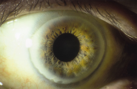
liver disease. A case report and literature review. Cornea 12:74–77, 1993.

16. Schilsky ML: Diagnosis and treatment of Wilson's disease. Pediatr Transplant 6:15–19, 2002.

17. Goyal V, Tripathi M: Sunflower cataract in Wilson's disease. J Neurol Neurosurg Psychiatry 69:133, 2000.

18. Lossner A, Lossner J, Bachmann H, et al: The Kayser-Fleischer ring during long-term treatment in Wilson's disease (hepatolenticular degeneration). A follow-up study. Graefes Arch Clin Exp Ophthalmol 224:152–155, 1986.

19. Esmaeli B, Burnstine MA, Martonyi CL, et al: Regression of Kayser-Fleischer rings during oral zinc therapy: correlation with systemic manifestations of Wilson's disease. Cornea 15:582–588, 1996.

20. Ferenci P: Diagnosis and current therapy of Wilson's disease. Aliment Pharmacol Ther 19:157–165, 2004.

21. Butler P, McIntyre N, Mistry PK: Molecular diagnosis of Wilson disease. Mol Genet Metab 72:223–230, 2001.

22. Fleming CR, Dickson ER, Wahner HW, et al: Pigmented corneal rings in non-Wilsonian liver disease. Ann Intern Med 86:285–288, 1977.

23. Lipman RM, Deutsch TA: A yellow-green posterior limbal ring in a patient who does not have Wilson's disease. Arch Ophthalmol 108:1385, 1990.

24. Alagille D: Alagille syndrome today. Clin Invest Med 19:325–330, 1996.

25. Oda T, Elkahloun AG, Pike BL, et al: Mutations in the human Jagged1 gene are responsible for Alagille syndrome. Nat Genet 16:235–242, 1997.

26. Alagille D, Estrada A, Hadchouel M, et al: Syndromic paucity of interlobular bile ducts (Alagille syndrome or arteriohepatic dysplasia): review of 80 cases. J Pediatr 110:195–200, 1987.

27. Hingorani M, Nischal KK, Davies A, et al: Ocular abnormalities in Alagille syndrome. Ophthalmology 106:330–337, 1999.

28. Nischal KK, Hingorani M, Bentley CR, et al: Ocular ultrasound in Alagille syndrome: a new sign. Ophthalmology 104:79–85, 1997.

29. Andrews W, Sommerauer J, Roden J, et al: 10 years of pediatric liver transplantation. J Pediatr Surg 31:619–624, 1996.

30. Bruckner R, Batschelet E, Hugenschmidt F: The Basel longitudinal study on aging. Doc Ophthalmol 64:235–310, 1986.

31. Chua BE, Mitchell P, Wang JJ, et al: Corneal arcus and hyperlipidemia: findings from an older population. Am J Ophthalmol 137:363–365, 2004.

32. Chambless LE, Fuchs FD, Linn S, et al: The association of corneal arcus with coronary heart disease and cardiovascular disease mortality in the Lipid Research Clinics Mortality Follow-up Study. Am J Public Health 80:1200–1204, 1990.

33. Crispin S: Ocular lipid deposition and hyperlipoproteinaemia. Prog Retin Eye Res 21:169–224, 2002.

34. Eye 3:240–250, 1989.

35. Winder AF: Relationship between corneal arcus and hyperlipidaemia is clarified by studies in familial hypercholesterolaemia. Br J Ophthalmol 67:789–794, 1983.

36. Civeira F: Guidelines for the diagnosis and management of heterozygous familial hypercholesterolemia. Atherosclerosis 173:55–68, 2004.

37. Barchiesi BJ, Eckel RH, Ellis PP: The cornea and disorders of lipid metabolism. Surv Ophthalmol 36:1–22, 1991.

38. Mahley RW, Rall SC: Type III hyperlipoproteinemia (dysbetalipoproteinemia): the role of apolipoprotein E in normal and abnormal lipoprotein metabolism. In: Scriver CR, Beaudet AL, Sly WS, Valle D (eds). The Metabolic and Molecular Bases of Inherited Disease. New York: McGraw-Hill, 1995:1953–1980.

39. Azoulay M, Henry I, Tata F, et al: The structural gene for lecithin: cholesterol acyl transferase (LCAT) maps to 16q22. Ann Hum Genet 51:129–136, 1987.

40. Idzior WB: Familial LCAT deficiency. Przegl Lek 58:919–923, 2001.

41. Carlson LA, Holmquist L: Evidence for deficiency of high density lipoprotein lecithin: cholesterol acyltransferase activity (alpha-LCAT) in fish eye disease. Acta Med Scand 218:189–196, 1985.

42. Carlson LA, Philipson B: Fish eye disease. A new familial condition with massive corneal opacities and dyslipoproteinaemia. Lancet 2:922–924, 1979.

43. Brooks-Wilson A, Marcil M, Clee SM, et al: Mutations in ABC1 in Tangier disease and familial high-density lipoprotein deficiency. Nat Genet 22:336–345, 1999.

44. Chu FC, Kuwabara T, Cogan DG, et al: Ocular manifestations of familial high-density lipoprotein deficiency (Tangier disease). Arch Ophthalmol 97:1926–1928, 1979.

45. Guffon N, Souillet G, Maire I, et al: Follow-up of nine patients with Hurler syndrome after bone marrow transplantation. J Pediatr 133:119–125, 1998.

46. Gullingsrud EO, Krivit W, Summers CG: Ocular abnormalities in the mucopolysaccharidoses after bone marrow transplantation. Longer follow-up. Ophthalmology 105:1099–1105, 1998.

47. Muenzer J: The Mucopolysaccharidoses: A heterogeneous group of disorders with variable pediatric presentations. J Pediatr 144(Suppl 5):S27–S34, 2004.

48. Kenyon KR: Ocular manifestations and pathology of systemic mucopolysaccharidoses. Birth Defects Orig Artic Ser 12:133–153, 1976.

49. Goldberg MF, Maumenee AE, McKusick VA: Corneal dystrophies associated with abnormalities of mucopolysaccharide metabolism. Arch Ophthalmol 74:516–520, 1965.

50. von Noorden GK, Zellweger H, Ponseti IV: Ocular findings in Morquio-Ullrich's disease with report of two cases. Arch Ophthalmol 64:585–591, 1960.

51. Kenyon KR, Topping TM, Green WR, et al: Ocular pathology of the Maroteaux-Lamy syndrome (systemic mucopolysaccharidosis type VI). Am J Ophthalmol 73:718–741, 1972.

52. Goldberg MF: A review of selected inherited corneal dystrophies associated with systemic diseases. Birth Defects 7:13–25, 1971.

53. Amir N, Zlotogora J, Bach G: Mucolipidosis type IV: clinical spectrum and natural history. Pediatrics 79:953–959, 1987.

54. Traboulsi EI, Maumenee IH: Ophthalmologic findings in mucolipidosis III (Pseudo-Hurler Polydystrophy). Am J Ophthalmol 102:592–597, 1986.

55. Libert J, van Hoof F, Farriaux JP, et al: Ocular findings in I-cell disease (mucolipidosis type II). Am J Ophthalmol 83:617–628, 1977.

56. Quigley HA, Goldberg MF: Conjunctival ultrastructure in mucolipidosis III (pseudo-Hurler polydystrophy). Invest Ophthalmol 10:568–580, 1971.

57. Emery JM, Green WR, Wyllie RG, et al: GM-1-gangliosidosis. Ocular and pathological manifestations. Arch Ophthalmol 85:177–187, 1971.

58. Newman NJ, Starck T, Kenyon KR, et al: Corneal surface irregularities and episodic pain in a patient with mucolipidosis IV. Arch Ophthalmol 108:251–254, 1990.

59. Kasmann-Kellner B, Weindler J, Pfau B, et al: Ocular changes in mucopolysaccharidosis IV A (Morquio A syndrome) and long-term results of perforating keratoplasty. Ophthalmologica 213:200–205, 1999.

60. Mullaney P, Awad AH, Millar L: Glaucoma in mucopolysaccharidosis 1-H/S. J Pediatr Ophthalmol Strabismus 33:127–131, 1996.

61. Quigley HA, Maumenee AE, Stark WJ: Acute glaucoma in systemic mucopolysaccharidosis I-S. Am J Ophthalmol 80:70–72, 1975.

62. Cahane M, Treister G, Abraham FA, et al: Glaucoma in siblings with Morquio syndrome. Br J Ophthalmol 74:382–383, 1990.

63. Kakkis ED, Muenzer J, Tiller GE, et al: Enzyme-replacement therapy in mucopolysaccharidosis I. N Engl J Med 344:182–188, 2001.

64. Levy HL, Brown AE, Williams SE, et al: Vitreous hemorrhage as an ophthalmic complication of galactosemia. J Pediatr 129:922–925, 1996.

65. Schweitzer KS: Early diagnosis of inherited metabolic disorders towards improving outcome: the controversial issue of galactosaemia. Eur J Pediatr 162(Suppl 1):S50–S53, 2003.

66. Beigi B, O'Keefe M, Bowell R, et al: Ophthalmic findings in classical galactosaemia—prospective study. Br J Ophthalmol 77:162–164, 1993.

67. Walter JH, Collins JE, Leonard JV: Recommendations for the management of galactosaemia. UK Galactosaemia Steering Group. Arch Dis Child 80:93–96, 1999.

68. Badawi N, Cahalane SF, McDonald M, et al: Galactosaemia—a controversial disorder. Screening and outcome. Ireland 1972–1992. Ir Med J 89:16–17, 1996.

69. Poggi-Travert F, Fournier B, Poll-The BT, et al: Clinical approach to inherited peroxisomal disorders. J Inherit Metab Dis 18(Suppl 1):S1–S18, 1995.

70. Folz SJ, Trobe JD: The peroxisome and the eye. Surv Ophthalmol 35:353–368, 1991.

71. Hittner HM, Kretzer FL, Mehta RS: Zellweger syndrome: lenticular opacities indicating carrier status and lens abnormalities characteristic of homozygotes. Arch Ophthalmol 99:1977–1982, 1981.

72. Cruysberg JR, Wevers RA, van-Engelen BG, et al: Ocular and systemic manifestations of cerebrotendinous xanthomatosis. Am J Ophthalmol 120:597–604, 1995.

73. Moghadasian MH, Salen G, Frohlich JJ, et al: Cerebrotendinous xanthomatosis: a rare disease with diverse manifestations. Arch Neurol 59:527–529, 2002.

74. Dotti MT, Rufa A, Federico A: Cerebrotendinous xanthomatosis: heterogeneity of clinical phenotype with evidence of previously undescribed ophthalmological findings. J Inherit Metab Dis 24:696–706, 2001.

75. Berginer VM, Salen G, Shefer S: Long-term treatment of cerebrotendinous xanthomatosis with chenodeoxycholic acid. N Engl J Med 311:1649–1652, 1984.

76. Caruso RC, Kaiser-Kupfer MI, Muenzer J, et al: Electroretinographic findings in the mucopolysaccharidoses. Ophthalmology 93:1612–1616, 1986.

77. Stanesu-Segal B, Evrard P: Zellweger syndrome, retinal involvement. Metab Pediatr Syst Ophthalmol 12:96–99, 1989.

78. Cohen SM, Brown FR 3rd, Martyn L, et al: Ocular histopathologic and biochemical studies of the cerebrohepatorenal syndrome (Zellweger syndrome) and its relationship to neonatal adrenoleukodystrophy. Am J Ophthalmol 96:488–501, 1983.

79. Mowat AP: Liver Disorders in Childhood 2nd ed. London: Butterworths, 1987:201–202.

80. Martinez M: Abnormal profiles of polyunsaturated fatty acids in the brain, liver, kidney and retina of patients with peroxisomal disorders. Brain Res 583:171–182, 1992.

81. Shimozawa N, Suzuki Y, Orii T, et al: Biochemical and morphologic aspects of peroxisomes in the human rectal mucosa: diagnosis of Zellweger syndrome simplified by rectal biopsy. Pediatr Res 24:723–727, 1988.

82. Moser HW, Moser AE, Singh I, et al: Adrenoleukodystrophy: survey of 303 cases: biochemistry, diagnosis and therapy. Ann Neurol 16:628–641, 1984.

83. Glasgow BJ, Brown HH, Hannah JB, et al: Ocular pathologic findings in neonatal adrenoleukodystrophy. Ophthalmology 94:1054–1060, 1987.

84. Lyons CJ, Castano G, McCormick AQ, et al: Leopard spot retinal pigmentation in infancy indicating a peroxisomal disorder. Br J Ophthalmol 88:191–192, 2004.

85. Aouburg P, Scotto J, Rocchiccioli F, et al: Neonatal adrenoleukodystrophy. J Neurol Neurosurg Psychiatry 49:77–86, 1986.

86. Rizzo WB, Leshner RT, Odone A, et al: Dietary erucic acid therapy for X-linked adrenoleukodystrophy. Neurology 39:1415–1422, 1989.

87. Miike T, Taku K, Tamura T, et al: Clinical improvement of adrenoleukodystrophy following intravenous gammaglobulin therapy. Brain Dev 11:134–137, 1989.

88. Aubourg P, Blanche S, Jambaque I, et al: Reversal of early neurologic and neuroradiologic manifestations of X-linked adrenoleukodystrophy by bone marrow transplantation. N Engl J Med 322:1860–1866, 1990.

89. Aubourg P, Adamsbaum C, Lavallard-Rousseau MC, et al: A two-year trial of oleic and erucic acids (Lorenzo's oil) as treatment for adrenomyeloneuropathy. N Engl J Med 329:745–752, 1993.

90. Shoulders CC, Brett DJ, Bayliss JD, et al: Abetalipoproteinemia is caused by defects of the gene encoding the 97 kDa subunit of a microsomal triglyceride transfer protein. Hum Mol Genet 2:2109–2116, 1993.

91. Bishara S, Merin S, Cooper M, et al: Combined vitamin A and E therapy prevents retinal electrophysiological deterioration in abetalipoproteinaemia. Br J Ophthalmol 66:767–770, 1982.

92. Duker JS, Belmont J, Bosley TM: Angioid streaks associated with abetalipoproteinemia. Arch Ophthalmol 105:1173–1174, 1987.

93. Crocker AC, Farber S: Niemann-Pick disease: A review of eighteen patients. Medicine—(Baltimore) 37:1–95, 1958.

94. Walton DS, Robb RM, Crocker AC: Ocular manifestations of group A Niemann-Pick disease. Am J Ophthalmol 85:174–180, 1978.

95. Bayever E, August CS, Kamani N, et al: Allogeneic bone marrow transplantation for Niemann Pick disease (type IA). Bone Marrow Transplant 10(Suppl 1):S85–S86, 1992.

96. Bembi B, Comelli M, Scaggiante B, et al: Treatment of sphingomyelinase deficiency by repeated implantations of amniotic epithelial cells. Am J Med Genet 44:527–533, 1992.

97. Sidransky E, Tsuji S, Martin BM, et al: DNA mutation analysis of Gaucher patients. Am J Med Genet 42:331–336, 1992.

98. Jaime S, Dalmas MF: A case of Gaucher's disease associated with peripheral retinal ischemia. J Fr Ophtalmol 12:461–463, 1989.

99. Tsai P, Lipton JM, Sahdev I, et al: Allogenic bone marrow transplantation in severe Gaucher disease. Pediatr Res 31:503–507, 1992.

100. Fallet S, Grace ME, Sibille A, et al: Enzyme augmentation in moderate to life-threatening Gaucher disease. Pediatr Res 31:496–502, 1992.

101. Barton NW, Brady RO, Dambrosia JM, et al: Replacement therapy for inherited enzyme deficiency: macrophage-targeted glucocerebrosidase for Gaucher's disease. N Engl J Med 324:1464–1470, 1991.

102. Fine RN, Wilson WA, Donnell GA: Retinal changes in glycogen storage disease type I. Am J Dis Child 115:328–331, 1968.

103. Traboulsi EI, Krush AJ, Gardner EJ, et al: Prevalence and importance of pigmented ocular fundus lesions in Gardner's syndrome. N Engl J Med 316:661–667, 1987.

104. Aiello LP, Traboulsi EI: Pigmented fundal lesions in a preterm infant with familial adenomatous polyposis. Arch Ophthalmol 111:302–303, 1993.

105. Burn J, Chapman PD, Delhanty J, et al: The UK Northern Region genetic register for familial adenomatous polyposis coli: use of age of onset, congenital hypertrophy of the retinal pigment epithelium and DNA markers in risk calculation. J Med Genet 28:289–296, 1991.

106. Olschwang S, Tiret A, Laurent PP, et al: Restriction of ocular fundus lesions to a specific subgroup of APC mutations in adenomatous polyposis coli patients. Cell 75:959–968, 1993.

107. Rhodes M, Bradburn DM: Overview of screening and management of familial adenomatous polyposis. Gut 33:125–131, 1992.

108. Shields JA, Shields CL, Eagle RC Jr, et al: Adenocarcinoma arising from congenital hypertrophy of retinal pigment epithelium. Arch Ophthalmol 119:597–602, 2001.

109. Wells AD, McDonnell PJ, Burnand KG: Purtscher's retinopathy in acute pancreatitis. Br J Surg 77:820, 1990.

110. Cohen SY, Gaudric A, Chaine G, et al: Rétinopathie des pancréatites. J Fr Ophtalmol 12:261–265, 1989.

111. Behrens-Baumann W, Scheurer G: Morbus Purtscher. Variationsbreite der klinischen Manifestationen bei 11 Patienten und Uberlegungen zur Pathogenese. Klin Monatsbl Augenheilkd 198:99–107, 1991.

112. Sanders RJ, Brown GC, Brown A, et al: Purtscher's retinopathy preceding acute pancreatitis. Ann Ophthalmol 24:19–21, 1992.

113. Hollo G, Popik E: Is retinopathy in pancreatitis caused by leukocyte emboli? Acta Ophthalmol (Copenh) 70:820–823, 1992.

114. Grey RH: Visual field changes following hepatic transplantation in a patient with primary biliary cirrhosis. Br J Ophthalmol 75:377–380, 1991.

115. Welsh BM, Smith AL, Elder JE, et al: Night blindness precipitated by isotretinoin in the setting of hypovitaminosis A. Australas J Dermatol 40:208–210, 1999.

116. Hopkins DJ, Horan E, Burton IL, et al: Ocular disorders in a series of 332 patients with Crohn's disease. Br J Ophthalmol 58:732–737, 1974.

117. Danzi JT: Extraintestinal manifestations of idiopathic inflammatory bowel disease. Arch Intern Med 148:297–302, 1988.

118. Kochhar R, Mehta SK, Nagi B, et al: Extraintestinal manifestations of idiopathic ulcerative colitis. Indian J Gastroenterol 10:88–89, 1991.

119. Greenstein AJ, Janowitz HD, Sachar DB: The extra-intestinal complications of Crohn's disease and ulcerative colitis: a study of 700 patients. Medicine—(Baltimore) 55:401–412, 1976.

120. Kaneko E, Nawano M, Honda N, et al: Ulcerative colitis complicated by idiopathic central serous chorioretinopathy with bullous retinal detachment. Dig Dis Sci 30:896–900, 1985.

121. Billson FA, De Dombal FT, Watkinson G, et al: Ocular complications of ulcerative colitis. Gut 8:102–106, 1967.

122. Pomonis E, Triantafillidis JK, Tjenaki M, et al: Report of Eales' disease and ulcerative colitis in the same patient. Am J Gastroenterol 87:1531–1532, 1992.

123. Lyons JL, Rosenbaum JT: Uveitis associated with inflammatory bowel disease compared with uveitis associated with spondyloarthropathy. Arch Ophthalmol 115:61–64, 1997.

124. Soukiasian SH, Foster CS, Raizman MB: Treatment strategies for scleritis and uveitis associated with inflammatory bowel disease. Am J Ophthalmol 118:601–611, 1994.

125. Orchard TR, Chua CN, Ahmad T, et al: Uveitis and erythema nodosum in inflammatory bowel disease: clinical features and the role of HLA genes. Gastroenterology 123:714–718, 2002.

126. Rosenbaum JT, Smith JR: Anti-TNF therapy for eye involvement in spondyloarthropathy. Clin Exp Rheumatol 20:S143–S145, 2002.

127. Hugot JP, Chamaillard M, Zouali H, et al: Association of NOD2 leucine-rich repeat variants with susceptibility to Crohn's disease. Nature 411:599–603, 2001.

128. Ogura Y, Bonen DK, Inohara N, et al: A frameshift mutation in NOD2 associated with susceptibility to Crohn's disease. Nature 411:603–606, 2001.

129. McKay DM: Intestinal inflammation and the gut microflora. Can J Gastroenterol 13:509–516, 1999.

130. Wahl C, Liptay S, Adler G, et al: Sulfasalazine: a potent and specific inhibitor of nuclear factor kappa B. J Clin Invest 101:1163–1174, 1998.

131. Hampe J, Cuthbert A, Croucher PJ, et al: Association between insertion mutation in NOD2 gene and Crohn's disease in German and British populations. Lancet 357:1925–1928, 2001.

132. Relman DA, Schmidt TM, MacDermott RP, et al: Identification of the uncultured bacillus of Whipple's disease. N Engl J Med 327:293–301, 1992.

133. Rickman LS, Freeman WR, Green WR, et al: Brief report: uveitis caused by Tropheryma whippelii (Whipple's 
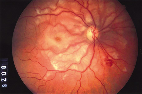
bacillus). N Engl J Med 332:363–366, 1995.

134. Collins ML, Traboulsi EI, Maumenee IH: Optic nerve head swelling and optic atrophy in the systemic mucopolysaccharidoses. Ophthalmology 97:1445–1449, 1990.

135. Harper CG, Giles M, Finlay-Jones R: Clinical signs in the Wernicke-Korsakoff complex: a retrospective analysis of 131 cases diagnosed at necropsy. J Neurol Neurosurg Psychiatry 49:341–345, 1986.

136. Eggspuhler AW, Bauerfeind P, Dorn T, et al: Wernicke encephalopathy—a severe neurological complication in a clinically inactive Crohn's disease. Eur Neurol 50:184–185, 2003.

137. Togay IC, Yigit A, Mutluer N: Wernicke's encephalopathy due to hyperemesis gravidarum: an under-recognised condition. Aust N Z J Obstet Gynaecol 41:453–456, 2001.

138. De La Paz MA, Chung SM, McCrary JA 3rd: Bilateral internuclear ophthalmoplegia in a patient with Wernicke's encephalopathy. J Clin Neuroophthalmol 12:116–120, 1992.

139. Lengyel D, Weissert M, Schmid L, et al: Eye movement abnormalities as a sign for the diagnosis in Niemann-Pick disease type C. Klin Monatsbl Augenheilkd 214:50–52, 1999.

140. Grover WD, Naiman JL: Progressive paresis of vertical gaze in lipid storage disease. Neurology 21:896–899, 1971.

141. Horikawa H, Juo K, Mano Y, et al: A case of neurovisceral storage disease with sea-blue histiocyte and severe horizontal supranuclear ophthalmoplegia. Rinsho Shinkeigaku 30:62–67, 1990.

142. Miller NR: Topical diagnosis of neuropathic ocular motility disorders. In: Miller NR (ed). Walsh and Hoyt's Clinical Neuro-Ophthalmology. 4th ed, vol 2. Baltimore: Williams & Wilkins, 1985:652–784.

143. Schwartz MA, Selhorst JB, Ochs AL, et al: Oculomasticatory myorhythmia: A unique movement disorder occurring in Whipple's disease. Ann Neurol 20:677–683, 1986.

144. Bradbury JA, Martin L, Strachan IM: Acquired Brown's syndrome associated with Hurler-Scheie's syndrome. Br J Ophthalmol 73:305–308, 1989.

145. Hoft RH, Pflugfelder SC, Forster RK, et al: Clinical evidence for hepatitis B transmission resulting from corneal transplantation. Cornea 16:132–137, 1997.

146. Khalil A, Ayoub M, el-Din-Abdel-Wahab KS, et al: Assessment of the infectivity of corneal buttons taken from hepatitis B surface antigen seropositive donors. Br J Ophthalmol 79:6–9, 1995.

147. Lee HM, Naor J, Alhindi R, et al: Detection of hepatitis C virus in the corneas of seropositive donors. Cornea 20:37–40, 2001.

148. Holland EJ, Bennett SR, Brannian R, et al: The risk of cytomegalovirus transmission by penetrating keratoplasty. Am J Ophthalmol 105:357–360, 1988.

Back to Top