Chapter 33
Systemic Infectious and Inflammatory Diseases
KRISTA M. RAMONAS and ALAN H. FRIEDMAN
Main Menu   Table Of Contents

Search

VIRAL DISEASES
BACTERIAL DISEASES
FUNGAL DISEASES
PARASITIC DISEASES
SELECTED INFLAMMATORY DISEASES
ACKNOWLEDGMENTS
REFERENCES

VIRAL DISEASES

HERPES SIMPLEX VIRUS

The herpes simplex virus (HSV) is a common cause of ocular, perioral, and genital infection in humans. The viral particle consists of a core of double-stranded DNA surrounded by a protein capsid and lipid envelope. After the primary infection, HSV survives in latent form inside sensory nerve ganglia and may reactivate at any time producing recurrent disease. HSV type 1 accounts for the majority of herpes labialis and ocular herpetic infections. HSV type 2 causes genital herpes infections.1

HSV has a worldwide distribution, and humans are the only known reservoir. HSV keratitis is a serious cause of blindness, and is second in incidence only to trachoma in terms of corneal-related causes of blindness. Transmission requires close personal contact. HSV type 1 is transmitted primarily by oral secretions and HSV type 2 by genital secretions. Primary exposure to HSV type 1 commonly occurs in childhood. By puberty, 60% to 85% of the population has serologic evidence of HSV type 1 infection. In contrast, the incidence of HSV type 2 cases does not begin to increase until approximately 14 years of age and ranges from 3% in sexually inactive populations to 70% in prostitutes.2

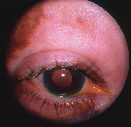
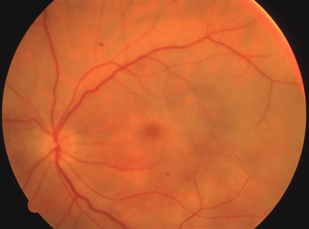
The incubation period for HSV is from 2 to 14 days. Primary infection with HSV type 1 is often asymptomatic but may produce oral, eye, or skin lesions (Fig. 1). In children younger than age 5, a painful gingivostomatitis or pharyngitis with fever and cervical adenopathy is the most common presentation. HSV type 2 infections usually involve the glans penis and penile shaft in men, and the vulva, perineum, buttocks, cervix, and vagina in women.1 Fever, malaise, anorexia, and tender inguinal adenopathy may accompany the typical vesicular skin eruption. The ulcerated lesions are exquisitely tender and may persist for several weeks before healing.

Fig. 1. Primary lesion of herpes simplex on the face of a child.

Congenital HSV infection is recognized at birth by jaundice, hepatosplenomegaly, central nervous system abnormalities, bleeding diatheses, microcephaly, seizure disorders, fever, chorioretinitis, and skin vesicles. It must be differentiated from similar syndromes caused by rubella, toxoplasmosis, and cytomegalovirus. Neonatal infection may be acquired in passing through the infected birth canal or by retrograde spread of the infection into the uterine cavity.

Less common presentations of HSV infection include primary infections of the skin of the fingers (whitlows). These are caused by HSV type 1 in medical and dental personnel exposed to oral secretions. In the general community this lesion is caused by HSV type 2 infection. HSV type 1 may also cause a skin infection that mimics herpes zoster, although the dermatomal distribution is not usually maintained and the pain is less severe. On rare occasions, herpes simplex can cause a necrotizing hemorrhagic encephalitis of the temporal lobes marked by rapid deterioration to coma and death.

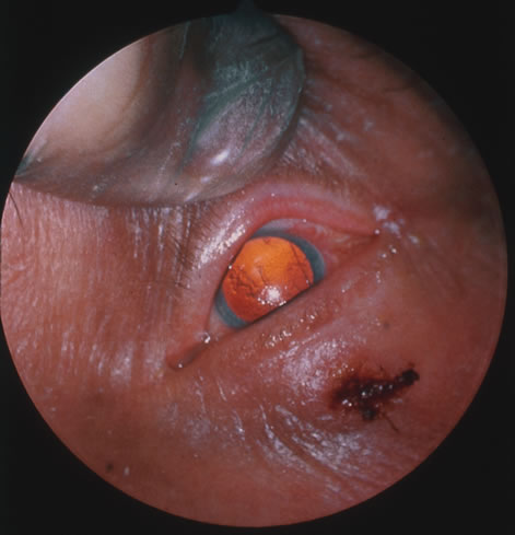
Reactivation of HSV may be precipitated by fever, trauma, menstruation, emotional stress, or systemic illness. Recurrent episodes of HSV type 1 and type 2 are generally preceded by several hours of burning, itching, and tingling before the skin vesicles appear. Systemic symptoms tend to be mild or absent, with the exception of recurrent adenopathy. The lesions appear similar to the primary infection and are frequently painful. The most frequent regional recurrences of HSV type 1 are on the vermilion border of the outer lip (herpes labialis) and in the eye (Fig. 2). Recurrences of HSV type 2 characteristically produce lesions on the penile shaft and glans in the male and on the labia and perineum in the female. Headache and aseptic meningitis are occasionally associated with a genital recurrence. Further recurrences may be seen at monthly or yearly intervals.

Fig. 2. Recurrent herpes simplex virus (HSV) type 2 on the face of a patient with aquired immune deficiency syndrome (AIDS).

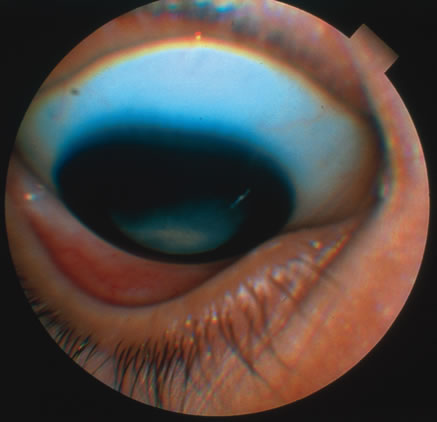
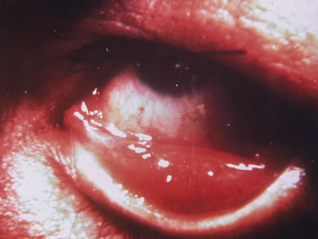
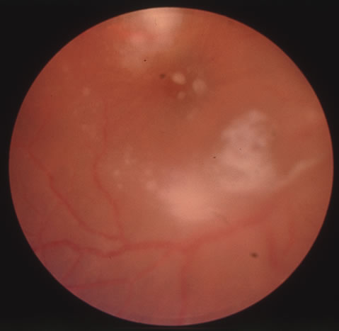
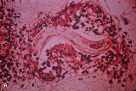
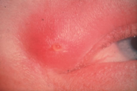
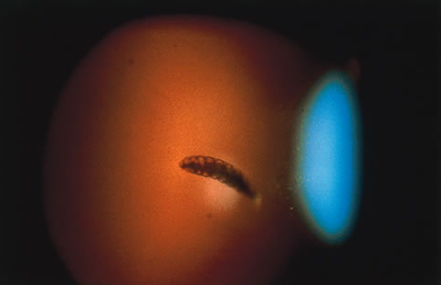
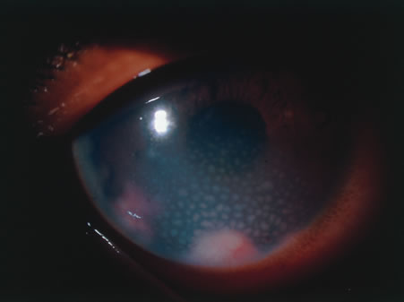
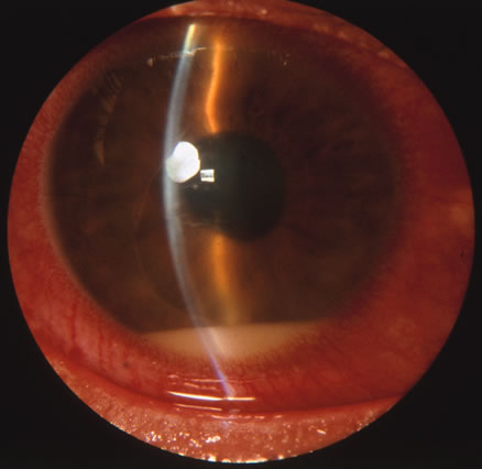
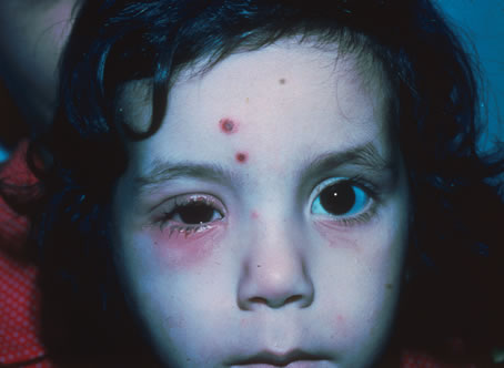
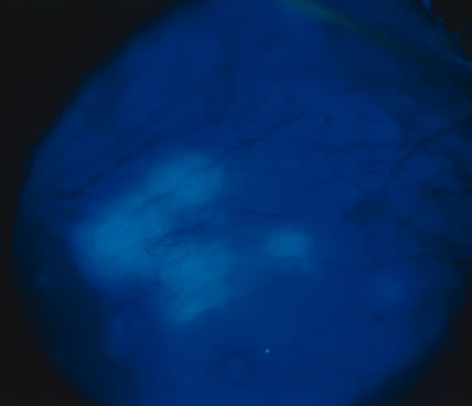
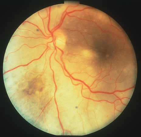
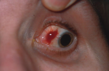
In primary ocular HSV infections, a severe follicular conjunctivitis with regional adenopathy is present. Vesicles may appear on the eyelid skin or lid margin, producing blepharitis (Fig. 3). Corneal involvement initially takes the form of a superficial punctate keratitis, which may progress to dendritic or geographic ulceration. Stromal infiltrates and uveitis are less common and relatively mild in primary disease. In uncomplicated cases, lesions usually heal completely in 2 to 3 weeks without scarring. Most cases of ocular HSV are recurrences. Recrudescent ocular herpes may take the form of dendritic or geographic ulcers, recurrent erosions, interstitial or disciform stromal keratitis, and anterior uveitis.3 HSV may also be an agent of retinitis and acute retinal necrosis (Fig. 4).

Fig. 3. Lesions of herpes simplex virus (HSV) type 1 on the surface of the eye after topical fluorescein administration.

Fig. 4. Acute retinal necrosis in the fundus. Note the widespread nature of the involvement.

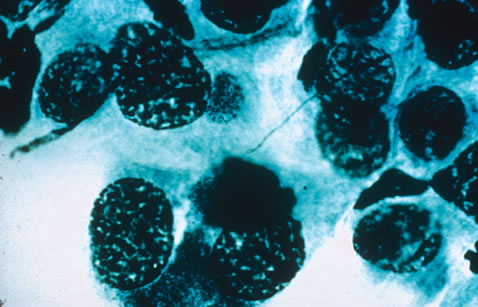
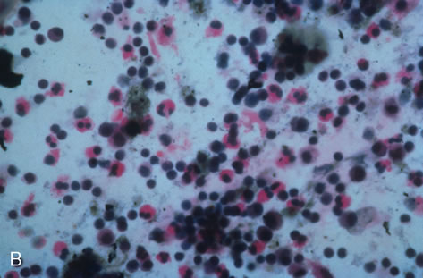
Clinical diagnosis of HSV infection can be made when the characteristic vesicular lesions are present. The diagnosis can be confirmed by isolation of the virus in tissue culture when necessary. Scraping the base of the lesions and Wright's, Giemsa, or Papanicolaou's stains will demonstrate the characteristic giant cells or intranuclear inclusions of herpesvirus infection.

Most infections with HSV are mild or asymptomatic and require no specific treatment; however, antiviral therapy can be life saving in cases of neonatal herpes, disseminated infections, and herpes encephalitis. Famciclovir and valacyclovir effective in reducing the pain, healing time, and duration of viral shedding in patients with first episode genital herpes and significantly shortens the clinical and virologic course in recurrent episodes. Immunocompromised patients with chronic progressive oral herpes have also demonstrated improved healing times and decreased viral shedding when treated with oral antivirals.4 Intravenous acyclovir is the drug of choice in the treatment of neonatal herpes infections and herpes encephalitis.

The corneal scarring caused by recurrent infection with HSV may be visually disabling and may require corneal transplantation. Over the course of the last decade, the Herpetic Eye Disease Study Group (HEDS) examined the short-term and long-term effects of oral acyclovir and topical steroids on epithelial keratitis, stromal keratitis and iritis. Long-term oral acyclovir was shown to reduce the rate of recurrent HSV epithelial keratitis and stromal keratitis, especially in those who have had multiple prior episodes of stromal keratitis.5,6 Topical steroids are beneficial in preventing progression of stromal inflammation and shortening duration of stromal keratitis. Topical steroids should be given in conjunction with topical Viroptic (King Pharmaceuticals, Bristol, TN) in those with stromal disease or disciform keratitis, but steroids should be avoided in those with epithelial disease. The HEDS trial showed a beneficial trend in the treatment of anterior herpetic uveitis with oral acyclovir, but the trend failed to reach statistical significance.7 Nevertheless, practitioners routinely use oral acyclovir in addition to topical steroids plus a cycloplegic for herpetic-related uveitis. Topical steroids are often difficult to discontinue and many patients require low-dose therapy for an indefinite amount of time.

VARICELLA AND HERPES ZOSTER

Varicella zoster virus (VZV) is an enveloped DNA-containing virus that is morphologically indistinguishable from other members of the herpesvirus family. Varicella is the primary infection. It usually occurs in children and is characterized by a generalized vesicular rash (chickenpox) with insignificant systemic manifestations. Herpes zoster is a reactivation of VZV in adults resulting in a dermatomal eruption of vesicles (shingles) which is usually accompanied by pain in the affected nerve distribution.8

Varicella occurs most commonly in the late winter and early spring. The highest attack rate is in the 2- to 8-year-old group. The infection is highly contagious and is presumably transmitted through direct person-to-person contact or by respiratory droplets. Approximately 96% of susceptible children will develop the disease within 1 month of exposure to the index case. Herpes zoster infections are seen in patients of all ages and occur with equal frequency throughout the year. Almost all infections occur with a history of prior exposure and are believed to be the result of reactivation of latent virus. The incidence of herpes zoster increases with age and is greater in immunocompromised persons.

Varicella usually begins 2 weeks after exposure to a preexisting case. There may be a short prodromal period before the rash develops marked by fever, headache, malaise, and lethargy. The exanthem usually starts on the scalp and the trunk and first appears as erythematous macules that progress to papules and then vesiculate. The lesions remain infectious until they crust over, which occurs in approximately 1 week. Successive crops of new lesions appear so that all stages and sizes of lesions may be present at the same time. Mucosal surfaces in the mouth and conjunctiva may be involved as well. The active course of an uncomplicated case is usually over in 7 to 10 days after which the virus remains in a latent state within the dorsal root ganglion of the sensory nerves.

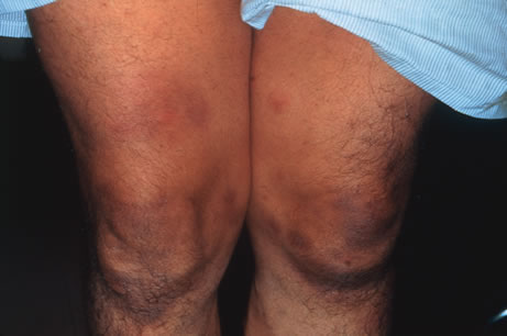
In the majority of cases of herpes zoster, pain and paresthesias precede the skin eruption by several days. The skin lesions morphologically resemble those of varicella; however, they tend to be larger and more bullous. The lesions conform to dermatomal distributions and do not cross the midline. The thoracic nerves are most frequently involved followed by the trigeminal and cervical nerves.

Herpes zoster occurs more commonly in patients with impaired immunity such as those with neoplastic disease, particularly lymphoproliferative disease, or human immunodeficiency virus (HIV).9 In uncomplicated herpes zoster the majority of patients younger than 60 will have complete resolution of their pain and skin lesions within 4 weeks. The persistence of pain beyond this point (postherpetic neuraglia) is more common in the elderly population.

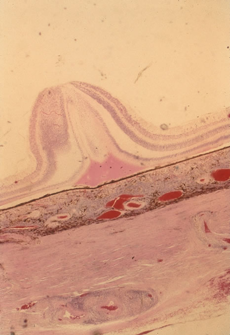
Most ophthalmic involvement by varicella is mild.10 Pocks on the eyelids and a nonspecific papillary conjunctivitis with a watery discharge are common findings. Foci of inflammatory cells may appear on the bulbar conjunctiva, limbus, or peripheral cornea either during the acute illness or weeks to months afterward. These pock-like lesions often ulcerate and are associated with conjunctival hemorrhage and a mucopurulent discharge containing primarily neutrophils. When on the cornea, they may leave faint superficial scars with neovascularization. Occasionally, varicella can cause a punctate, dendritic, geographic, or disciform keratitis associated with a mild to moderate iridocyclitis. Unusual ocular complications of varicella include necrosis of the eyelids, interstitial keratitis, corneal melting, cataract, and optic neuritis. Congenital infection may result in microphthalmia, cataract, chorioretinitis, microcephaly, deafness, and cardiac abnormalities.

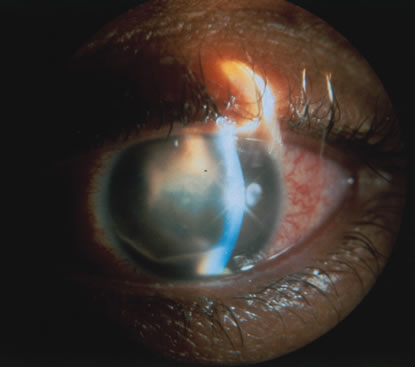
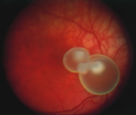
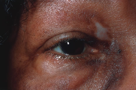
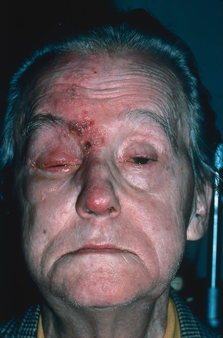
Involvement by VZV of the first division of the trigeminal nerve is referred to as herpes zoster ophthalmicus regardless of whether the eye is inflamed (Fig. 5). Eighty-five percent of patients with cutaneous eruptions on the side of the tip or midportion of the nose (Hutchinson's sign) will have ocular involvement. Varicella zoster virus can cause blepharitis, canaliculitis, episcleritis, scleritis, conjunctivitis, keratitis, iridocyclitis, uveitis, retinal vasculitis, retinitis, choroiditis, and optic neuritis. Cataracts and glaucoma are late complications related to the uveitis and long-term use of topical corticosteroid therapy.

Fig. 5. Facial involvement in a case of zoster. Note the characteristic strict segmental distribution of the lesions.

Corneal complications occur in the majority of patients with herpes zoster ophthalmicus. A punctate epithelial keratopathy with pseudodendrite formation is the earliest and most common lesion that occurs. Stromal infiltrates, disciform reactions, and rarely interstitial keratitis are also observed. Scarring and contracture of the lid margin often results in trichiasis, distichiasis, entropion, or ectropion. Corneal sensation is reduced, and neurotrophic ulceration, corneal melting, or perforation may ensue. Extraocular muscle involvement with facial paresis has been observed.11

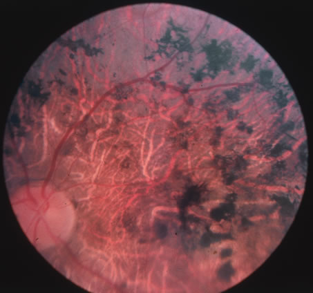
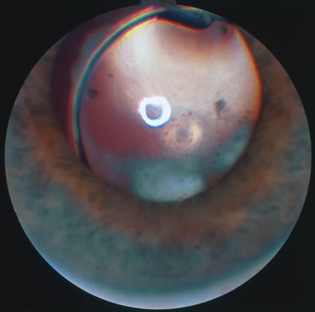
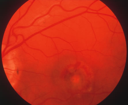
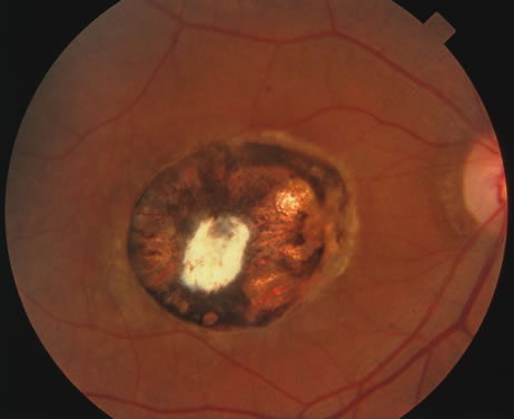
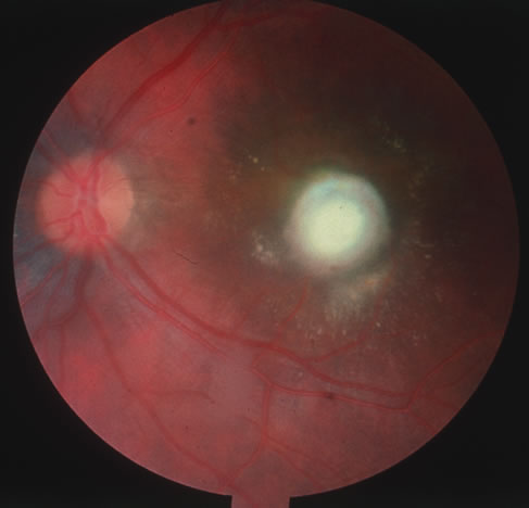
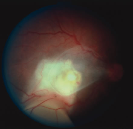
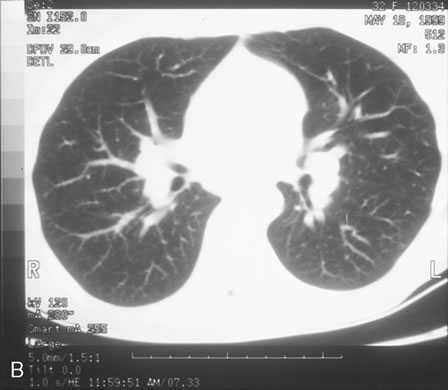
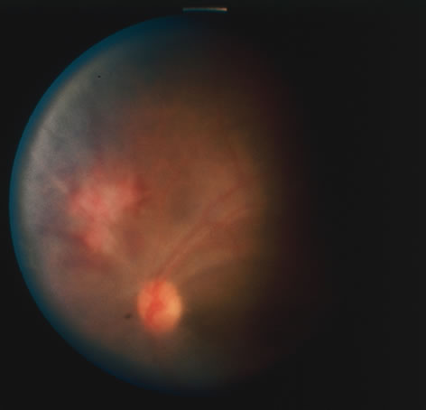
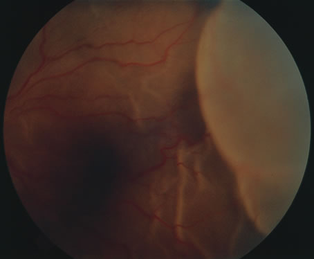
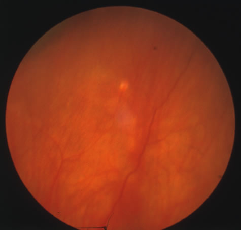
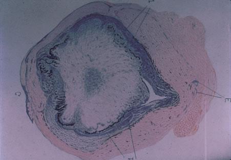
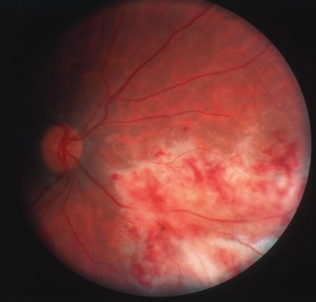
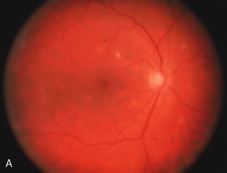
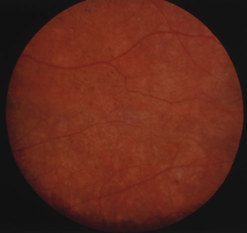
The acute retinal necrosis (ARN) syndrome is most often caused by the herpes zoster virus, although occasionally it may be a result of herpes simplex infection.12 It is typically described in healthy patients although association with immunosuppressed patients has also been described. Granular, nonhemorrhagic areas of retinal necrosis may be observed in the fundus, often rapidly coalescing with resulting blindness often caused by retinal detachment. There is often an associated vasculitis and vitritis. In immunocompromised patients VZV or HSV retinitis may also take the pattern of progressive outer retinal necrosis (PORN) (Fig. 6). PORN differs from ARN in that the former is multifocal, localized to the outer retina, and is less often associated with vasculitis and vitritis.

Fig. 6. Fundus lesions of progressive outer retinal necrosis (PORN). Note the multifocal lesions.

Diagnosis of varicella is usually not difficult, given the frequently obtainable history of exposure to a preexisting case and the characteristic rash. Serologic confirmation can be obtained by demonstrating increasing anti-VZV antibody titers. Multinucleated giant cells and eosinophilic intranuclear inclusions can be demonstrated by Papanicolaou's or other stains of smears from acute skin lesions.

The clinical diagnosis of herpes zoster can be made in the presence of the characteristic vesicular lesions in a dermatomal distribution. Scrapings of the lesions demonstrate giant cells and intracellular inclusions. It is important to note that although most patients with herpes zoster are otherwise healthy, the possibility of an underlying systemic illness should be considered if the patient is young (i.e., younger than 50 years old) or the history suggests other systemic problems.

The treatment of varicella is primarily supportive, consisting of analgesia, antipyretics, antipruritics, and good skin hygiene to prevent secondary infection. The use of aspirin in children is to be avoided in view of the belief that it may predispose to Reye's syndrome following varicella. Most eye disease is mild and self-limited and does not require treatment.

Oral acyclovir (Zovirax) given five times daily early in the course of zoster infection has been shown to reduce the duration of viral shedding and the severity of acute pain. The greatest benefit is seen when medication is begun within 72 hours of onset of skin lesions. Two newer antiviral agents available for the treatment of herpes zoster are valacyclovir (Valtrex) and famcyclovir (Famvir). These agents have the advantage of a simpler dosing regimen, three times daily rather than five times daily with acyclovir, and a more favorable pharmacokinetic profile.13 It is thought that famcyclovir may also help speed resolution of postherpetic neuralgia. Valacyclvoir and famcyclovir are now preferred to acyclovir as first-line therapy for the treatment of zoster. They are well-tolerated drugs with few systemic side effects other than a requirement for reduced dosing in patients with renal failure. Oral corticosteroids also seem to have a beneficial effect in reducing acute pain and speeding cutaneous healing. They have not been shown to have an effect on postherpetic neuralgia.

Topical corticosteroids and cycloplegics are commonly used to treat the inflammation associated with herpes zoster ophthalmicus.10 Tear supplements, bandage contact lenses, surgical correction of lid abnormalities, and tarsorrhaphy may be necessary to promote the healing of epithelial defects. Vision-threatening pathologic processes such as chorioretinitis and optic neuritis should be treated aggressively with intravenous acyclovir and high doses of prednisone.

Treatment of postherpetic neuralgia is often disappointing. Routine analgesic drugs are generally ineffective. Cimetidine, colchicine, phenothiazines, and tricyclic antidepressants have been used with some success. Capsaicin cream and lidocaine patches may also be applied for local relief. The pain tends to slowly diminish and disappear over the course of 1 to 2 years but may be prolonged, especially in elderly patients. For those with severe intractable pain, intrathecal injection of methylprednisolone acetate once weekly for 4 weeks has recently been shown to provide relief.14

CYTOMEGALOVIRUS INFECTION

Cytomegalovirus (CMV) is a herpes virus responsible for severe congenital malformations, a mononucleosis syndrome in young adults, and disseminated lesions in the immunosuppressed, especially retinal infection and pneumonia in patients with acquired immunodeficiency syndrome (AIDS). Patients with AIDS with CD4 counts between 50 to 100 cells per microliter are particularly susceptible to CMV retinitis. If the retinitis is untreated the prognosis is very poor often resulting in full-thickness retinal necrosis and blindness. Despite treatment, CMV retinitis often recurs in up to 50% of cases.15

In the United States, 20% of people are seropositive for CMV by age 20. Approximately 1% of infections are congenital and 5% perinatal. Anti-CMV immunoglobulin (Ig) G is present in greater frequency in homosexual men, on the order of 80% to 90%. The virus may be transmitted via blood, cervical secretions, semen, and breast milk and as an intrauterine infection. Serologic testing for CMV is not routinely performed because of this frequent exposure in the general population.

Approximately 10% of congenitally infected newborns have permanent sequelae. The full syndrome is manifest as hepatosplenomegaly with hepatitis and cirrhosis, maculopapular rash, purpura, hemolytic anemia, pneumonia, microcephaly, microgyria, encephalitis, chorioretinitis, pathologic bone fractures, and growth retardation. Otherwise asymptomatic children may develop late sensorineural hearing loss or mental retardation.

In normal older children and adults, the most common manifestation is an atypical mononucleosis, comprising fever and lymphocytosis with atypical lymphocytes. Lymphadenopathy and pharyngitis may be present, and rubelliform rashes occur. Heterophil antibodies are negative.

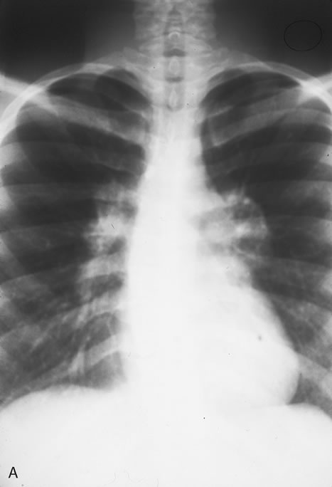
In the immunocompromised, especially organ transplant recipients and patients with AIDS, CMV commonly is responsible for serious clinical syndromes. It causes an interstitial pneumonia, resulting in dyspnea and dry cough, with interstitial infiltrates on the roentgenogram. Subacute encephalitis, enterocolitis, adrenalitis, and adrenal necrosis may be noted.

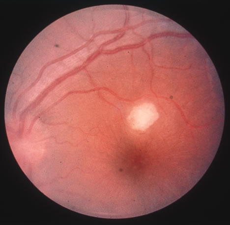
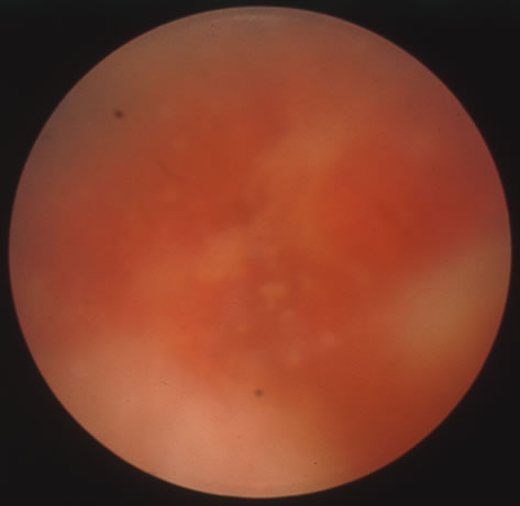
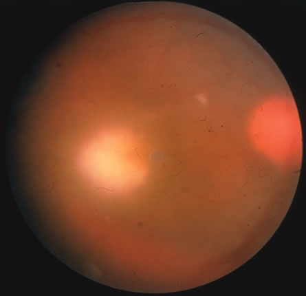
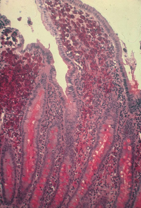
CMV retinitis is common in patients with AIDS. Perivascular granular white or yellowish infiltrates with hemorrhage are typical (Fig. 7). Minimal vitritis or anterior uveitis may be present. The posterior pole, including optic nerve, or peripheral retina may be involved. Optic neuritis may be retrobulbar. Resolution of retinal lesions reveals local retinal atrophy and pigment dispersion. Iridocyclitis may occur secondary to infection of ciliary processes. CMV retinitis in patients with AIDS is considered a poor prognostic sign in terms of life expectancy. Congenital CMV infection may also cause retinitis, as well as anterior uveitis, cataract, and optic atrophy. New foci of retinochoroiditis can develop in later years of congenital infection, and such infants require periodic reexamination as long as virus is shed in the urine.

Fig. 7. Cytomegalovirus retinitis in a patient with aquired immune deficiency syndrome (AIDS). Note the large area of retinal necrosis along with the perivascular distribution of the lesions.

In the immunocompromised patient, other pathogens must be excluded, even in the presence of positive CMV cultures or serology. Tissue culture methods may take as long as 4 weeks. IgM antibody develops in primary infection and then disappears over 6 to 9 months, but it may reappear with reactivation of infection. The IgM test has unfortunately low specificity and sensitivity in infants and is less helpful than urine culture obtained at birth or in the first weeks of life.

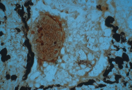
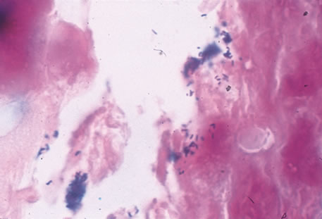
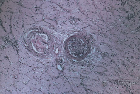
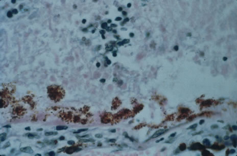
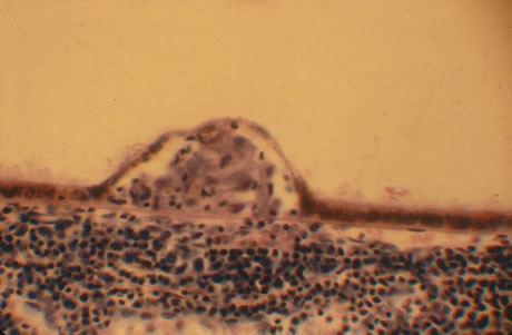
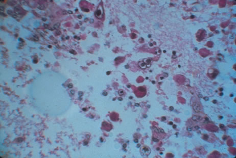
Any cell type may be infected, appearing larger than normal (cytomegalic) and demonstrating eosinophilic intranuclear “owl's eye” and smaller intracytoplasmic viral inclusion bodies, which are better seen with Giemsa or Papanicolaou's stains (Fig. 8). Histologic examination of tissue specimens, including bronchoalveolar lavage fluid and urine, may be useful for diagnosis.

Fig. 8. Typical lesion of cytomegalovirus (CMV) produces retinal necrosis with large cells possessing owl's eye inclusions.

Gancyclovir, or dihydroxypropoxymethyl guanine (DHPG), prevents DNA synthesis in infected cells. It is recommended for consideration in immunocompromised patients with retinitis, pneumonitis, enterocolitis, encephalitis; in infants with severe congenital or acquired disease; and in otherwise healthy patients with acute severe primary cytomegalovirus infection. It is effective in treatment of various CMV lesions, resulting in clinical stabilization or improvement. However, relapse almost always occurs when the drug is discontinued. An exception to this rule has been noted of late in patients who sustain elevated CD4 counts through the aid of highly active antiretroviral therapy (HAART).16 HAART has dramatically increased the life expectancy of patients with AIDS with low CD4 counts, and it has also been shown to improve the outcome of patients with CMV retinitis.17 Gancyclovir is limited to intravenous use, and neutropenia occurs in approximately 20% of patients. The newer agent valgancyclovir (Valcyte; Roche, Basel, Switzerland) is an oral formulation that is considered more efficacious than its original intravenous counterpart. It is being used for induction as well as maintenance therapy. Zidovudine, a reverse transcriptase inhibitor often used in the treatment of HIV, also results in neutropenia and should not be used in association with gancyclovir.18

Intravenous foscarnet, cidofovir and fomivirsen are other anticytomegaloviral agents that are currently available.19,20 The primary toxicity of foscarnet and cidofovir is renal and requires strict monitoring of blood, urea, and nitrogen (BUN) and creatinine levels during treatment. After high-dose induction therapy, most patients require life-long maintenance therapy for all anticytomegaloviral agents. In those who cannot tolerate the systemic side effects associated with intravenous therapy, intravitreal injection or intravitreal implantation of gancyclovir is a consideration.21

ACQUIRED IMMUNODEFICIENCY SYNDROME

AIDS is a debilitating and fatal disease caused by HIV, characterized by T-cell lymphocytopenia, and development of secondary infections and tumors. CMV retinitis and other ocular manifestations of HIV-related disease are common.

For purposes of reporting and surveillance, the Centers for Disease Control (CDC) defines AIDS in terms of specific indicator diseases and laboratory evidence of HIV infection.22 In general, a case of AIDS is a reliably diagnosed opportunistic disease at least moderately indicative of underlying cellular immunodeficiency and with no other known cause of underlying cellular immunodeficiency or reduced resistance reported to be associated with an opportunistic disease, including secondary immunodeficiencies associated with immunosuppressive therapy, lymphoreticular malignancy, or starvation.

According to the CDC, as of the year 2000 approximately 800,000 people currently living in the United States have been infected with HIV, with approximately 40,000 newly diagnosed cases per year. Worldwide incidence of HIV infection is a much more menacing burden because more than 30 million people have been reportedly infected with the virus. The majority of these individuals are African American men, but during the last decade women have represented increasingly larger percentages of those afflicted. Homosexual intercourse is still the most common route of infection. However transmission of the virus is now more common in heterosexual sex, as well as through the sharing of contaminated needles in intravenous drug users. The mid to late 1990s showed a decline in the number of AIDS-related deaths as more potent antiretroviral medication became available. More recently there has been a leveling off in terms of the number of AIDS-related fatalities. Some experts postulate that as resistance begins to develop among patients on the newer, more potent drug regimens, there will again be a resurgence of deaths because of AIDS.23

Transplacental transmission from an infected mother is estimated to occur in 50% of cases. Breast milk may be a source of infection. There is currently no evidence for vector-borne transmission or infection via casual household contacts. The health care worker's risk for infection via needle stick is approximately 1 in 1000 punctures. The virus is very sensitive to heat, lipid solvents, and common antiseptics.

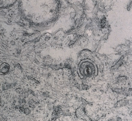
HIV was previously known as human, T-cell lymphotropic virus type 3 (HTLV-III) and lymphadenopathy-associated virus (LAV). There are two known types of virus: HIV type 1, which causes AIDS, and HIV type 2, the disease spectrum of which is uncertain. HIV type 2 has been found in some African cases of AIDS. HIV is a retrovirus, meaning it contains a reverse transcriptase enzyme that produces a double-stranded DNA copy (cDNA) from the virus RNA genome (Fig. 9).24 HIV infection involves attachment of viral envelope glycoprotein to the CD4 molecule present on T-helper (T4, OKT4, or CD4+) cell surfaces. The virus then fuses with the cell membrane, enters cytoplasm, loses its envelope, and reverse transcription of RNA to DNA occurs. Viral DNA becomes integrated into host cell DNA as latent provirus, in which state it may go unrecognized by the immune system. The proviral DNA can then be transcribed and translated, producing viral proteins and viral genome RNA, which assemble and bud through the host cell plasma membrane as new virions. Antigenic stimulation of lymphocyte activity appears to be important in conversion from latency to progressive infection.

Fig. 9. An electron micrograph of the human immunodeficiency virus type 1 (HIV-1) in the retina.

HIV infection results in depletion of T4 cells, which are central to various forms of immune responsiveness. Risk of developing AIDS is inversely related to the absolute T4 cell count. Antigen-responsive T cells become selectively depleted or functionally impaired early in the course of AIDS. There is diminished helper function for B-cell responses, depressed cytotoxic and natural killer cell responsiveness, decreased blast transformation, and decreased lymphokine production. Infected patients' B-lymphocytes spontaneously produce immunoglobulin, resulting in hypergammaglobulinemia, but are unable to produce specific antibody in response to new infections. Anergy is common. HIV also infects other cells that express the CD4 antigen, such as monocytes and macrophages, but is not cytopathic in these cells. HIV has been identified in brain capillary endothelium, oligodendrocytes, astrocytes, lymph node dendritic cells, Langerhans' cells, intestinal mucosa, myocardium, tears, corneal epithelium, aqueous, vitreous, and retina.

Clinical manifestations of HIV are protean. Seropositive persons may be asymptomatic. Various acute HIV infection syndromes have been described. An acute mononucleosis-like syndrome, with fever, malaise, rash, lymphadenopathy, pharyngitis, myalgias, and arthralgias, has been noted after recent HIV exposure. Acute neurologic manifestations resembling aseptic meningitis, encephalitis, or focal lesions may be seen.25 AIDS-related complex is a somewhat ambiguous term encompassing features such as chronic fever, night sweats, weight loss, fatigue, generalized lymphadenopathy, chronic diarrhea, and laboratory abnormalities. AIDS-related complex implies an intermediate stage in the spectrum of HIV disease.

CDC-defined indicator diseases include HIV encephalopathy, HIV wasting syndrome, CMV disease not involving liver, spleen or lymph nodes (26), progressive multifocal leukoencephalopathy, recurrent Salmonella septicemia, multiple or recurrent serious bacterial infections in children, extrapulmonary myobacterial infection, disseminated histoplasmosis, disseminated coccidioidomycosis, chronic isosporiasis, lymphoid interstitial pneumonia or pulmonary lymphoid hyperplasia in children, primary brain lymphoma, and various non-T–cell non-Hodgkin's lymphomas. Less specifically associated with HIV infection are manifestations of papillomavirus, molluscum contagiosum, VZV, Epstein-Barr virus, hepatitis B virus, chlamydia, gonorrhea, syphilis, campylobacter, pityrosporum, trichophyton, aspergillus, acanthamoeba, entamoeba, giardia, strongyloides, basal cell carcinoma, squamous cell carcinoma, idiopathic thrombocytopenic purpura, cerebrovascular infarction, and intracranial hemorrhage.27–30

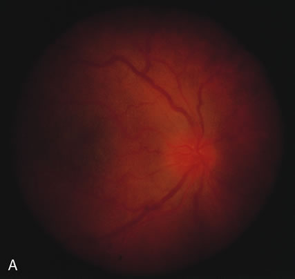
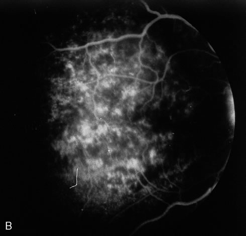
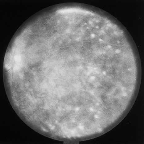
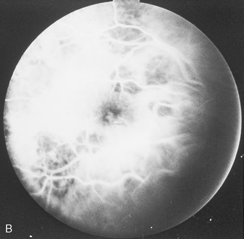
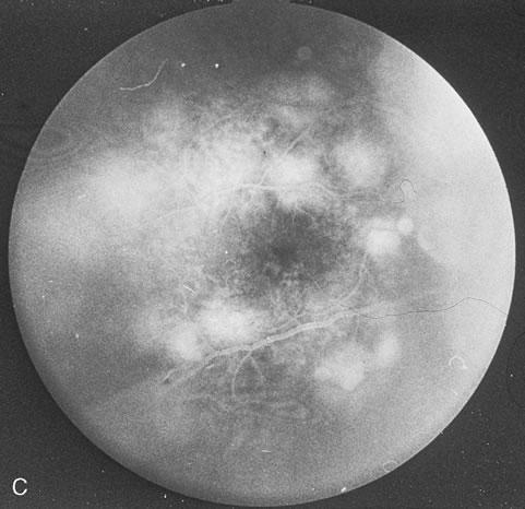
Pneumocystis carinii pneumonia once was the most common presentation of AIDS. It is typically of insidious onset, with features including dry cough, dyspnea, and a diffuse bilateral interstitial infiltrate evident on a chest roentgenogram. Typical multifocal fundus lesions have been described. They are yellow-white and have a characteristic pattern on fluorescein angiography (Fig. 10A, 10B, and 10C). Diagnosis is made by demonstrating the protozoon via sputum induction, bronchioalveolar lavage, and transbronchial or open-lung biopsies. Treatment modalities include trimethoprim-sulfamethoxazole (TMP-SMX).31

Fig. 10. A. The retinal involvement of Pneumocystis carinii is manifested by multifocal, yellow-white lesions. B and C. On fluorescein angiography, the lesions fail to delineate in the early phase (B) but stain in the late phase (C).

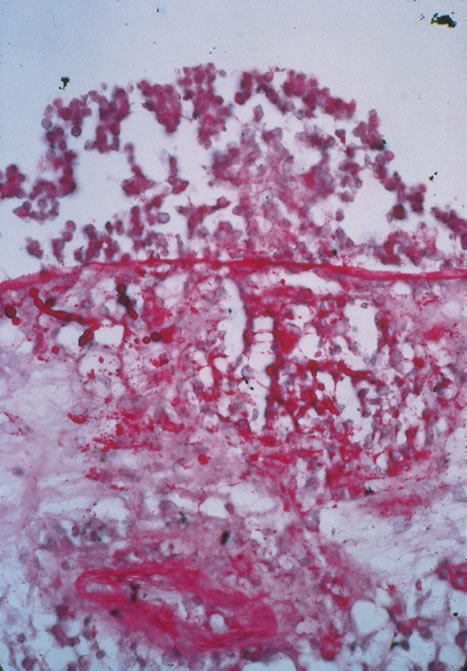
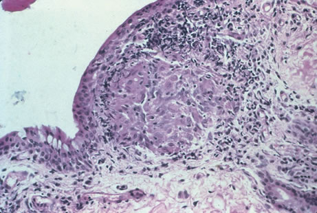
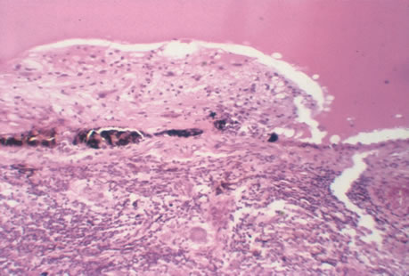
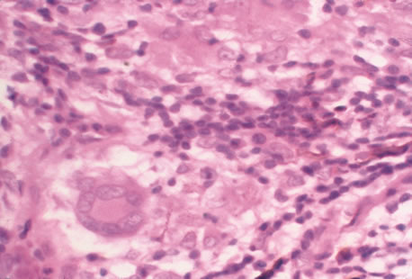
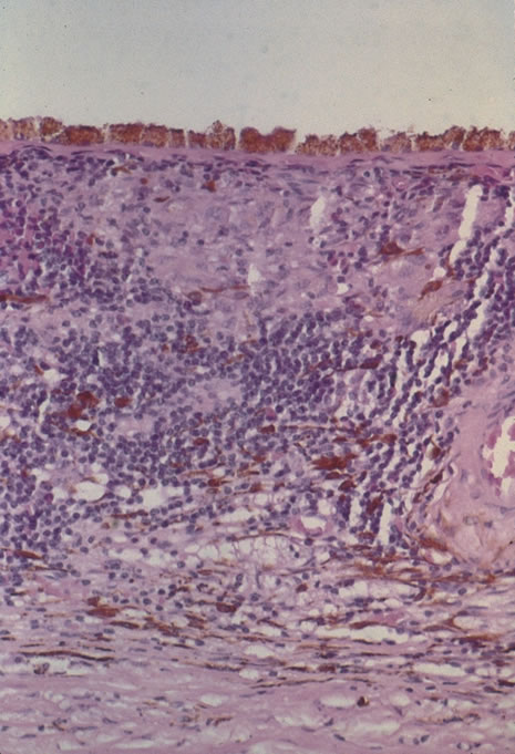
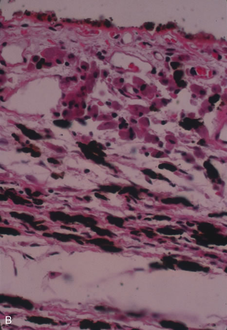
Kaposi's sarcoma is much more frequent in male homosexuals and bisexuals than in other patients with AIDS. Cutaneous lesions are usually red or violaceous, palpable, and nonblanching papules, nodules, or plaques, often appearing on the hard palate, nose, eyelid, and conjunctiva (Fig. 11). Visceral and lymph node involvement is common and often clinically silent. Biopsy specimens show proliferation of small, incompletely formed blood vessels lined by unusually large endothelial type cells, with extravasated red blood cells and infiltrates of spindle-shaped cells. Chemotherapy with etoposide, Vinca alkaloids, or interferon-α may be indicated in advanced cases.28

Fig. 11. The typical lesion of Kaposi's sarcoma on the conjunctiva.

Mycobacterium avium-intracellulare (MAI), M. kansasii, and other atypical mycobacteria most commonly present as disseminated infections in patients with AIDS, with fever, sweats, chills, malaise, weight loss, and pulmonary and gastrointestinal symptoms. Signs of malabsorption, jaundice, skin lesions, and oral ulcers may be noted. Blood cultures, stool examination, and biopsy of various sites including lymph nodes, liver, gastrointestinal tract, skin and bone marrow may be useful for diagnosis by smear or culture. Antituberculosis therapy may improve symptoms, but such infections are generally regarded as incurable in those with AIDS. The fundus lesions of MAI consist of discrete yellow-white choroidal foci (Fig. 12A and 12B).32 Extrapulmonary M. tuberculosis is an indicator disease and is covered elsewhere in this chapter.

Fig. 12. A. Mycobacterium avium-intracellulare (MAI) in the fundus produces multiple, discrete yellow intrachoroidal lesions. B. Histopathology of the choroidal infiltrate shows histiocytes laden with acid fast bacilli.

Cryptosporidium species and Isospora belli are intestinal protozoa causing severe diarrhea in the immunosuppressed. Among patients with AIDS, it is more common in homosexual males and children. Stool specimens and intestinal and other biopsies may yield the organisms for diagnosis. Disease is usually self-limited but may be life-threatening in the compromised patient. Therapy is mainly supportive. Spiramycin, α-difluoromethylornithine, and furazolidone have been used to treat cryptosporidiosis, and TMP-SMX for isosporiasis, all with limited success. Giardiasis and amoebic colitis are also seen in patients with AIDS.33

Progressive multifocal leukoencephalopathy is a chronic central demyelinating disease caused by the papovavirus JC (Jacob-Creutzfeldt virus). The disease may begin with subtle encephalopathic signs, later demonstrating various focal deficits. Visual loss is frequently reported. Computed tomography typically shows multiple hypodense nonenhancing lesions in white matter, and cerebrospinal fluid is usually normal or shows elevated protein. Diagnosis is made by brain biopsy. There is no effective therapy.25

Lymphocytic interstitial pneumonitis (LIP) occurs in approximately half of children with AIDS. Pulmonary lymphoid hyperplasia (PLH) is a nodular variant of LIP. The LIP/PLH complex is yet another AIDS-related chronic pulmonary disease presenting with diffuse bilateral interstitial infiltrates. Biopsy demonstrates either nodular peribronchiolar lymphoid aggregates, or diffuse alveolar and peribronchiolar lymphocytic and plasma cell infiltrates. Exacerbations may indicate superimposed infection. Corticosteroid therapy is controversial.30,31

CMV, herpes simplex and zoster, candidiasis, cryptococcosis, toxoplasmosis, and other AIDS-related infections are described in detail elsewhere in this chapter.

The ophthalmic findings associated with AIDS are reviewed in another chapter of this section. Approximately 80% of HIV-positive patients will have ocular manifestations during the course of their illness. Commonly encountered anterior segment manifestations include keratoconjunctivtis sicca, infectious keratitis, iritis, herpes-zoster ophthalmicus, Kaposi's sarcoma of the lid or conjunctiva, molluscum contagiosum and conjunctival microvasculopathy. Posterior segment manifestations include cytomegalovirus retinitis, VZV retinitis, toxoplasmic retinochoroiditis, infectious choroiditis, bacterial and fungal retinitis and the more recently reported chronic multifocal retinal infiltrates.32–39 Neuroophthalmic manifestations include papilledema, cranial nerve palsies, ocular motility disorders and visual field defects. They occur in 10% to 15% of patients with HIV infection.

Serologic diagnosis of HIV infection uses enzyme-linked immunosorbent assay (ELISA) kits for serum antibody detection. These assays are highly sensitive and may demonstrate false-positive results. Positive tests are therefore confirmed by repeat ELISA, followed by the Western blot test, which relies on the banding pattern of serum antibody to electrophoretically separated viral antigens.

Seroconversion usually occurs within 2 to 3 months, but may take much longer. Assay results will occasionally be read as indeterminate, indicating a reaction pattern that does not meet criteria for positivity, and retesting in several months is advised. Because of placental transfer of antibodies, a seropositive child younger than 15 months old must have other evidence of infection to support a diagnosis, such as clinical disease, positive HIV cultures, or HIV antigen testing. Serologic testing for HIV requires written informed patient consent in some locales, at least in part because of social stigmata associated with AIDS. Testing is now mandatory in the United States for blood donors, federal prisoners, military recruits and active-duty personnel, foreign-service officers, and immigrants to the United States.

In addition to HIV serodiagnosis, absolute T4 lymphocytopenia is the most useful laboratory finding related to HIV infection. A count of less than or equal to 400 cells/mm3 increases the risk of progression to AIDS.

Zidovudine (azidothymidine [AZT]) is a nucleoside analogue that inhibits viral reverse transcriptase. Zidovudine is virostatic, having no effect on latent virus. Taken orally, it has been shown to decrease symptoms, opportunistic infections, mortality rate, and HIV antigen values and to improve immunologic function in infected patients. The ability of HIV to undergo rapid antigenic variation of envelope glycoproteins, its low immunogenicity, and our incomplete understanding of what constitutes a protective immune response have contributed to the difficulty in developing an effective vaccine.

Several other antiretroviral agents known by their classification as nucleoside analogues, nonnucleoside analogues, or protease inhibitors have been introduced. Nucleoside analogues inhibit transcription of the HIV virus by blocking the enzyme reverse transcriptase, thereby inhibiting replication of the virus. Nucleoside analogues include Didanosine (ddI), Zalcitabine (ddC), Stavudine, (d4T) and Lamivudine (3TC). Combivir, a combination of AZT and 3TC, is a popular choice in initial HIV therapy. Drugs known as nonnucleoside analogues, nevirapine and delavirdine, may be used in combination with nucleoside analogues. Due to high levels of resistance to AZT monotherapy, various combinations usually consisting of three different drugs are typically employed. This new armamentarium of medications against the human immunodeficiency virus has dramatically lengthened life expectancy and improved quality of life.36

In 1996, the clinical introduction of the protease inhibitor (PI) class of antiretrovirals, promised a dramatic breakthrough in the ability to demonstrate a significant impact on the amount of detectable virus in both blood plasma and lymphoid tissue (LT), the principal site of HIV replication and storage. It has now become standard practice to initiate HAART (highly active antiretroviral therapy) with combinations of PIs and other antiretrovirals to more completely and durably suppress viral replication. Some practitioners wait to introduce the protease inhibitors into the cocktail of medications until later stages of immunosuppression. The most commonly used protease inhibitors are saquinavir, indinavir, and ritonavir.

EPSTEIN-BARR VIRUS

Epstein-barr virus (EBV) is a ubiquitous DNA virus that belongs to the human herpesvirus family. In early childhood the infection is usually subclinical; however in adolescence and early adulthood it is the principal cause of infectious mononucleosis.

By adulthood more than 90% of the population demonstrates antibody to the virus. EBV is often transmitted by intimate contact with asymptomatic carriers. Live virus has been found to persist in the oropharynx for up to 18 months after clinical recovery from the initial infection. Infectious mononucleosis has also occurred after blood transfusions and open heart surgery.

EBV infection is characterized by the classic triad of fever, sore throat and lymphadenopathy. Cervical adenopathy is present in 80% to 90% of patients. Splenomegaly occurs in 50% of patients. Other signs include hepatomegaly, jaundice, palatal petechiae, and rash. A wide spectrum of complications have been observed with EBV infection. These include anemia, thrombocytopenia, hepatitis, pericarditis, myocarditis, meningitis, encephalitis, optic neuritis, Guillain-Barré syndrome, and rarely, splenic rupture. Most patients recover spontaneously over a 2- to 3-week period, although rare fatalities have been reported.

Conjunctivitis is the most commonly reported of all ocular manifestations. Other manifestations include Sjögren's syndrome, conjunctival lymphocytic infiltrates,40 dacryoadenitis,41 keratitis, oculoglandular syndrome, uveitic, choroiditis, retinitis, acute posterior multifocal placoid pigment epitheliopathy (APMPPE), papillitis, and ophthalmoplegia. Even in cases of retinal involvement, EBV rarely if ever leads to permanent loss of vision. Resolution of the posterior segment inflammation usually leads to return of normal acuity.42,43

EBV infection may be related to neoplasia in several settings. This includes Burkitt's lymphoma in patients of African descent and nasopharyngeal carcinoma in Asians.

Diagnosis of EBV may be made clinically and with serologic demonstration of heterophil antibodies. Serum antibody titers to EBV capsid antigen can also be specifically tested. Treatment of EBV is largely supportive. Corticosteroids may be useful in the treatment of severe thrombocytopenia or hemolytic anemia, but are usually of limited value. Antiviral therapy such as acyclovir or interferon-α may be considered for slowly resolving EBV retinitis because both have been shown to inhibit EBV replication in vitro.

MUMPS

The mumps virus is an encapsulated RNA virus in the paramyxovirus family that causes acute parotitis in children and young adults. The virus is transmitted in saliva and respiratory secretions. Most cases of mumps occur in the winter and early spring. The primary clinical features are fever and nonsuppurative salivary gland inflammation. Orchitis is present in 20% to 30% of postpubescent males. Meningitis, meningoencephalitis, pancreatitis, oophoritis, myocarditis, and sensorineural hearing loss are less common complications of mumps infection.44

A nonspecific conjunctivitis is a relatively common finding in patients with mumps infection and is occasionally hemorrhagic. Dacryoadentitis, episcleritis, scleritis, keratitis, optic neuritis, and more recently reported acute posterior multifocal placoid pigment epitheliopathy45 and neuroretinitis46 after mumps.

The diagnosis of mumps can usually be made based on clinical findings. The virus can be cultured from saliva or spinal fluid, and seroconversion can be documented by a variety of assays. Efforts at preventing infection are primarily to avoid the complications of orchitis and encephalitis. Vaccination with live attenuated virus successfully produces immunity in approximately 90% of subjects. Since the licensing of the vaccine in 1967 there has been a more than 99% decline in the annual incidence of mumps in the United States.44 Prior to institution of the vaccine, epidemics of mumps occurred every 2 to 5 years. Mumps is now so rare that only 751 cases were reported to the CDC in 1996. Treatment of mumps is symptomatic and supportive with analgesics and antipyretics.

MEASLES

Measles is a highly contagious viral exanthem caused by an encapsulated single-stranded RNA virus in the paramyxovirus family. The illness is characterized by a prodromal period of malaise, with conjunctival and respiratory irritation followed by a maculopapular rash. Measles has a worldwide distribution. Its incidence has diminished in developed countries as a result of vaccination programs. Between 1990 and 1996, fewer than 1000 cases per year in the United States were reported to the CDC. However, measles still remains a serious cause of childhood illness, blindness, and mortality in developing countries.

Measles is transmitted by direct contact with infectious respiratory droplets. The attack rate exceeds 90% among susceptible persons. The prodromal symptoms develop 10 to 14 days after exposure and precede the rash by three to four days. Bright red spots with a bluish white center (Koplik's spots) appear on the buccal mucosa 1 to 2 days before the exanthem and are considered pathognomonic of measles. The rash consists of reddish, maculopapular lesions that first appear at the hairline and subsequently descend over the face, neck, and upper trunk, reaching the lower extremities on the third or fourth day.47

Pneumonia, laryngotracheitis, otitis media, mastoiditis, lymphadenitis, diarrhea, and keratoconjunctivtis are common complications of measles infection. Although rare, measles encephalitis and hemorrhagic thrombocytopenia lead to significant morbidity and mortality. Subacute sclerosing panencephalitis (SSPE) is believed to be caused by a chronic infection of the brain by the measles virus, leading to a slow and progressive degeneration of intellect and motor function that culminates in decorticate rigidity and death. Chorioretinitis is a common finding among patients with SSPE.48–50 Nystagmus, optic atrophy, papilledema, involuntary eye movements, and cortical blindness also occur with this condition.

Other ocular findings in measles include a nonspecific conjunctivitis with follicles or papillae and conjunctival hyperemia. An occasional Koplik's spot may be seen. In severe cases, punctate erosions of the bulbar conjunctiva are accompanied by a mucopurulent discharge. Subepithelial corneal infiltrates that stain with fluorescein may also appear transiently during the illness.51 Patients with poor nutritional status or vitamin A deficiency may develop corneal ulceration or perforation. Measles encephalitis is associated with optic neuritis, neuroretinitis,52 and extraocular muscle palsies.

A clinical diagnosis of measles can be made in patients with the characteristic prodrome and morbilliform rash. Laboratory confirmation is obtained by demonstrating seroconversion.

Treatment of patients with measles is supportive. Secondary bacterial infections and dehydration should be treated aggressively. The live attenuated virus vaccine gives a 98% conversion rate under ideal conditions. After immunization, fever and a mild rash may develop; rare cases of encephalitis and uveitis have occurred.53 No therapy is required for the conjunctivitis.

RUBELLA

Rubella (German measles) is a childhood exanthem caused by an encapsulated RNA virus of the togavirus group. The viral illness may be totally asymptomatic in adults or present with a mild self-limited fever, morbilliform rash, and lymphadenopathy. The most significant aspect of rubella infection is in its effect on the fetus. Prenatal infection may result in miscarriage, stillbirth or the congenital rubella syndrome.

Since 1969, the year of rubella vaccine licensure, the incidence of rubella infection and congenital rubella have declined 99% in the United States. Most cases occur in the spring, the highest incidence being among children. Patients are most contagious in the week prior to the eruption of the rash and transmit the virus through respiratory droplets. Infants with congenital rubella shed large quantities of virus in their body fluids for months and may retain viable virus in the lens for years after the initial infection.54

Despite immunization programs, 10% to 20% of young adults remain susceptible to rubella infection and limited local outbreaks continue to occur in settings such as schools and military bases where groups of susceptible persons are in close contact.

Typically there are no prodromal symptoms of rubella infection in children: however, adults may experience malaise, fever, and anorexia for several days before the rash erupts. Ocular features generally include conjunctivitis and epithelial keratitis. Lymphadenopathy occurs most commonly in the posterior auricular, posterior cervical, and suboccipital lymph nodes; generalized adenopathy and splenomegaly occasionally are seen. The exanthem of rubella begins on the forehead and face and spreads downward to the trunk and limbs. The lesions are discrete and maculopapular but may coalesce to form a diffuse erythema. Petechial lesions may erupt on the soft palate (Forschheimer spots). In young women, a self-limited arthritis involving the wrists, fingers, and knees may accompany the rash and occasionally persists after the other manifestations of rubella have disappeared.55

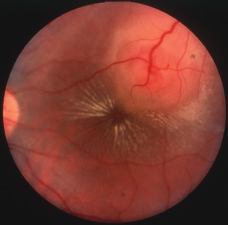
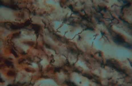
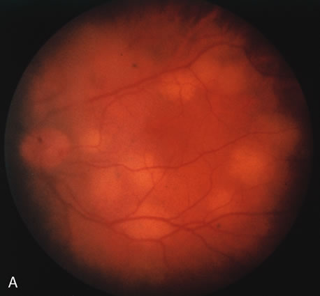
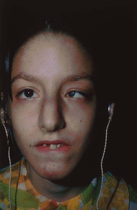
Rubella infections that occur early in gestation are associated with the greatest risk of fetal damage, however, infection in the third trimester may still cause damage. Maternal infection in the first 2 months of pregnancy results in a 40% to 60% chance of multiple congenital anomalies or fetal death. Congenital heart disease, cataracts, and deafness are the classic triad of the congenital rubella syndrome; however, there are many other commonly recognized complications of prenatal rubella infection (Fig. 13). Encephalitis, microcephaly, mental retardation, intrauterine growth retardation, thrombocytopenic purpura, hepatosplenomegaly, obstructive jaundice, interstitial pneumonitis, myocarditis, and radiolucent bone lesions are all possible sequelae of congenital rubella infection.54 The classic salt-and-pepper retinopathy occurs less commonly (Fig. 14), but may be noted to show progression. Other ocular findings include congenital cataracts, microphthalmia and glaucoma. Ocular disease and hearing loss frequently occur together, and glaucoma is frequently associated with microphthalmos.56

Fig. 13. A patient with the congenital rubella syndrome. Note the hearing aids, left esotropia, left microphthalmos and cataract.

Fig. 14. The classic salt-and-pepper fundus of congenital rubella.

The devastating fetal damage caused by maternal rubella infections can only be prevented through rigorous immunization programs. All susceptible women of childbearing age should be vaccinated 3 months before a planned pregnancy or in the postpartum period. As a result of immunization programs there have been no major outbreaks of rubella in the last 35 years.

Postnatal rubella infection may go undetected because of its mild and nonspecific symptomatology. The diagnosis can be made by culturing virus from the throat or by demonstrating rising antibody titers to rubella specific IgG. When suggestive birth defects are present, the diagnosis of congenital rubella requires either viral isolation from the throat, urine, or other body secretions or serologic evidence of infection.

Back to Top
BACTERIAL DISEASES

CHLAMYDIA TRACHOMATIS INFECTION

Chlamydiae are gram-negative intracellular parasites dependent on host cells for energy-related molecules. Chlamydia trachomatis infection is the most common sexually transmitted disease in industrialized countries and the most common cause of eye infection worldwide. It is also an important cause of infant pneumonia and may be related to Reiter's syndrome.

Nongonococcal urethritis (NGU) is the most common manifestation of genital C. trachomatis infection in males, characterized by mucoid or mucopurulent discharge, dysuria, and pruritus. Urethral infection is often mild and may be asymptomatic. Twenty percent of male cases of gonococcal urethritis have simultaneous chlamydial infection. Postgonococcal urethritis due to chlamydia may develop 2 to 3 days after successful treatment of gonorrhea. Chlamydia proctitis is not uncommon in male homosexuals. Epididymitis and Reiter's disease may be complications.

Cervicitis is the most common genital manifestation in female patients. Most women are asymptomatic. Mucopurulent discharge occurs in 40%. Chlamydia infections cause one third of cases of acute salpingitis (pelvic inflammatory disease), often with only mild symptoms of abdominal pain and only 5% positive cervical smears. Four to 21% of pregnant women may have C. trachomatis infection of the cervical canal, thus exposing the newborn at birth. Inclusion conjunctivitis may occur after an incubation period of 6 to 19 days. Thirty to 40% of pneumonias in the early months of life are because of Chlamydia, and approximately half of such children also have conjunctivitis.57

Lymphogranuloma venereum (Nicolas-Favre disease) is a sexually transmitted infection of C. trachomatis serotypes L1, L2, and L3, which are more invasive strains causing disease primarily in lymphatic tissue and rarely associated with Parinaud's oculoglandular syndrome. The primary lesions of lymphogranuloma venereum is a small painless papule, vesicle, or ulcer of the penis or vulvar or anorectal areas occurring 1 to 6 weeks after intercourse. Painful massive regional lymphadenopathy becomes suppurative, and draining fistulas develop within weeks of the primary lesion. Elephantiasis occurs as lymphatic vessels are obstructed.

Trachoma is a chronic infectious keratoconjunctivitis caused by C. trachomatis serotypes A, B, and C. It affects approximately 400 million persons, mainly in parts of developing Africa, the Middle East, and Asia, but also may be seen in natives of the American Southwest. Trachoma may present as an acute conjunctivitis, but classic findings include conjunctival follicles, tarsal scarring and Herbert's pits.59 C. trachomatis serotypes D through K are the most common cause of infectious neonatal conjunctivitis in the United States and the cause of adult inclusion conjunctivitis as well as nonlymphogranuloma venereum genital infections. Adult and neonatal inclusion conjunctivitis and trachoma are reviewed elsewhere in these volumes.

Gram's stain does not detect chlamydia. Tissue culture techniques are most sensitive. These cultures require special transport medium and refrigeration of the specimen and 48 to 72 hours of incubation with the cell culture prior to specific immunofluorescence staining for diagnosis. The McCoy cell culture using fluorescent antibody test for staining has become the gold standard.57 Direct Giemsa stained smears have a relatively low yield, with sensitivity varying from 50% to 90%, demonstrating dark blue or purple intracytoplasmic inclusions in a field of polymorphonuclear leukocytes, lymphocytes, and other cell types (Fig. 15). The microimmunofluorescent antibody stain for smears has approximately 90% sensitivity and 100% specificity compared to cultures. Serum antichlamydial IgM may aid diagnosis in neonatal pneumonia, and high titers of antichlamydial IgG may be found in complications of genital infection such as acute salpingitis. In addition to the McCoy cell culture, polymerase chain reaction may also aid in detection of C. trachomatis DNA.

Fig. 15. A positive Giemsa stain in a case of chlamydial conjunctivitis.

Therapy for genital chlamydia infection is administration of tetracycline, doxycycline, or erythromycin for 2 to 3 weeks. Asymptomatic sexual contacts should be treated as they are likely to harbor genital chlamydial infections. Treatment failures are rare. The recommendation has been made to test conjunctival scrapings three weeks following completion of the course of antibiotics as some patients may require two to three courses of antibiotic therapy.59

TUBERCULOSIS

Mycobacterium tuberculosis is a bacterium that causes infection through inhalation of aerosolized droplets filled with the organism. An asymptomatic pulmonary granuloma may develop and reactivate at any point, particularly when accompanied by diseases such as HIV, malignancy, and diabetes mellitus. Rates of reactivity have been found to be highest in non-white, older men of lower socioeconomic status. In the last decade a resurgence of tuberculosis (TB), particularly in crowded urban settings such as New York City, has been noted.60

Ocular involvement most commonly occurs after reactivation in the lung or alimentary tract and is thus referred to as secondary TB (Fig. 16). Primary ocular involvement of TB is rare and is almost always limited to the cornea and conjunctiva. Conjunctival TB has been reported as a chronic unilateral conjunctivitis with a conjunctival mass or ulceration associated with regional lymphadenopathy. Although also rarely seen, conjunctival phlyctenulosis has been found to be associated with tuberculoprotein hypersensitivity. Biopsy and culture proven scleral TB was reported in the literature in 1976 and 1989. Other ocular manifestations of TB include anterior uveitis, either granulomatous or nongranulomatous, choroiditis, and choriodal tubercles/tuberculoma. A recent case series demonstrated the protean ocular findings associated with intraocular TB in addition to a variable response to antitubercular treatment.61

Fig. 16. A choroidal tubercle in a patient with miliary tuberculosis.

The gold standard for diagnosis of intraocular tuberculosis is demonstration of acid-fast bacilli in fluid or tissue sample either by direct smear and culture, polymerase chain reaction (PCR) or ELISA (Fig. 17). A screening test such as the Mantoux test is useful but is less specific and must be interpreted based on the size of induration in correlation with socioeconomic status, immune status, and contact history of the patient. The test consists of an intradermal injection of purified protein derivative (PPD) from tubercle bacilli. Systemic sensitization to this protein occurs several weeks after primary infection. At 48 to 72 hours postinjection, skin induration larger than 5 mm in an HIV-positive patient or patients with a positive chest x-ray that are previously untreated, larger than 10 mm in other high-risk populations, and larger than 15 mm for healthy patients with low risk factors are considered significant. A positive reaction may persist in the absence of clinically active disease and therefore must be correlated with chest radiography and symptomatology.

Fig. 17. Acid fast bacilli are present in the biopsy of a lesion.

Classic histopathology consists of caseating granulomatous inflammation in a zonal distribution. The noncaseating tubercle may be present but is less specific (Fig. 18). The organism is acid-fast, meaning that it does not decolorize with acid alcohol after staining with basic fuchsin, appearing as bright red, long thin rods. A definitive laboratory diagnosis of intraocular tuberculosis may be made via anterior chamber tap, vitreous or chorioretinal biopsy depending on the site of inflammation. The PCR may be helpful in making the diagnosis.

Fig. 18. Histopathologic examination of an enucleated eye demonstrates a caseating focus in the choroid.

Treatment of tuberculosis consists of at least two drugs because of the increasing incidence of resistance. First-line agents include isoniazid, rifampin, pyrazinamide, ethambutol, and streptomycin. Treatment for adults and children invariably consists of a 2-month treatment with INH, rifampin and pyrazinamide followed by four months of isoniazid (INH) and rifampin. Pyridoxamine may be added to the regimen to prevent INH-induced peripheral neuropathy. Monitoring the response to treatment should be done with sputum cultures. By the end of the third month of treatment most patients are culture-negative. Those that remain culture-positive at the end of 6 months should be considered treatment failures. In a uveitic patient with a positive PPD, a 2-week therapeutic trial of INH may aid in the diagnosis. If ocular inflammation decreases a full course of anti-TB treatment is used.64

Prevention of tuberculosis is through rapid diagnosis, bacille Calmette-Guérin (BCG), vaccination and preventative chemotherapy. BCG vaccine is not used in the United States, but is recommended in countries where the prevalence of TB is high. The vaccine is from a strain of live attenuated bovine tubercle bacilli used to induce an immune response to M. tuberculosis. The vaccine varies greatly in efficacy from 0% to 80%. Patients who have received the BCG vaccine will have a false positive PPD skin test reactivity.

LYME DISEASE

Borrelia burgdorferi is a tick-borne spirochete that produces Lyme disease. Lyme disease was originally named in 1977 when arthritis was observed in a group of children in Lyme, Connecticut. B. burgdorferi is now considered endemic in more than 15 states in the United States, as well as Europe and Asia. In 1999, 16,273 cases of Lyme disease were reported to the CDC, more than 90% from the states in the northeastern and mid-Atlantic region of the United States. While B. afzelii and B. garinii also produce Lyme disease, only B. burgdorferi has been found in the United States.65

Since 1982 when the CDC began surveying for Lyme disease much has been learned regarding diagnosis and treatment of the disease. Although a vaccine against Lyme disease is now available, it requires a series of three injections resulting in only 76% efficacy, and must be followed by booster injections every 1 to 3 years to maintain immunity. The vaccine is only recommended for those who are in frequent or prolonged contact with Ixodes ticks. For everyone else prevention remains the key aspect in controlling the disease. This includes awareness of disease-infested areas particularly in summer months and appropriate use of repellent, protective clothing, and if possible, avoidance of the area.

The vector for the transmission of the bacterium B. burgorferi to humans is the black legged Ixodes tick. Ixodes scapularis are most often found in the northeastern United States and I. pacificus is more commonly found in the western United States. The Ixodes tick is very small, said to be the size of a pinhead, and the tick bite therefore often goes unnoticed. The disease process can be classified into three stages. Stage one occurs several days to weeks after the bite as a slowly expansile area of erythema with an area of central clearing known as the bulls-eye rash or erythema migrans appears. The size of the lesion varies from 5 to 15 cm. Nonspecific symptoms of malaise, fever, myalgias, and arthralgia accompany the rash in the initial stages of infection. While often difficult to diagnose because of the subtlety of findings, this stage is most susceptible to treatment with antibiotic therapy. Stage two occurs as spirochetes spread from the site of initial infection via cutaneous, lymphatic, or blood-borne routes. Signs of disseminated infection include development of multiple smaller annular skin lesions, and involvement of the cardiac, central nervous, and musculoskeletal systems. Cardiac involvement is rare, involving less than 5% of untreated cases, and include atrioventricular block, pericarditis, and cardiomegaly. Neurologic involvement, also known as neuroborreliosis, is slightly more common occurring in 15% of untreated patients. Manifestations include meningitis with neck stiffness and headaches, encephalitis, cranial neuropathy particularly facial nerve palsy, cerebellar ataxia, and myelitis. Stage three consists of late or persistent infection. A late neurologic syndrome called Lyme encephalopathy has been defined in the United States manifesting as subtle difficulty with mentation.66 The most common late manifestation of Lyme disease is arthritis. Sixty percent of untreated patients develop arthritis usually involving the large weight-bearing joints such as the knee.

Lyme disease can involve nearly all structures of the eye. The most common early manifestation is conjunctivitis, occurring in approximately 11% of patients; however, most ocular manifestations of Lyme disease are associated with late stages of the disease. Follicular conjunctivitis, episcleritis, trigeminal neuralgia, and retinal vasculitis have been reported in a recent observational case series of 10 patients with Lyme borreliosis.67 Severe panophthalmitis resulting in blindness was reported in a patient found to have B. burgdorferi in the vitreous.68 Other findings include anterior uveitis, multifocal choroiditis, optic disc edema, and intermediate uveitis (Fig. 19).69

Fig. 19. Intermediate uveitis in a case of Lyme borreliosis.

The diagnosis of Lyme disease is made by clinical presentation and serology. It is, however, appropriate to treat based on clinical findings and a history of a known exposure. CDC recommendations for serologic testing include an ELISA followed by a Western blot. Antibodies are often present for years after exposure to infection, regardless of treatment of the disease. Therefore, seropositivity must not be mistaken for active disease. Treatment recommendations for Lyme disease include a 2- to 3-week course of doxycycline, with substitution with amoxicillin for children and pregnant women.70 Patients with evidence of neurologic abnormalities should undergo a more intensive 2- to 4-week course of intravenous ceftriaxone.

SYPHILIS

Syphilis is a sexually transmitted disease caused by the bacterium Treponema pallidum. A primary skin lesion known as a chancre is followed 3 weeks to 6 months later by hematogenous dissemination of the organism, a stage known as secondary syphilis. Secondary disease manifestations are broad and nonspecific ranging from a maculopapular rash and generalized mucocutaneous lesions to malaise, fever, and painless lymphadenopathy. Less commonly gastrointestinal, hepatic, and renal disease have been reported. Secondary syphilis generally resolves within months but may recur, often within the same year. Late sequelae involving the central nervous system and the aorta is referred to as the tertiary stage of syphilis. Approximately 30% of untreated patients develop tertiary syphilis, but less than 1% develop clinical signs of neurosyphilis. Neurosyphilis can result in a cerebral vasculitis producing symptoms of transient ischemia or infarction, general paresis, or chronic encephalitis producing progressive dementia and tabes dorsalis. Tabes dorsalis is a progressive spinal cord lesion involving the posterior columns and dorsal nerve roots resulting in the classic broad-based foot slapping gait. The average interval from infection to central nervous system involvement is 5 to 10 years for meningovascular disease, 20 years for general paresis, and 25 to 30 years for tabes dorsalis. Progression to neurosyphilis may be accelerated in patients with AIDS.71

The clinical manifestations of congenital syphilis are as broad as those of acquired syphilis. The first sign is often rhinitis, followed by mucocutaneous lesions resembling secondary syphilitic disease in adults. Inflammatory skeletal lesions are common, as are lymphadenopathy, hepatosplenomegaly, jaundice, anemia, thrombocytopenia, and leukocytosis. Neonatal death may occur from pulmonary hemorrhage, secondary bacterial infections, or severe hepatitis. Stigmata include widely spaced peg-shaped teeth (Hutchinson's teeth), frontal facial bossing, saddle nose, maxillary hypoplasia, saber shins, and linear scarring around body orifices known as rhagades.

Ocular involvement may occur in all three stages of syphilis. Conjunctival chancre formation may occur in primary syphilis. Nonspecific conjunctivitis, episcleritis and scleritis may occur in secondary syphilis. Syphilitic interstitial keratitis is usually associated with congenital infection but may also be acquired. Iritis, dilated iris capillaries, iris nodules, and chorioretinitis including the classic salt-and-pepper pigmentary degeneration of the fundus seen in congenital disease (Fig. 20).72 Acute syphilitic posterior placoid chorioretinitis in association with HIV positivity describes fundus findings of large solitary placoid yellowish lesions at the level of the pigment epithelium.73 Neuroophthalmic findings include cranial nerve involvement and Argyll-Robertson pupils occur in all cases of general paresis and often with tabes dorsalis. A recent review of 552 patients over a 5-year period noted that syphilis was a more common etiology of uveitis than had been previously reported with 4.3% incidence.74

Fig. 20. Fundus pigmentary changes in a case of secondary syphilis.

Despite near eradication of the disease with the introduction of penicillin in the 1950s, a reemergence was noted in the 1980s and early 1990s. A shift in the demographics of syphilis has occurred from the primarily homosexual male in the 1980s to the young to middle-aged heterosexual African American (75%) or Hispanic male. In the United States in 1999, approximately 35,600 cases of syphilis were reported to health officials, a marked decrease from the 128,000 cases reported in 1991.75

Diagnosis of syphilis is difficult at times and often requires a high level of suspicion because of the varied manifestations of the disease. Syphilis can be diagnosed by examination of material from infectious sores by dark field microscopy (Fig. 21). The organism may be demonstrated with silver stains or direct fluorescent antibody microscopy. Pathology specimens show mononuclear infiltration particularly around blood vessels. The easier and more commonly used route of testing is serologic. Serologic tests can be categorized as either treponemal or non-treponemal. Venereal Disease Research Laboratory (VDRL) and rapid plasma regain (RPR) are nontreponemal tests that detect antibodies to cardiolipin cholesterol antigen. These tests are best suited for general screening, but a positive result must be confirmed with a treponemal test such as fluorescent treponemal antibody absorption test (FTA-ABS).76

Fig. 21. Histopathologic examination of silver stained conjunctival lesion revealed spirochetes.

Penicillin G is the drug of choice for syphilis. Primary or secondary syphilis of less than 1 year's duration may be treated with a one-time intramuscular injection. The CDC recommends treatment of syphilis of longer than 1 year's duration with penicillin G, 2.4 million units intramuscularly weekly for 3 weeks. Neurosyphilis requires treatment with intravenous penicillin every 4 hours for 10 to 14 days as does congenital syphilis. Oral doxycyline or tetracycline has been recommended for those with a documented penicillin allergy.76

LEPTOSPIROSIS

Leptospirosis is a waterborne disease common to tropical climates that is caused by the spirochete Leptospira interrogans. Wild animals and rodents are considered reservoirs of this disease. Animals are infected via water or soil that is contaminated with the spirochete. Humans in turn acquire disease through contact with animal urine or infected animal tissue.77

The first phase of the illness is characterized by frontal or retro-orbital headache, severe myalgia, and high fever. Leptospira organisms are present in the blood and cerebrospinal fluid during this period, which lasts 4 to 9 days. Defervescence and a decrease in symptoms occur over the next few days. A second (immune) phase follows, with return of fever and symptoms. Ocular involvement occurs during the second, or immune, phase of the disease. Conjunctival injection and subconjunctival hemorrhages may be present. Unilateral or bilateral uveitis, occasionally with hypopyon, has been reported with leptospirosis.78 The prognosis for this uveitis is generally good. About 90% of patients develop relatively mild systemic disease. Less commonly, patients develop jaundice, hepatomegaly, and hemolytic anemia that occasionally results in death. Overall mortality is 5% to 7%. Patients older than 50 years are at much greater risk of fatal disease.

The gold standard for diagnosis of leptospirosis is the serologic microagglutination test. This test determines a rise in antibody titer that is considered diagnostic of infection. Treatment of systemic leptospirosis is with penicillin or tetracycline.

RELAPSING FEVER

Relapsing fever is a syndrome of cyclic febrile and afebrile intervals, caused by various species of the spirochete Borrelia. The two major types of relapsing fever are named by their vectors, lice and ticks. Louse-borne relapsing fever is caused by B. recurrentis, transmitted by the human body louse.79 It is endemic to parts of Africa, South America, and the Far East and occurs as epidemics in wartime and famine. Tick-borne relapsing fever is caused by many species of Borrelia and occurs mainly in the western mountain states in the United States but has also been reported in Africa, Europe, Asia, and South America. It is transmitted by the bite of a soft-bodied tick belonging to the genus Ornithodoros.

Spirochetemia produces an abrupt onset of headache, fever, rigors, nausea, myalgia, and arthralgia. Petechiae, epistaxis, and microscopic hematuria occur frequently, and more severe bleeding manifestations may be seen. Hepatosplenomegaly is common. Arrhythmias, heart failure, jaundice, and neurologic signs may be features. Several days after onset, a chill and an abrupt rise in temperature and in blood pressure, heart, and respiratory rates occur, described as a “crisis.” The organisms are then cleared from the bloodstream by immobilizing antibodies, and the patient becomes diaphoretic and hypotensive. Cardiovascular collapse may occur at this time, but usually blood pressure and temperature normalize and the patient becomes comfortable. About 1 week later, relapse occurs with similar findings. Louse-borne fever tends to be more severe and relapse only once, whereas tick-borne fever is a milder disease with more numerous cycles.

Ocular manifestations have only been reported during tick-borne relapsing fever. Ocular involvement usually occurred only after several cycles of febrile episodes and led to rapidly devastating visual loss. Iritis is the most common ocular manifestation. Optic neuritis, choroiditis, retinal venous congestion, hemorrhage, and exudate have been reported. Cranial nerve palsies may cause motility disturbances or ptosis.80

Diagnosis is made by demonstrating organisms on Giemsa or Wright's-stained blood smears during the febrile phase. Phase-contrast microscopy of wet mounts may be helpful. Blood specimens may also be injected into laboratory animals and their blood subsequently examined for spirochetemia. Anemia and thrombocytopenia are common. Leukopenia occurs during the crisis.

Tetracycline and long-acting penicillins have been recommended as treatment for tick-borne and louse-borne disease, respectively. A single intramuscular injection of penicillin is effective in treatment of louse-borne disease.81 Tick-borne disease, on the other hand, requires treatment for several days and tetracycline appears to be the drug of choice. It is most effective when administered during the first febrile episode, and it is continued for 5 to 10 days. Mortality rates may be significant in epidemics of louse-borne disease.

RICKETTSIOSIS

Rickettsiae are gram-negative coccobacilli borne by insect vectors and causing disease characterized by fever, headache, and rash. In the United States, Rocky Mountain spotted fever is the most commonly reported rickettsial disease. While rickettsiae are transmitted by ticks, mites, fleas, and lice, it is the bite of the tick Dermacentor (imlils) variabilis and D. andersoni that transmits Rickettsia rickettsii, which is responsible for Rocky Mountain spotted fever. The states most commonly cited as having the highest incidence of reported cases are the Carolinas, Oklahoma, and Tennessee, however, an outbreak was reported in New York City in 1988.82 Reports vary from 600 to 1200 cases annually and peak in the spring and summer months.

The classic triad of symptoms includes fever, rash, and exposure to a tick bite. The exanthem that begins approximately 1 week after exposure is the most characteristic sign of the disease, although it may not develop in up to 10% of confirmed cases.83 A macular rash appears around the wrists and ankles eventually spreading to involve the trunk, face, palms, and soles. Delay in diagnosis and treatment may result in a widespread vasculitis resulting in death from disseminated intravascular coagulation.

Ocular signs of rickettsiosis include conjunctivitis varying from mild to granulomatous and ulcerative, conjunctival petechiae, keratitis, anterior and posterior uveitis, vitreous opacities, retinal venous engorgement, arteriolar and venous occlusions, cotton-wool spots, retinal hemorrhage and exudates, perivasculitis, and optic nerve head and retinal edema.

Other than symptomatology, diagnosis is made using serology. Antibodies may be detected 7 to 10 days after onset of illness. The Weil-Felix assay involves agglutination to various strains of Proteus vulgaris and is a nonspecific test that is no longer recommended. Although rarely used, the PCR technique to detect R. ricketsii antigen and direct immunofluorescent testing of skin biopsies is available.84

Antibiotic treatment of choice is tetracycline for 5 to 7 days or 48 hours after resolution of fever. Oral chloramphenicol is substituted in children and intravenous chloramphenicol is preferred in patients with central nervous system disease.

CAT-SCRATCH DISEASE

Cat-scratch disease is an infectious disorder caused by a small gram-negative bacterium classified in the early 1990s as Bartonella henselae.85 The disease is commonly transmitted by the scratch of a domestic cat. Patients frequently develop flu-like symptoms and regional lymphadenopathy in the nodes that drain the inoculation site. Cat-scratch disease occurs throughout the United States and the world. The incidence of cases is greatest in the second half of the year.

The organism that causes cat-scratch disease can enter the body through the conjunctiva or any break in the skin, including an insect bite, abrasion, or puncture wound. A probable inoculation site can be found in 92% of patients. It first appears as a pink or red papule that, over the next 3 days, successively becomes vesicular, pustular, and finally a crusted lesion. A remaining macule may be observed for as long as 2 to 3 months. Within 1 or 2 weeks of exposure, lymphadenopathy develops. Because most contact with cats is with the hands and arms, axillary and epitrochlear adenopathy is the most common. This is followed in decreasing prevalence by cervical, submandibular, inguinal, preauricular, femoral, clavicular, and postauricular involvement. Suppuration occurs in approximately 11% to 48% of cases. Patients also generally experience body aches, fever, malaise, anorexia, and rarely nausea and abdominal pain. In a small percentage of cases inoculation occurs through the conjunctiva and results in a granulomatous conjunctivitis and preauricular adenopathy (Parinaud's syndrome). The inoculation site can at times be recognized as an area of soft granulation tissue that causes little erythema of the surrounding tissue. Other uncommon manifestations include encephalitis (1% to 2%), osteomyelitis (less than 1%), and erythema nodosum.

Ocular involvement occurs in fewer than 10% of patients with cat-scratch disease (Fig. 22).86 In addition to Parinaud's oculoglandular syndrome, other manifestations include neuroretinitis, focal or mutilfocal retinochoroiditis, branch retinal artery and vein occlusions, serous macular detachments and optic disc edema associated with peripapillary serous retinal detachments.87 B. henselae is thought to be the most common cause of neuroretintis, which is characterized by optic disc edema and a macular exudates in the form of a star.88 Other possible etiologies of neuroretinits include toxoplasmosis, syphilis, leptospirosis and Lyme disease, and multiple sclerosis to name a few.

Fig. 22. A culture-proven lesion of Bartonella on the ocular surface.

Histologically, lymph nodes contain nonspecific reactive hyperplasia with granuloma formation and occasional central necrosis. B. henselae organisms may be demonstrated in involved lymph nodes using the Warthin-Starry silver stain or electron microscopy (Fig. 23). The diagnosis can be made clinically in the majority of patients who have regional adenopathy, an identifiable inoculation site, and exposure to cats. Diagnosis is best supported by the presence of high anti-B. henslae serum antibody titers. Pyogenic infections, atypical mycobacterial infection, tularemia, early Hodgkin' s disease, and early tuberculosis can produce a similar clinical picture. Biopsy of enlarged lymph nodes should be reserved for cases in which the diagnosis is in doubt and delay might hinder the treatment of malignancy.

Fig. 23. Histopathologic study employing the Warthin-Starry stain confirms the presence of typical Bartonella sp. The organisms are argyrophilic and stain positively with the silver stain.

Infection of immunocompetent patients with B. henselae is usually self-limited. Antibiotics have no clear effect on the duration of the disease or the suppuration of lymph nodes. The lymphadenopathy usually regresses completely over several months. Conjunctival granulomas usually disappear over a few weeks without scarring. Immunocompromised patients on the other hand show dramatic improvement when treated with either erythromycin or doxycyline for 4 months after infection.

GONORRHEA

Neisseria gonorrhea causes the most common reportable venereal disease in the United States. Sites of primary infection include the urethra, anal canal, endocervix, pharynx, and conjunctiva.

More than 1 million cases are reported yearly, the peak incidence occurring at ages 18 to 24, with as many as two thirds of cases going unreported.

In males, symptoms of urethritis include purulent discharge, meatal erythema, dysuria, and urinary frequency. Epididymitis, local lymphadenitis, periurethral abscess, and fistula may occur. Pharyngeal gonorrhea may be asymptomatic or produce an exudative tonsillitis. Rectal infection may be asymptomatic or produce pain, pruritus, tenesmus, and discharge. In females, exudative endocervicitis causes dysuria, urinary frequency, increased discharge, abnormal bleeding, and anorectal irritation. Extension occurs in at least 15% of cases, causing endometritis, abnormal bleeding, abdominal pain, and acute salpingitis, the major complication of gonorrhea. Peritonitis, pelvic abscess, or perihepatitis (Fitz-Hugh–Curtis syndrome) may follow. During birth, infection may involve the pharynx, respiratory tract, anal canal, or conjunctiva of the newborn. In childhood, infection may result from accidental exposure or sexual abuse by an adult.

Dissemination of infection may occur shortly after infection or later, as with menstruation. Most patients with gonococcemia lack urethral, anorectal, or pharyngeal symptoms. Onset of hematologic spread is characterized by fever, rash, and arthralgias. Skin lesions usually appear on the extremities and may appear as petechiae, papules, pustules, or necrotic areas. Septic arthritis may occur even without prior systemic symptoms. Myopericarditis and toxic hepatitis are common. Infrequently, meningitis or endocarditis may ensue.

N. gonorrhea produces a hyperacute conjunctivitis with lid edema, chemosis, hyperemia, and copious mucopurulent discharge. Preauricular lymphadenopathy is prominent. The organism may invade corneal epithelium, with rapidly progressive corneal ulceration, perforation, and endophthalmitis.89 In adults, eye disease usually is secondary to self-inoculation from genital infection. Iridocyclitis may occur rarely in association with later disease manifestations, such as arthritis or prostatitis.

A smear demonstrating gram-negative bean-shaped diplococci within leukocytes is considered diagnostic if the specimen is obtained from urethral or endocervical exudate. Culture on chocolate agar and antibiotic sensitivity testing may be helpful in detection of penicillinase-producing N. gonorrhea (PPNG) and multidrug-resistant strains. Smears and cultures of conjunctival discharge, synovial fluid, skin lesion aspirate, and blood are helpful when indicated. Serologic testing for syphilis is recommended at the time of diagnosis. Careful interviewing to identify sexual contacts is mandatory for the public health.

Intramuscular ceftriaxone is probably the current drug of choice for therapy for gonorrhea in the United States, with the emergence of PPNG. Other cephalosporins, penicillin, spectinomycin, tetracycline, erythromycin, quinolones, and monobactams may be useful. The use of topical antibiotic drops plus systemic therapy in both adult and neonatal gonococcal conjunctivitis has not been proven more effective than systemic antibiotics plus buffered saline irrigation to remove purulent discharge. Hospitalization is indicated for gonococcal ophthalmia. Topical povidone-iodine, silver nitrate solution, tetracycline, and erythromycin are effective prophylactic medications for newborns. A recent study emphasized the use of povidone-iodine in prophylaxis against ophthalmia neonatorum as more effective, less toxic, and less costly than its other antibacterial counterparts.90 The presence of vaginitis in the mother increases the likelihood of development of gonococcal ophthalmia neonatorum in the infant.91

Most recurrent infections after recommended treatment are because of reinfection. Postgonococcal urethritis and endocervicitis may occur 2 to 3 weeks after treatment, often because of Chlamydia trachomatis.

BACTERIAL SINUSITIS

Most sinus infections follow common colds. Bacteria are normally effectively cleared by nasal mucociliary transport. Viral infection disrupts this transport, allowing access to the usually sterile sinuses via the ostia. Orbital cellulitis is the most common serious complication of sinusitis. In children, orbital cellulitis is most commonly secondary to infection of the ethmoid sinuses.92

Hemophilus influenzae and Streptococcus pneumoniae are responsible for approximately 70% of acute community-acquired sinusitis in adults and children. Mixed anaerobic infection in adults reflects dental disease. Hospital-acquired infections are usually caused by gram-negative bacilli, often Pseudomonas aeruginosa.

A history of cold symptoms usually precedes complaints of facial pain, pressure, or headache associated with purulent nasal discharge. Recurrent acute infection leads to permanent impairment of the sinus clearance mechanisms. Chronic sinusitis symptoms include persistent nasal and postnasal discharge, cough, change in voice quality, fetid breath, and headache.

Epistaxis may result from ethmoid venous thrombosis. Nasal polyps and mucoceles may develop after scarring from infection and may erode bone and cause intracranial complications. Osteomyelitis occurs infrequently.

Involvement of any sinus frequently produces orbital pain. Ethmoid infection may extend into the orbit via the lamina papyracea or by venous channels. Orbital cellulitis may then progress intracranially to acute bacterial meningitis, brain abscess, or venous sinus thrombosis.92

Local tenderness to percussion and erythema are often not present. Rhinoscopy can reveal pus exiting the sinus ostium. Transillumination may be helpful. Sinus aspiration for Gram's stain and culture is indicated in patients with severe disease, treatment failure, or nosocomial infection. Roentgenograms demonstrate sinus opacity, air-fluid levels, and mucosal thickening.93

Antimicrobials effective against both S. pneumoniae and H. infiuenzae are indicated in community-acquired cases. Therapy for chronic sinusitis is directed toward acute infectious episodes and may require surgical drainage. Obstructive neoplastic lesions should be considered in cases of recurrent acute or chronic sinusitis.

ENDOGENOUS BACTERIAL ENDOPHTHALMITIS

Endogenous endophthalmitis is an intraocular infection caused by hematogenous spread of organisms from extraocular sites. It is a serious, vision threatening disease often associated with life-threatening infection. Endogenous fungal endophthalmitis is reviewed elsewhere in these volumes.

Endogenous bacterial endophthalmitis is uncommon. Any age group may be affected. Predisposing factors include diabetes mellitus, intravenous drug abuse, recent surgery or trauma, cardiac valvular disease, hematologic and other malignancies, renal failure, corticosteroid therapy, or other cause for immunodeficiency such as AIDS.94 Right eyes are involved more often than left, presumably because of a more direct course of blood from the heart. Approximately 25% of cases are bilateral.

The clinical presentation depends on the nature of the extraocular disease. Meningitis, endocarditis, urinary tract and hepatobiliary infection are the most common causes of bacteremia and septic embolization to the eye.95

Ocular involvement may be focal or diffuse within either or both the anterior or posterior segments. Signs include decreased vision, pupillary abnormalities, motility disturbances, lid edema, conjunctival injection, corneal edema, corneal infiltrates, aqueous flare and cells, hypopyon, iris abscess, vitreous cells and opacification, vitreous abscess, retinal arteriolar emboli and perivasculitis, retinal hemorrhage, retinal necrosis and disc edema (Fig. 24). Gas bubbles in the anterior chamber have been reported with Escherichia coli infection. Bilateral involvement is often simultaneous and of similar severity in the two eyes.

Fig. 24. Early diagnosis and prompt treatment failed to halt the devastation caused by endogenous endophthalmitis. Note the multifocal fundus involvement and vitreous abscess.

The most common organisms responsible for acute onset endogenous endophthalmitis in the United States are gram-positive bacteria. Streptococcus species are the most common group, but Staphylococcus aureus is the single most common organism.94 Bacillus cereus is associated with intravenous drug abuse. Meningococcal and Hemophilus infections occur with meningitis in otherwise healthy young patients. S. aureus and streptococci are associated with skin lesions and chronic debilitating disease such as malignancies, diabetes, and renal failure. The gram-negative bacilli are agents of urinary tract and gastrointestinal infections, often in diabetics. Nocardia causes pulmonary infections in the immunosuppressed. Actinobacillus may be responsible for endocarditis. Klebsiella pnemoniae has recently emerged as a more commonly identified cause of gram-negative endophthalmitis than previously reported.96

Definitive diagnosis is made by recovery of the organism from the eye. Anterior chamber paracentesis is more helpful in cases of endogenous endophthalmitis than in postoperative endophthalmitis. Vitreous biopsy may not be necessary in all cases, such as in patients with known sites of extraocular infection. Blood and urine cultures are always indicated, and other specimens may be helpful, depending on the clinical picture. Antigen studies such as counter immunoelectrophoresis or latex agglutination tests may aid rapid diagnosis.

Intravenous antibiotic therapy is mandatory in doses comparable to those for meningitis or similarly serious infection. Drugs of choice should be appropriate to the likely organisms at the primary infectious focus. In cases of intravenous drug abuse, clindamycin plus an aminoglycoside must be considered for treatment of B. cereus. Intravenous antibiotics combined with vitrectomy and intravitreal antibiotics achieved the best outcome in terms of visual acuity in a recent retrospective study.97 It is postulated that because the eye is involved via hematogenous spread, infection is established in perivascular foci, local vascular permeability is increased, and thus the intravenous route of therapy is most effective. Later use of corticosteroids may be considered.

Vision of finger counting or better is retained in 40% of eyes. A better result is achieved in patients who are diagnosed early and receive appropriate treatment. Patients with N. meningitis or H. infuenzae endophthalmitis do relatively well. B. cereus and enteric gram-negative rod infections tend toward very poor outcomes. Death is a common result in septic, chronically debilitated patients.

PROPIONIBACTERIUM ACNES

Propionibacterium acnes is an anerobic bacterium that is most commonly associated with late onset postoperative endophthalmitis.97 Classic presenting signs include chronic low-grade uveitis which may or may not be granulomatous, accompanied by a white fibrous plaque on the posterior lenticular surface. This plaque is thought to harbor the bacteria (Fig. 25). Disruption of the plaque through the use of a yttrium aluminum garnet (YAG) laser may disperse the organisms, further increasing the degree of intraocular inflammation. Patients may also present with a hypopyon.

Fig. 25. Note the white plaque on the intraocular lens due to Propionibacterium acnes.

Because of the chronic indolent nature of the disease process, the diagnosis is often missed. The diagnosis is made based on clinical suspicion and can be confirmed by vitrectomy (Fig. 26). Organisms may also be demonstrated by capsulectomy. P. acnes often takes up to 2 weeks to grow in culture and therefore suspicion of this diagnosis should be conveyed to laboratory personnel.

Fig. 26. Histopathologic study of the surgically-removed “bag” shows innumerable gram-positive Propionibacterium acnes.

Several treatment options exist in terms of management of patients with P. acnes endophthalmitis. These include intraocular antibiotic injection alone, combined with pars plana vitrectomy, additionally combined with capsulectomy, and most definitively combined with lensectomy. Two recent retrospective case series demonstrated the use of intraocular antibiotics, pars plana vitrectomy, capsulectomy and lensectomy to be the definitive treatment in those with recurrent inflammation. Intraocular lens exchange did not appear to be necessary in the initial management of this disease process.98,99

DIPHTHERIA

Diphtheria is caused by the exotoxin of Corynebacterium diphtheriae, a gram-positive club-shaped rod. It usually results in an upper respiratory tract infection and a toxic reaction involving the heart and peripheral nerves occasionally leading to death. Ocular lesions include keratoconjunctivitis and oculomotor paralysis. Transmission occurs by direct contact with infected respiratory secretions.100

Diphtheria is common in some temperate areas outside the United States, with occasional outbreaks accounting for most cases in this country. A recent study noted that despite immunization with the diphtheria vaccine, a significant number of adults (63%) did not have protective levels of antibody against diphtheria.101 A recent outbreak of diphtheria in adults occurred in 1990 in Russia.

Pharyngeal diphtheria is most typical, causing sore throat, mild fever, and leukocytosis. An adherent gray membrane of necrotic epithelium, inflammatory cells, fibrin, and bacteria forms in the throat, leaving a bleeding raw surface if removed. Cutaneous infections occur at sites of skin wounds, producing a “punched-out” ulcer.102 Diphtheria exotoxin produced at the local lesion is a polypeptide chain that is absorbed into the bloodstream, binds cell surface receptors, passes into the cytoplasm, and inhibits protein synthesis.

Toxic demyelinating neuritis involves 10% of patients. Any peripheral motor nerve can be affected. Soft palate, posterior pharyngeal, and ocular accommodative paralysis may occur within the first few days of illness. Neuritis usually occurs 2 to 6 weeks after onset but may be delayed as long as 3 months. Cranial nerves III, VI, VII, IX, and X are most commonly involved. Neuropathies usually resolve completely. Respiratory muscle failure may be fatal. Approximately 10% of patients develop clinical myocarditis.

Ocular infection occurs alone or with other organ involvement and is rare. Membranous or pseudomembranous conjunctivitis with discharge, lid edema, and regional lymphadenopathy is typical. Full-thickness lid involvement is possible, with areas of necrosis and scarring.

C. diphtheriae may penetrate intact corneal epithelium, causing ulceration. Eye and lid movement disorders follow cranial neuropathies. Paralysis of accommodation is also described.

Diagnosis is made by culture on Loeffler's medium or blood agar. Staining with fluorescein-labeled antitoxin may be helpful. Methylene blue-stained smears are reported to be positive in up to 85% of cases.103

Antimicrobials do not alter the course of systemic disease. The specific treatment of diphtheria is intravenous antitoxin, a horse serum product requiring patient sensitivity testing. Once the toxin has entered a cell, antitoxin cannot bind it. Mortality increases directly with delay in treatment. Some persons remain asymptomatic carriers, with respiratory tract colonization by C. diphtheriae persisting long after the acute illness, and should be treated with erythromycin, penicillin, clindamycin, or rifampin.

Diphtheria is largely preventable by immunization with toxoid, accounting for its low incidence in the United States. The overall mortality rate in the United States is approximately 10%, with death being most frequent in the very young and elderly and when the larynx is affected.

BOTULISM

Botulism is a progressive descending paralysis caused by exotoxins of Clostridium botulinum, an anaerobic gram-positive rod, spores of which are widely distributed in soil and along shorelines. The most common presenting symptoms are ocular.

Botulism is rare in the United States. Periodic outbreaks occur in association with ingestion of contaminated canned foods, most commonly vegetables, potatoes, and fish.104 Infants occasionally develop disease in association with intestinal colonization by the bacillus. Wound contamination may also cause botulism. The toxins are proteins that block presynaptic release of acetylcholine at peripheral nerve terminals. Moist heat at 120°C for 30 minutes destroys spores, and boiling for 10 minutes destroys the toxin. Botulinum toxin is one of the most potent toxins known to man. Botulinum toxin has been used in the treatment of strabismus.

Clinical manifestations vary from mild complaints to fulminant life-threatening disease. Symptoms usually begin within 12 to 36 hours of exposure. Symmetric extremity weakness occurs, and respiratory muscle paresis may be fatal. Impaired cholinergic autonomic transmission causes gastrointestinal complaints and urinary retention. Manifestations of wound botulism are similar to those of food-borne botulism. An increasingly common mode of development of wound botulism involves infection at sites of injection drug use.105

Cranial nerves are affected early. Diplopia, blurred vision from accommodative paralysis, and photophobia from pupillary dilation are often the first complaints. Ptosis, decreased lacrimation, dysarthria, and dysphagia occur.106

Infant botulism is characterized by an alert but lethargic child with generalized weakness, constipation, and poor feeding. Infant botulism has now become the most common type of botulism occurring in the United States. Contaminated honey is the most commonly cited source of colonization. For this reason infants younger than 1 year of age should not be fed honey products. Botulinum toxicity may be a cause of sudden infant death syndrome.

Presence of C. botulinum toxin is demonstrated by injecting extracts of suspected serum or feces intraperitoneally into mice. ELISAs have been developed to detect toxin in the last two decades, but the mouse bioassay still remains the gold standard. If botulinum toxin is present, animals protected by antiserum live and the others die within 24 hours. Food, stool, and wound specimens should be sent for anaerobic culture.

Therapy is primarily supportive, often involving intubation and mechanical ventilation in cases of severe botulism. After testing for horse serum sensitivity, antitoxin may be administered within 24 hours of symptom onset to patients with food-borne or wound botulism. The adult dose is one 10-ml vial. Serum antitoxin is not recommended for infant botulism. Guanidine hydrochloride may benefit some patients by enhancing acetylcholine release from nerve terminals. Antibiotics are of limited value.

The disease tends to progress over several days then stabilize, and gradual recovery takes place over days to months. Many patients recover completely, but residual weakness may remain, including ocular motor disturbances. The mortality rate of food-borne botulism is approximately 10%, and that of infant botulism approximately 3%. Survival, however, provides no immunity against future infection.

Botulinum toxin has recently been approved by the Food and Drug Administration (FDA) for cosmetic purposes such as the treatment of fine lines and wrinkles. It has also been used for several other problems such as essential blepharospasm and spasmodic dysphonia, to name a few.107 The development of botulinum toxin deployed in SCUD missiles has also unfortunately been linked to organizations involved with bioterrorism.106

TULAREMIA

Tularemia, originally described as a plague-like disease of rodents, is caused by the small but potent gram-negative coccobacillus Francisella tularensis. The number of reported cases in the United States each year is low, varying from 150 to 300, and are most common in males living in Arkansas, Missouri, and Oklahoma.77 The disease presentation depends on the route of infection, which can vary from tick bite, ingestion of infected meat, inhalation of aerosolized organisms, and direct contact with infected animal tissues such as skinning of wild rabbits.

Tularemia presents with acute onset of high fevers, shaking chills, headache, and general malaise. The incubation period of 2 to 10 days is followed by formation of an ulcer at the site of penetration. The various manifestations of tularemia are frequently categorized by systems and include ulceroglandular, typhoidal, pneumonic, oropharyngeal and oculoglandular. The most common portal of entry is the skin, but in 1% of patients F. tularensis penetrates the conjunctiva. Oculoglandular tularemia is the least common form of tularemia (1% to 4% of cases), and typically presents with a painful purulent conjunctivitis and preauricular lymphadenopathy.108

Treatment is based on clinical suspicion as laboratory testing remains non-specific. A 10- to 14-day course of an aminoglycoside is the therapy of choice, preferably streptomycin, but gentamicin may be substituted.109 The mortality rate from untreated infection can be as high as 30%, but decreases to less than 1% with treatment. Death usually results from systemic spread of organisms resulting in septic shock.110 Immunity to tularemia after infection is life-long. A vaccine is available from the CDC for laboratory workers but is not currently recommended as prophylaxis.

BRUCELLOSIS

Infection caused by species of Brucella, a gram-negative rod, is characterized by the often insidious onset of nonspecific constitutional symptoms. Iritis and other ocular manifestations may occur.

Species associated with disease include B. melitensis, B. abortus, B. suis, and occasionally B. canis, associated, respectively, with goats, cattle, hogs, and dogs. Human disease results from exposure to infected animal tissue such as carcasses or unpasteurized milk products. Approximately 200 cases of brucellosis are reported in the United States yearly, most often in slaughterhouse workers. Human infection with Brucella organisms has declined predominately because of the proper handling of animal carcasses and pasteurization of milk products.111 Veterinarians may develop disease from live vaccine used for animal inoculation.

The organisms may localize in regional lymph nodes or cause a bacteremia and spread to other reticuloendothelial tissues; focal findings are unusual. Complaints include low-grade fever, sweats, chills, malaise, anorexia, weight loss, and various body aches and pains. If this acute phase goes untreated the disease may become chronic and unremitting. Later signs include lymphadenopathy, hepatosplenomegaly, spinal tenderness, and orchitic or arthritic manifestations. Pulmonary infection, cholecystitis, suppurative arthritis, osteomyelitis, nephritis, meningoencephalitis, and infectious endocarditis occur rarely.

Granulomatous and nongranulomatous anterior uveitis, disc hyperemia, nodular choroiditis, nummular keratitis, and optic neuritis have been reported.112,113

In most cases, diagnosis is made by serology. Chronic active brucellosis is suggested by titers of 1:160 or greater. Fifteen to 20% of cases may be confirmed by culture. Blood cultures should be kept for 4 weeks, and growth requires a 5% to 10% carbon dioxide environment. Tissue biopsy demonstrates granulomatous inflammation. Tissue specimens may be useful for culture.

Recommended treatment is oral tetracycline plus intramuscular streptomycin. Debilitated patients with acute febrile illness may benefit from a short course of prednisone. The mortality rate is less than 2%.

Back to Top
FUNGAL DISEASES

CANDIDIASIS

Candidiasis is a fungal infection that is often encountered as a commensal of the mouth, stool, vagina, and conjunctiva that may become a disseminated deadly infection, particularly in the immunosuppressed. The clinical importance of early suspicion and rapid identification of these organisms has become of increasing importance as the incidence of reported candidemia continues to increase. Candida albicans is the most commonly reported of the species, but other species of pathogenic importance continue to be identified. Donahue and colleagues114 identified the following species in a prospective review of intraocular candidiasis in patients with candidemia: C. albicans, C. tropicalis, Torulopsis glabrata also known as C. glabrata, and C. parapsilosis, which is particularly noted for its ability to cause endocarditis.

Oropharyngeal thrush is a common neonatal condition presenting as discrete white plaques on the oropharyngeal mucosa. Oral thrush is also common in HIV infection and often involves the distal portion of the esophagus. Cutaneous candidiasis manifests in the intertriginous areas presenting as a red macerated rash. Vulvovaginitis can develop in females and similarly urethritis or balanitis may develop in the male genital tract.

Disseminated candidiasis is most common in patients with AIDS, intravenous drug users and hospitalized patients being treated with intravenous broad spectrum antibiotics, particularly diabetics, neonates, and burn patients. Dissemination may cause pulmonic infiltrates, endocarditis, meningitis, brain abscess, arthritis, osteomyelitis, choroiditis, or retinitis (Fig. 27). Extension of the infection into the vitreous causing endophthalmitis also occurs in candidemia. The ocular sequelae can have devastating consequences resulting in total loss of vision (Fig. 28). Funduscopic examination is therefore advised in all patients with candidemia, particularly in patients who have visual complaints, are immunosuppressed or have multiple positive blood cultures. Other ocular findings include corneal ulceration often in the presence of preexisting corneal disease. Two recent case reports of such fungal keratitis demonstrate coinfection with Steptococcus species suggesting a synergistic relationship.115

Fig. 27. A solitary focus of intraretinal involvement by Candida sp. in an intravenous drug abuser.

Fig. 28. The enucleated autopsy eye shows extensive retinal necrosis with many viable organisms despite intensive antibiotic therapy.

Scrapings of mucocutaneous lesions will demonstrate pseudohyphae on wet smear. Culture of vitrectomy specimens may yield organism in smear and culture. Diagnosis should be confirmed by culture on Sabouraud's medium or blood agar as well. Blood cultures are often negative with the exception of cases involving endocarditis.

Cutaneous candidiasis is treated with topical clotrimazole, miconazole or nystatin powder. Intravenous amphotericin B is the drug of choice in disseminated candidiasis. Flucytosine is synergistic with amphotericin and given at a dose of 150 mg/kg per day allows for a decreased dosing rate of amphotericin. Ocular penetration of intravenous amphotericin is poor, therefore, pars plana vitrectomy and intravitreal injection of amphotericin should be considered, in addition to intravenous therapy. Vitrectomy may be useful for diagnosis as well as therapy. Amphotericin B has severe systemic toxicity as well as retinal toxicity often requiring discontinuation of therapy. It has been suggested that oral fluconazole may be used as initial treatment or in cases refractory to intraocular amphotericin.116

ASPERGILLOSIS

Aspergillus is a common mold found throughout the world. It is most often associated with soil and/or decaying vegetation. Spores become aerosolized and enter human alveoli through inhalation of contaminated air. In a normal immunocompetent host the spores rarely cause disease. However, immunocompromised individuals such as those with leukemia, HIV, endocarditis or patients with heart, lung and liver transplants are at risk for devastating invasive disease caused by aspergillus. The most common species causing disease in humans is Aspergillus fumigatus, followed by A. flavus, A. niger, and A. nidulans.117 Diseases from infection with invasive Aspergillus include pneumonitis, sinusitis and rarely endophthalmitis.

Endophthalmitis most commonly occurs through spread from adjacent sinus infection or via blood borne spread in intravenous drug users. Fungal endophthalmitis typically presents with rapid onset of pain, visual loss and a yellow macular infiltrate in the subretinal space which may progress to full-thickness retinal necrosis (Fig. 29). Aspergillosis is the second most common cause of endogenous endophthalmitis after Candidiasis.118,119 Other ocular manifestations of aspergillus infection include fungal keratitis, orbital abscess, orbital apex syndrome, and optic neuropathy.120 Aspergillus species have a tendency to invade blood vessels resulting in thrombosis, infarction, and necrosis of involved tissue.

Fig. 29. In this case of Aspergillus sp. endophthalmitis there are multiple retinal lesions.

Diagnosis is made histopathologically through demonstration of large septate branching hyphae. Blood cultures are of limited value because they have characteristically low yield. In cases of fungal endophthalmitis vitrectomy is both diagnostic and therapeutic. Vitrectomy is accompanied by intravitreal injection of amphotericin B as well as intravenous administration. Amphotericin B is also thought to be synergisitic with flucytosine or rifampin. In cases of orbital aspergillosis surgical debulking is crucial to treatment. Visual prognosis is typically poor and overall mortality rates are high in those with invasive aspergillosis.

HISTOPLASMOSIS

Histoplasma capsulatum causes a spectrum of disease ranging from skin test positivity to rapidly fatal pulmonary or disseminated infection. Histoplasmin skin test surveys indicate that the Ohio and Mississippi River Valleys of the central United States are endemic regions.

Inhalation of spores causes infection. Most infected persons are asymptomatic. Typical illness involves abrupt onset of fever, chills, cough, chest discomfort, headache, arthralgias, and myalgias. Erythema nodosum and erythema multiforme may accompany arthralgias. Healed pulmonary lesions usually resolve completely. Calcified granulomas may later be seen in lung parenchyma, hilar and mediastinal lymph nodes, and spleen. More than 90% of normal hosts recover uneventfully.121

Chronic pulmonary infection is more likely seen in male smokers over age 40 and resembles chronic pulmonary tuberculosis, with progressive cough, weight loss, and apical infiltrates with cavitation and retraction seen on the chest roentgenogram. Progressive disseminated histoplasmosis occurs more often in young children and patients with AIDS. In acute stages, these patients have high fever, hepatosplenomegaly, variable lung findings, and pancytopenia. Mucosal ulcers, granulomatous hepatitis, Addison's disease, endocarditis, aortic aneurysms, intracranial mass lesions, and chronic meningitis have been described.

The presumed ocular histoplasmosis syndrome comprises chorioretinal “punched-out” lesions in the posterior pole and mid-periphery, peripapillary scarring, and disciform maculopathy, with or without choroidal neovascularization (Fig. 30). The vitreous is clear. Bilateral disease occurs in 50%, and bilateral maculopathy is noted in 20% to 30%. A dreaded sequelae is the development of subfoveal choroidal neovascularization. Histoplasmin skin testing is positive in 95%, and prevalence of eye disease is greater in areas of endemic histoplasmosis.121

Fig. 30. A fundus photograph of the posterior pole in case of presumed ocular histoplasmosis syndrome, showing peripapillary scarring and a choroidal neovascular membrane in the macula.

Pulmonary histopathology demonstrates granuloma formation, and caseation necrosis and calcification may mimic tuberculosis. The fungus can be seen on periodic acid-Schiff or silver stains. Bone marrow biopsy is the diagnostic method of choice in progressive disseminated disease. Sputum culture is positive in fewer than 10% of all cases, and culture results may take up to 30 days. Serum anti-Histoplasma antibodies are detectable 3 to 6 weeks after exposure. Serology is negative in 30% with acute disease and up to 50% with progressive disseminated histoplasmosis. Histoplasmin skin test reactivity is extremely common in endemic areas by adulthood but again may be negative with progressive disseminated lesions. Macular lesions can be aggravated in association with skin testing.

Most patients are asymptomatic or have rapidly resolving acute disease requiring no treatment. In severe acute histoplasmosis and progressive disseminated disease, treatment is amphotericin B. With chronic disease, oral ketoconazole or intravenous amphotericin may be used. Surgery may be required for intrathoracic complications. The use of corticosteroids for ocular histoplasmosis is controversial. The risk of visual loss from macular subretinal neovascularization may be significantly reduced by laser photocoagulation for extrafoveal and juxtafoveal lesions.122,123 No benefit was shown for lesions located at the foveal center.

COCCIDIOIDOMYCOSIS

Coccidioides immitis is a soil saprophyte causing systemic disease that primarily involves the lungs. It is endemic to the southwestern United States, particularly the San Joaquin Valley, infecting approximately 50,000 persons yearly. Two thirds of infections are asymptomatic and recognized only by skin testing.

Symptomatic infection typically results in cough with scanty sputum production, fever, chest pain, and malaise. Alveolar infiltrates, ipsilateral hilar adenopathy, and pleural effusion may be present on roentgenographic examination. Sequelae include granulomatous nodules, cavities, miliary disease, and chronic progressive pneumonia resembling tuberculosis. Miliary coccidioidomycosis is associated with immunosuppression and is fatal in 50% of cases. Destructive extrathoracic lesions rarely involve the skin, joints, bones, and meninges. The valley fever syndrome consists of erythema nodosum or erythema multiforme with arthritis, arthralgia, and occasionally necrotizing granulomatous conjunctivitis.124

Ocular coccidiomycosis is rare, and generally only occurs in patients with disseminated infection. Ocular lesions described include conjunctivitis, episcleritis, phlyctenules, superficial keratitis, Parinaud's oculoglandular syndrome, granulomatous anterior and posterior uveitis, chorioretinal punched-out lesions and scars, retinal hemorrhage and exudate, serous macular detachment, papilledema, vitreous opacities, and endophthalmitis.125 Asymptomatic chorioretinal scars and positive C. immitis urine cultures are present in 10% of untreated patients with resolved infection. Travel history may be important as coccidiomycosis becomes higher on the differential list for patients with a granulomatous iritis who have traveled through endemic areas of the Southwest.126

Sputum culture is positive in 40% to 60% of patients with primary infection. Pulmonary biopsy or thoracentesis may afford a diagnosis. Serologic testing for IgM is positive in over 90% of primary infections by 2 weeks and for IgG by 8 weeks. A positive coccidioidin or spherulin skin test is helpful only if recently negative. Coccidiomycosis grows as a nonbudding spherule in host tissue inciting a pyogenic, granulomatous reaction.

Intravenous amphotericin B and oral fluconazole are the drugs of choice. Fluconazole has better central nervous system penetration than amphotericin B and may therefore be substituted. Optimal duration of therapy has not been established. Treatment is usually not indicated for primary infection. Immunocompromised patients or immunocompetent patients with more severe infection including intraocular involvement are exceptions. Uveitis and other eye lesions can improve with systemic antifungal therapy. Cavitary lung disease may require surgical resection.

CRYPTOCOCCOSIS

Cryptococcus neoformans is a yeast-like fungus found in high concentration in pigeon feces. Human infection is acquired by inhalation of contaminated particles producing the primary pulmonary infection. The organism is most virulent when it spreads hematogenously to the central nervous system causing a meningitis or meningoencephalitis.127 Patients most susceptible to this devastating consequence of disease are patients with AIDS,128,129 diabetes, sarcoidosis, and lupus.

Headache, nausea, and neck stiffness are findings commonly associated with meningitis. Approximately 40% of patients with cryptococcal meningitis have ocular involvement (Fig. 31). Papilledema is the most commonly reported ophthalmic manifestation of cryptococcal meningitis. The precise reason for visual loss is not clear, but several mechanisms have been cited including direct invasion of the optic nerve, increased intracranial pressure and ocular toxicity caused by amphotericin B treatment. Orbital spread of disease may manifest as a multifocal choriditis, retinitis, or endophthalmitis, but is much more rare and often subclinical in presentation.

Fig. 31. The fundus in a patient with chronic renal disease and acute endophthalmitis due to Cryptococccus neoformans.

The diagnosis of cryptococcal meningitis requires identification of the organism in CSF by India ink preparation revealing encapsulated yeast, culture, or serology. An ELISA for cryptococcal antigen is also available. Treatment is with intravenous and possibly intraocular amphotericin B alone or in combination with flucytosine,127 followed by fluconazole. Untreated cases are fatal, but survival rate in non-Patients with AIDS with treatment can be as high as 70%.130 Therapy for cryptococcal meningitis is continued until culture results are negative, occurring at an average of 6 weeks of treatment.

MUCORMYCOSIS

Mucormycosis is a rare but potentially fatal fungal infection generally afflicting immunosuppressed patients. The three genera most commonly noted for causing disease in the order Mucorales are Rhizopus, Mucor, and Absidia. The fungi have a propensity to invade blood vessels, more often arteries than veins, causing an arteritis, ischemia and tissue necrosis. Severe visual loss from mucormycosis results from fungal invasion of vessels such as the central retinal artery, ophthalmic artery, posterior ciliary artery or the internal carotid artery resulting in optic nerve ischemia, infarction and blindness.131

Mucorales is a ubiquitous airborne pathogen that is absorbed by the oral and nasal mucosa. Immunosuppressed patients fail to adequately control the pathogen resulting in spread of infection to paranasal sinuses, the orbits and ultimately the brain producing stroke and coma. Patients typically affected are the poorly controlled diabetic and HIV-positive patients, although those with leukemia, lymphoma, renal disease, and patients receiving deferoxamine therapy132 are noted to be at higher risk as well.133

Mucor sinusitis presents with fever, facial pain and numbness, with or without nasal congestion or thin bloody nasal discharge. Decreased vision, ophthalmoplegia, headache and change in mental status are also noted as early findings (Fig. 32). Various cranial nerve palsies have been reported as extravascular routes of orbital spread of mucor have been noted to be along orbital nerves.134

Fig. 32. Histopathologic study of an extenterated orbit shows many viable Mucormycosis organisms in and around the central retinal artery.

Biopsy of nasal lesions is recommended for accurate diagnosis. Histopathology demonstrates broad nonseptate hyphae with right angle branching associated with an acute inflammatory reaction. Inclusion of arterial vessels in biopsy specimens may be important in diagnosis.

Treatment consists of aggressive debridement of involved orbital tissue, often requiring orbital exenteration which may be life saving. The drug of choice in treatment is intravenous amphotericin B. Common side effects of amphotericin include fever, nausea, vomiting and reversible nephrotoxicity. Patients may be premedicated with acetaminophen, Benadryl, and hydrocortisone to help diminish these reactions. Treatment with liposomally coated amphotericin B also decreases side effects. Local packing of nasal mucosa with amphotericin B has been studied,135 with reported success. Fungistatic effect has also been reported with the use of hyperbaric oxygen136 and should be considered in initial management of all patients, especially those at high risk for poor outcome.

Back to Top
PARASITIC DISEASES

PROTOZOA

Toxoplasmosis

Toxoplasmosis is a parasitic infection that is caused by the obligate intracellular protozoan Toxoplasma gondii. All infections are ultimately related to cats, the definitive host of this organism. Toxoplasmosis occurs worldwide. Although serologic evidence indicates a high rate of human exposure, most infections are benign and go undetected.

T. gondii reproduces in the small intestine of cats, and oocytes are shed in cat feces. Children can ingest the infectious oocytes while playing in contaminated soil or sand. Adults usually become infected by consuming raw or undercooked meat from infected sheep, goats, pigs or cattle that have acquired the infection from the soil. In the acute phase of infection, actively proliferating tachyzoites destroy the cells they parasitize and invade adjacent cells, producing progressively enlarging focal lesions. Eventually, the replication process is arrested by the immune response of the host and quiescent tissue cysts are formed. The cysts can remain latent for years.

Acquired Toxoplasma infections are usually asymptomatic. The most common clinical manifestations are lymphadenopathy (89%), accompanied by fever, headache, hepatosplenomegaly, and a rash. Less common features include central nervous system involvement, myocarditis and pericarditis, and atypical pneumonia.137

Toxoplasmosis is transmitted to the fetus when the mother becomes acutely infected during her pregnancy. Fetal infections in the first trimester are likely to be severe, resulting in prematurity, fetal death, or neonatal infection. Children born with congenital toxoplasmosis commonly have hepatosplenomegaly, jaundice, fever, anemia, lymphadenopathy, and chorioretinitis. Other complications include pneumonia, rash, convulsions, intracranial calcifications, microcephaly, hypothermia, eosinophilia, and abnormal bleeding time. Affected infants have a 12% mortality rate, and the survivors frequently have permanent and severe neurologic damage, including mental retardation, convulsions, spasticity and palsy, severely reduced vision (63%), hydrocephalus or microcephaly, and deafness.

Most cases of congenital infection with T. gondii are asymptomatic. Despite this many children later develop retinal disease or neurologic abnormalities. Newborns in most states are now screened for T. gondii with an IgM immunoassay in the hope that early detection of disease will limit later sequelae.138

The most common eye findings in cases of congenital toxoplasmosis are peripheral chorioretinal scars, followed by macular scars (Fig. 33). The active lesions appear as large (3 to 4 disc diameters), ill-defined yellow areas underlying a dense vitritis. The choroid may become necrotic, revealing white sclera beneath the lesion. When the lesion heals, it leaves behind large, atrophic chorioretinal scars with hyperpigmented borders. A recent study showed that ocular toxoplasmosis causes retinitis, retinal necrosis and choroidal inflammation leading to irreversible damage to the retina.139 Despite this, visual acuity may vary from 20/20 to 20/400 in children born with toxoplasmosis. Poor acuity is usually the result of macular scarring, dragging of the macula, optic atrophy, amblyopia, and retinal detachment.140

Fig. 33. Fundus photograph of a typical inactive lesion of congenital Toxoplasmosis in the posterior pole.

Almost all cases of toxoplasma chorioretinitis in adults are reactivations of congenital infection. Repeat attacks often develop in the same eye at the edge of old atrophic lesions and are believed to be caused by the breakdown of Toxoplasma cysts in the retina with renewed proliferation of the tachyzoite form. The acute lesions are elevated with indistinct borders and most often are located in the posterior pole. Ophthalmoscopy is frequently obscured by dense vitreous exudates, giving the classic “headlight-in-a-fog” appearance (Fig. 34). Retinal detachments or retinal breaks are more common in patients with myopia and severe intraocular inflammation and have a poor prognosis.141

Fig. 34. This photograph shows the typical “headlight in the fog” appearance of a necrotic focus in the posterior pole due to toxoplasmosis.

Uveitis occurs in approximately 3% of patients with acquired toxoplasmosis and is most common among patients with central nervous system disease (16%). A focal chorioretinitis is the typical finding: however, cases of panuveitis and exudative retinitis have also been described. Optic nerve involvement is rare, but may present as an anterior optic neuropathy142 associated with papillitis and optic atrophy.

In the immunocompromised patient, central nervous system involvement is a devastating complication of Toxoplasma infection, leading to altered mental status, seizures, meningoencephalitis, cerebral mass lesions, focal neurologic deficits and if untreated, death (Fig. 35). Patients with AIDS have the poorest prognosis, with a mortality rate approaching 60% to 100% despite treatment. Patients with AIDS often display multifocal, bilateral central nervous system lesions.

Fig. 35. Histopathologic examination of an enucleated eye shows a large area of retinal necrosis with typical toxoplasma organisms present.

Diagnosis of toxoplasmosis is by serology. A variety of specific IgG and IgM tests are available. PCR has also been used on vitreous samples to aid in diagnosis.143 Treatment is recommended in patients with clinically active systemic disease, congenital toxoplasmosis, vision-threatening lesions, and immunocompromised patients. Despite treatment, recurrence of disease remains extremely common.144

The combination of sulfadiazine and pyrimethamine (Bactrim; Hoffmann-LaRoche, Basel, Switzerland) is effective against the actively proliferating tachyzoites but does not eliminate the chronic infection caused by encysted organisms. The optimal duration for treatment has not been determined, but the initial period of therapy is 4 to 6 weeks. Treatment is continued until signs of active disease have ceased. Folinic acid three times weekly may prevent the thrombocytopenia and anemia that can complicate the use of these drugs. In patients who are allergic to sulfas, clindamycin is an effective alternative; however long-term use of this drug may produce pseudomembranous colitis. More recently atovaquone, 750 mg four times daily for 3 months, has been reported to be an effective alternative to conventional therapy.145 In cases in which central vision is threatened by an active lesion or if there is heavy vitreous infiltration, a short course of corticosteroids may be prescribed along with the antimicrobial therapy to reduce the inflammatory component.

Malaria

Malaria is a parasitic infection that is transmitted by an infected Anopheles mosquito. Despite eradication from North America and Europe, malaria remains a major cause of morbidity in the tropical and subtropical areas of the world causing 1 to 3 million deaths per year.146 Malaria has also been cited as causing death in 5% of children younger than 5 years of age in Africa. Plasmodium vivax and P. falciparum account for 95% of infections and have the widest distribution. The remainder of infections are caused by P. malariae and P. ovale.

Almost all deaths are caused by falciparum malaria. Cerebral malaria is the most severe manifestation of infection with P. falciparum. Ocular fundus findings in cerebral malaria occur because of sequestration of red blood cells in the microvasculature resulting in a clinically unique white and orange coloration of the retinal vasculature. This represents dehemoglobinization of blood and portends a poor prognosis.147 Death from falciparum malaria results from severe anemia, renal failure, diffuse encephalopathy, and circulatory collapse.

Other ocular complications of malaria include conjunctival hyperemia, subconjunctival hemorrhage, and yellow pigmentation of the conjunctiva caused by erythrocyte breakdown products. Papilledema, optic neuritis, paralytic strabismus, and pupillary abnormalities are also possible complications. As mentioned previously, abnormalities in the retinal vasculature may result in vasoocclusive events. Recent fundoscopic photographic documentation.148 demonstrates retinal hemorrhages, white centered hemorrhages, diffuse retinal whitening, and the characteristic unusual appearance of the retinal vasculature. The typical whitening is patchy and most prominent along the vasculature. It often resolves within weeks to months leaving no residual deficits.149

The life cycle of malaria is complex. Transmission of the infective sporozoite to humans occurs from the bite of the anopheles mosquito. During the incubation period of 1 to 2 weeks, the patient is usually asymptomatic as the protozoa mature and multiply in the liver (preerythrocytic phase). In the second phase of the life cycle, merozoites infect red blood cells (erythrocytic phase) and establish a cycle of hemolysis and reinfection that corresponds to the periodic febrile paroxysms experienced by the patient. These paroxysms are a classic feature of malaria infection and consist of alternating chills and fever. Other common clinical features of malaria are splenomegaly, hepatomegaly, jaundice and constitutional symptoms of lethargy, nausea, and vomiting.150

Diagnosis of malaria is made by demonstration of the parasite in peripheral blood smears. Both thick and thin blood smears should be examined using Giemsa or Wright's stains.146 Chloroquine remains the drug of choice for prophylaxis and treatment of malaria. When there is resistance to chloroquine, sulfadoxine-pyrimethamine, quinine plus tetracycline/doxycycline, or mefloquine may be used.

Leishmaniasis

Leishmaniasis is a tropical disease caused by protozoa of the genus Leishmania. Transmission occurs through the bit of an infected sandfly. The disease may primarily affect the reticuloendothelial system as visceral leishmaniasis or the skin (cutaneous leishmaniasis). The disease has worldwide distribution, occurring more frequently in the tropics and subtropics, and occurring only rarely in the United States. Most cases of visceral leishmaniasis are in India (particularly in Bihar, India), Bangledesh, Nepal, the Sudan, and Brazil. The number of cases worldwide reported to the CDC each year of visceral leishmaniasis is 500,000 compared to the 1.5 million new cases annually of cutaneous leishmaniasis.151 Visceral leishmaniasis is the most serious form of leishmaniasis as patients that do not receive treatment die within a few years.

Visceral leishmaniasis is also known as kala-azar or black fever. Infective promastigotes enter the host when the female sandfly takes a blood meal. The protozoa multiply in the macrophages of their host until they penetrate the cell membrane and are engulfed again by a fresh macrophage. This process leads to granuloma formation in the liver, spleen and bone marrow. Clinically, patients rapidly develop anemia, pancytopenia, and hypersplenism.

In cutaneous leishmaniasis the manifestations are varied and include single or multiple skin nodules, ulcers, and lepromatous lesions. Cutaneous leishmaniasis may produce scarring and mutilation of the face and eyelids. Lesions close to the eyes may cause an accompanying papillary conjunctivitis, ptosis, ectropion,152 and rarely scleral perforation.153 The most common ocular abnormality seen in visceral leishmaniasis is intraretinal hemorrhage. Although these may be due to hematogenous spread of parasites to the eye, they more likely are a response to the hematologic abnormalities present in this condition.

The diagnosis of leishmaniasis can be made by identifying the parasite in a biopsy of skin, lymph gland, liver, spleen or bone marrow. ELISA serology, immunofluorescent antibody tests, and PCR testing is possible, but are not readily available.

The majority of cutaneous lesions in immunocompetent individuals caused by Leishmania will heal spontaneously. Oral fluconazole has also been used to treat cutaneous leishmaniasis with success. For non-resolving lesions, therapy with interferon-γ has been used.154 The current treatment for visceral leishmaniasis consists of injections of amphotericin B. This regimen is not ideal as it often requires extended hospitalization and the drug itself is fraught with side effects. Therefore many of those infected currently go untreated. A recent report155 noted the high cure rate (94%) of visceral leishmaniasis with the oral agent miltefosine, a drug originally developed to treat breast cancer. The pill is taken one to two times per day for approximately 1 month.155

LARVAL

Toxocariasis

Toxocariasis is a nematodal infection caused by roundworms typically found in the intestines of dogs and cats. It has two clinical manifestations: visceral larva migrans and ocular toxocariasis (ocular larva migrans). Visceral larva migrans is caused by systemic dissemination of Toxocara larvae. In ocular toxocariasis, the organisms are typically limited to the posterior segment of the eye.

Toxocara is found worldwide. Infection rates vary geographically. In the United States, approximately 10,000 cases are reported annually to the CDC.156 Positive serologies have been reported in 14.3% of English schoolchildren, 5% of children in New York, and 86% of children on the Caribbean island of St. Lucia.

The causative organism in humans is Toxocara canis or Toxocara cati, which puppies or cats157 usually contract transplacentally. The larvae mature in the intestine of the animal and after 3 to 4 weeks begin producing eggs that are shed into the environment in feces. Children are usually infected by ingesting soil contaminated with the eggs. In humans, ingested ova mature in the small intestine and then migrate to other sites in the body where they elicit a granulomatous reaction. Lesions are usually found in the liver but also occur in the lungs, heart, striated muscle, brain, and eyes. Symptoms are caused by movement of worms throughout the body causing fever, asthma, abdominal pain, hepatomegaly, and headache.158

Ocular larval migrans occurs when the worm enters the eye commonly causing decreased vision, uveitis, and a subretinal granuloma eventually leading to a chorioretinal scar (Fig. 36).159 In the early stages the granuloma is elevated and may resemble a neoplasm, which must be differentiated from retinoblastoma. Less frequently, exudative endophthalmitis with retinal detachment is seen. Of the 10,000 cases reported annually, approximately 700 report a visual deficit.156

Fig. 36. The typical fundus lesion, a subretinal granuloma, in a case of ocular toxocariasis.

Histologically, the tracks of migrating larvae are marked by hemorrhagic necrosis and infiltration by eosinophils and lymphocytes. Remains of larvae are found within the granulomas, which contain closely packed eosinophils and histiocytes (Fig. 37).

Fig. 37. A. Histopathological study of an enucleated eye showed larvae with a large area of granulomatous inflammation. B. Note the large number of eosinophils in the inflammatory focus.

Both visceral larva migrans and ocular toxocariasis are clinical diagnoses based on signs and symptoms together with a history of exposure to ascarid infected pets or pica. The demonstration of larvae in biopsied material is difficult and rarely achieved. Toxocara ELISA serology and eosinophil count are the only useful laboratory tests in establishing the diagnosis of toxocariasis.160 Although the eosinophil count is usually elevated, a normal eosionphil count does not preclude the diagnosis. The ELISA test is both sensitive and specific in active cases of visceral larva migrans, but may not be as reliable in cases of ocular toxocariasis and long-standing visceral larval migrans.

Visceral larval migrans is treated with antiparasitic drugs such as thiabendazole, usually in combination with oral corticosteroids to control inflammation caused by the death and necrosis of the larvae. Treatment of ocular toxocariasis includes antihelminthics, corticosteroids, laser photocoagulation, pars plana vitrectomy, and subretinal surgery.161 Prevention of infection through regular administration of antihelminthic agents to pets is essential.

Trichinosis

Trichinosis is caused by larvae of the nematode of the genus Trichinella. Trichinella spiralis is the most prevalent subtype, but T. pseudospiralis, T. nativa, T. nelsoni, and T. britovi are now also recognized. Periorbital edema occurs in approximately 90% of patients, and the extraocular muscles are commonly involved. The disease occurs worldwide, and is most common in the Untied States and Europe. Although the cases of trichinosis are steadily declining, prevalence of infection remains at approximately 2% in the United States. A recent case review from the CDC from 1991 to 1996 reported 3 deaths in 230 cases reported from more than 31 different states in the United States.162

Trichinosis is associated with the ingestion of undercooked pork, with sausage as the most commonly implicated pork product. Occasionally wild game meats such as bear, walrus, and cougar meat have also been associated with trichinosis. After ingestion of meat containing cysts, larvae are released from the cysts and invade the small bowel mucosa where they further develop into adult worms. Females then release larvae that migrate to skeletal muscle fibers where they encyst and later calcify.163 Extramuscular larvae die and may simulate granulomatous inflammation.

The patient first experiences nausea, abdominal pain, and diarrhea lasting 1 to 2 days. A maculopapular rash, fever, and prostration may occur. Myositis ensues over the next weeks, presenting with muscle pain, tenderness, and weakness. The most well-perfused muscles are particularly affected including the diaphragm, intercostals, tongue, extraocular muscles, and the heart. Pericardial effusion is the most commonly reported manifestation with resolution of all cases at 6-month follow-up after treatment.164 Widespread nervous system disease may occur, presenting variably as polyneuritis, focal or diffuse paresis, meningitis, or encephalitis.

Periorbital edema and pain with eye movements is common during extraocular muscle involvement. Conjunctivitis and subconjunctival hemorrhage also may occur.

Diagnosis is based on clinical symptomatology, epidemiologic history, detection of Trichinella larvae in infected meat, serologic testing, and muscle biopsy. Eosinophilia is common in serologic testing and the rapid detection of T. spiralis larvae is now possible with polymerase chain reaction testing.165 Muscle biopsy may demonstrate viable larvae within the myofibrils or myositis with a predominance of eosinophils. Counts of 50 to 100 larvae per gram are associated with severe disease.

Treatment is with oral albendazole or thiabendazole. Both are equally effective, but albendazole is often better tolerated.166 Patients with angioedema, urticaria, and myocardial or central nervous system involvement usually respond to prednisone. Analgesics and supportive measures are indicated.

Prevention is achieved by storing meat in the freezer and thoroughly cooking the meat until it is no longer pink, which involves heating all portions of the meat to 60°C. The mortality rate is approximately 1% overall and less than 10% with central nervous system involvement. Focal deficits may persist.

Echinococcus

Echinococcus, also known as hydatid disease, is caused by the larval stage of the canine tapeworm Echinococcus granulosus, which produces characteristic tissue cysts throughout the body. The dog is the definitive host that becomes infected by ingesting the cyst-containing organs of the intermediate host, usually sheep, cattle, or pigs. Human infection usually occurs in children who ingest the eggs while playing in contaminated soil or by contact with infected dogs. The eggs hatch in the small intestine, and emerging oncospheres (larvae) penetrate the intestinal wall and are carried by the bloodstream to various organs where they develop into hydatid cysts. The liver is the most commonly affected organ, followed by the lung, spleen, brain and eye.

The cysts typically grow slowly and do not produce symptoms for many years unless they are located in the eye or the central nervous system. Damage is caused by the mechanical mass effect of the growing cyst resulting in portal hypertension, pneumothorax, bone fractures, focal neurologic deficits and increased intracranial pressure. Spontaneous rupture of a cyst can cause serious allergic sequelae, including fatal anaphylactic reactions.

E. granulosus occurs worldwide, and more frequently in rural, grazing areas where dogs ingest organs from infected animals. E. multilocularis occurs in the Northern Hemisphere, including central Europe and the northern parts of Europe, Asia, and North America. E. vogeli and E. oligarthrus occur in Central and South America.167

The overall incidence of ocular involvement in hydatid disease is small, even in endemic areas. Reports range from 0.63% to 2.9% of cases. Intraorbital cysts may produce proptosis and limitation of motility (Figs. 20 to 22 Enlarging cysts may lead to the development of papilledema. The cysts may also appear in the vitreous or subretinal space leading to a retinal detachment. A recent case demonstrated vitreoretinal surgery as treatment for such a subretinal hydatid cyst.168

Serologic assays exist for E. granulosus that are both sensitive and specific, and eosinophilia will also likely be present. The hydatid cyst can be identified on imaging such as ultrasonography or magnetic resonance imaging as a well circumscribed cystic mass. High-dose mebendazole or albendazole may cause regression of the cyst. Surgical removal of the cyst is the definitive and preferred treatment modality, however, spillage of the contents of the cyst may lead to a severe anaphylactic reaction and incomplete removal. Percutaneous removal of hydatid cysts has been performed for liver,169 lung,170 and more recently orbital disease171 with satisfactory results as a safer alternative to traditional surgical removal.

Cysticercosis

Cysticercosis is a parasitic infection caused by human infestation with Taenia solium, the pork tapeworm. Infection is caused when the organism enters the body and forms cysts, or cysticerci. Central nervous system and ocular involvement are common and produce most of the clinical complications in cysticercosis.

Cysticercosis is found worldwide, but is most common in rural, developing countries and wherever raw or inadequately cooked pork is eaten. The disease is prevalent in the southwestern United States, and more common in Mexico and Central America. The human is the definitive host and the pig is the intermediate host in the life-cycle of T. solium.

Two forms of disease are encountered in infection by T. solium. In taeniasis adult tapeworms are limited to the intestine resulting in generally benign disease. In contrast, cysticercosis occurs when larval forms invade tissue. Once inside the small intestine, the eggs hatch and the emerging oncospheres penetrate the intestinal wall and are disseminated throughout the body where they develop into mature larvae after 60 to 70 days. The most frequent location of involvement by the larvae is subcutaneous and intramuscular tissue, followed by that of the eye and brain. Lesions in the heart, liver, lungs, and abdominal cavity have also been reported.172

Signs and symptoms of cysticercosis depend on the location and number of cycsticerci in the body. Cysticerci in the muscles are usually asymptomatic, but may produce small subcutaneous nodules. Symptoms of neurocysticercosis manifest as headaches and seizures. Focal or diffuse neurologic deficits, increased intracranial pressure, hydrocephalus, meningitis, and transverse myelitis may result from meningeal, parenchymal, ventricular, or spinal cysts. Neurologic involvement is the most dangerous and potentially fatal form of the disease.

Cysticerci may be found anywhere in the eye or orbital adnexa. Intraocular lesions caused by cysticercosis occur subconjunctivally, intramuscularly and in the vitreous or subretinal space (Figs 38 and 39).173 Extraocular muscle involvement may produce proptosis, restricted motility, and ptosis. Visual disturbances may initially be referable to the shadows cast by the moving larvae in front of the retina, or the patient may present with severe loss of vision, marked uveitis, retinal detachment, or leukocoria. The lesion in the retina appears as a whitish or grayish vesicle up to 20 mm in diameter. At times, a protruding scolex or the characteristic undulations and contractions of the vesicle may be seen. Bilateral and multifocal involvement is rare.

Fig. 38. Clinical photograph shows cysticerci in the vitreous. Note the scolices.

Fig. 39. Clinical photograph demonstrates subretinal cysticercus.

Definitive diagnosis of cysticercosis is usually based on excision of larvae and their examination under the microscope (Fig. 40). Plain roentgenograms may locate calcified lesions in the muscles and subcutaneous tissue which can then be biopsied. Ultrasonography may identify the cystic structure of the larva (Fig. 41). An ELISA to detect antibody to cystercerci is now available. A positive ELISA test confirms the diagnosis, but a negative ELISA does not rule out the diagnosis as the detection of antibody in neurocysticercosis is positive in only 61% to 79% of cases.174 In cases of cysticercosis a computed tomography scan is always performed to rule out neurologic involvement. A cystic structure with a scolex may be visible on computed tomography and this confirms the diagnosis of cysticercosis. The presence of a cystic lesion without a scolex, but with a positive ELISA test is also considered diagnostic.

Fig. 40. A gross photograph demonstrates the scolex of a cysticercus removed from the vitreous.

Fig. 41. A B-scan ultrasonogram shows the cystic structure of the larva in the vitreous.

Praziquantel and albendazole175 are effective for the treatment of cysticercosis. Taeniasis or intestinal infection is treated with a single dose of Praziquantel (5 to 10 mg/kg). Cystic extraocular muscle lesions with a scolex are treated with oral albendazole (15 mg/kg) for 4 weeks. The disappearance of subcutaneous and cystic brain lesions has been observed after Praziquantel therapy, along with clinical improvements in the majority of cases. Oral corticosteroids are frequently used in addition to the cysticidal agents to control the exacerbation of symptoms that occurs with treatment. The inflammation elicited by the dying cyst can be severe and fatal with high levels of infection.176 Cystic lesions without a scolex are treated with corticosteroids and observed. If there is no resolution oral albendazole is administered.177 Subconjunctival cysts should be excised.

Ophthalmomyiasis

Ophthalmomyiasis refers to invasion of the human eye by the larval form (maggots) of flies of the order Diptera. Fewer than 5% of cases of myiasis in humans manifest with ophthalmic involvement. Other forms of myiasis produce lesions of the skin, nasal cavity, ear, anus, vagina, intestine and bladder. Myiasis is more common in rural laborers who live in poor, crowded, unsanitary living conditions. The organisms are generally attracted to previously diseased and purulent tissue.178 The flies that infect humans are also responsible for diseases seen in cattle, sheep, deer, and horses.179 Myiasis has a worldwide distribution.

Flies may deposit their eggs directly on the lid margins, or they may be transmitted by the patient's hand or a secondary vector such as a tick or mosquito. Ophthalmomyiasis may be subdivided into external and internal disease. External disease refers to involvement of periorbital tissue, including the palpebral region, cornea,180 and conjunctiva (Fig. 42). The eggs hatch, and the larvae burrow beneath the conjunctiva of the upper and lower fornices, producing small undulating nodules with concomitant conjunctival irritation.

Fig. 42. A skin mass produced by a fly larva.

Internal ophthalmomyiasis is less common and occurs when the larvae penetrate the sclera and migrate into the anterior chamber, vitreous, or subretinal space (Fig. 43). When confined to the uveal tissue the larvae leave subretinal migratory tracts that are hypopigmented and hyperfluoresce on angiography.181 Vision is usually not disturbed unless the organism crosses the macula. The infiltrating larvae may invade the macula and optic nerve head resulting in total loss of vision.182 Rarely, the larvae invade the orbit and cause massive inflammation and destruction of its contents. The potential for bony erosion with fatal intracranial extension should not be overlooked.183,184

Fig. 43. A fly larva in the vitreous.

Early removal of fly larvae is essential to limiting intraocular damage and potential loss of vision (Fig. 44). In cases of posterior involvement this can be achieved through pars plana vitrectomy185,186 and retinectomy. Laser photocoagulation has also been used to destroy the organism obviating the need for invasive surgery.187 In cases of advanced disease enucleation or orbital exenteration may be required.

Fig. 44. The larva after removal by vitrectomy.

Back to Top
SELECTED INFLAMMATORY DISEASES

REITER'S SYNDROME

Reiter's syndrome is classically described as the triad of arthritis, urethritis, and conjunctivitis, although broader definitions have been proposed. It may occur as a reactive arthritis following gram-negative dysenteries or nongonococcal urethritis. Chlamydia trachomatis and Ureaplasma urealyticum are the most commonly associated nongonococcal urethritis organisms. Enteric pathogens related to reactive arthritis include Shigella, Salmonella, and Brucella species, Yersinia enterocolitica, and Campylobacter jejuni.188

Reiter's syndrome occurs most commonly in men 20 to 30 years of age. Between 1% and 3% of patients with nongonococcal urethritis develop reactive arthritides, and 20% of those patients with HLA-B27 positive disease do so. Reiter's syndrome may follow 1.5% of cases of Shigella enterocolitis and 25% of HLA-B27–positive Shigella cases.

Presenting signs and symptoms include, in order of frequency, polyarthritis, urethritis or cervicitis, back or heel pain, eye disease, stomatitis, keratoderma, and diarrhea. There is typically a latent interval from onset of urethritis or diarrhea to rheumatologic disease. The arthritis usually involves weight-bearing joints, especially the knees and ankles. Plantar fasciitis and achilles tendinitis is common. The arthritic manifestations may be acute and short lived but often are recurrent or chronic. Secondary ankylosing spondylitis is not uncommon.

Keratoderma blennorrhagica begins as discrete vesicles that thicken and become hyperkeratotic nodules, histologically and clinically resembling psoriasis. Lesions occur most commonly on plantar surfaces, toes, and the glans penis. Mucocutaneous lesions are notably absent in primary ankylosing spondylitis. Cardiac arrhythmias and aortic valve disease may be evident.

Conjunctivitis occurs in 30% to 60% of cases and is not usually chronic. Superficial keratitis with multifocal subepithelial and anterior stromal infiltrates has been described.189 Nongranulomatous anterior uveitis occurs in 3% to 12% of patients and may be severe and acute, recurrent, or chronic.190

Radiologic examination demonstrates various arthritic changes. Synovial fluid aspirates and synovial biopsy show a nonspecific mixed inflammatory reaction. The erythrocyte sedimentation rate may be elevated. Rheumatoid factor and antinuclear antibodies are negative. Smears of urethral discharge may be sent for immunofluorescent antichlamydial staining. Routine urine cultures will be negative. Stool cultures can be helpful for enteric pathogens. HLA-B27 antigen testing is not diagnostic but may be of interest.

Nongonococcal urethritis and other infections can be treated specifically with antibiotics, but otherwise management is symptomatic. Physical therapy, nonsteroidal antiinflammatory drugs, and intralesional corticosteroids may be helpful for joint, tendon or fascial inflammation.188 Keratolytics or topical corticosteroids may improve cutaneous lesions, and topical corticosteroids are indicated for iritis.

Many patients have chronic persistent or recurrent disease over many years, including arthritis, urethritis, uveitis, and skin lesions.

SARCOIDOSIS

Sarcoidosis is a chronic granulomatous disorder of unknown etiology involving virtually all organ systems. It is postulated that the etiology of sarcoidosis is an exaggerated immune reaction to an unknown antigen, a reaction which may be caused by an inherited or acquired deficiency of the immune system. In the United States, the disease is more common in middle-aged adult blacks. Outside of the United States, however Scandinavian and Irish people have a notably higher incidence of sarcoidosis. Blacks often present acutely with severe disease in contrast to whites who are more often asymptomatic. However, the final outcome of disease in terms of morbidity and mortality is similar.191

Most patients with sarcoidosis present with respiratory difficulties in addition to constitutional complaints of fever, weight loss and fatigue. Patients may also be asymptomatic and discovered based on incidental findings of hilar adenopathy on chest radiography performed for other reasons. The lungs are most frequently involved in sarcoidosis, followed by the lymph nodes, eyes and skin (Fig. 45A and 45B). Skin involvement consists of a nonspecific manifestation of erythema nodosum and lupus pernio which is specific for sarcoidosis (Fig. 46).

Fig. 45. Chest x-ray (A) and computed tomography scan (B) help to demonstrate typical finding of pulmonary sarcoidosis.

Fig. 46. The manifestation of erythema nodosum on the leg of a patient with sarcoidosis.

Eye disease is present in 25% to 50% of histologically proven cases of sarcoidosis.192 The most common manifestations are anterior granulomatous uveitis, followed by posterior uveitis and conjunctival nodules. Uveitis is a chronic problem in 50% of patients with sarcoid associated uveitis that is difficult to treat and may lead to significant reduction in vision (Fig. 47). Koeppe's nodules on the pupillary margin and Busacca's nodules on the iris stromal surface are nodules of inflammatory debris that are characteristic of, but not specific for sarcoidosis. Other ophthalmic manifestations include keratitis sicca, lacrimal gland enlargement, infiltrates surrounding retinal veins known as “candle-wax drippings,” chorioretinal granulomas, chorioretinitis, or most rarely orbital sarcoidosis involving pain, proptosis and limitation of motility.193 Two syndromes associated with sarcoidosis include Heerfordt's syndrome, which consists of fever, parotid swelling, uveitis, and occasionally facial palsy. Lofgren's syndrome is occasionally associated with anterior uveitis, but more frequently with erythema nodosum, arthritis, and hilar adenopathy.

Fig. 47. Clinical photograph of a patient with granulomatous anterior uveitis due to sarcoidosis. Note the large number of keratic precipitates on the corneal endothelial surface.

Pediatric sarcoidosis is rare and more commonly occurs in children ages 8 to 15, with a similar presentation of lung, ocular, and skin involvement, although it often may be asymptomatic. Children younger than 4 years of age present more frequently with a uveitis that is associated with a rash and arthritis, causing misdiagnosis of juvenile rheumatoid arthritis. These patients have a poor prognosis, with a high incidence of blindness.194,195

Diagnosis of sarcoid requires use of various modalities because no single examination, including tissue biopsy, is pathognomonic for sarcoid. Screening tests include the measurement of serum angiotensin converting enzyme (ACE) levels, lysozyme levels and chest x-ray. ACE levels are elevated in patients with tuberculosis, diabetes, leprosy, and in childhood. Increased levels of ACE in the aqueous chamber of patients with sarcoid has been noted, although the utility of this for diagnostic purposes has not been determined.196 Other more specific tests for sarcoid include chest computed tomography, gallium scanning, and tissue biopsy of lung, conjunctiva, lacrimal gland, skin, and lymph nodes (Fig. 48). The Kveim-Siltzbach test, which used tissue obtained from the spleen of a patient with known sarcoid to induce granuloma formation, is no longer used.

Fig. 48. A conjunctival biopsy reveals a noncaseating granuloma in the conjunctival substantia propria.

Treatment is with systemic steroids and is indicated for patients with symptomatic pulmonary disease or significant involvement of any organ system. Treatment is not indicated for asymptomatic hilar adenopathy. Steroids provide symptomatic relief but to date are not known to prevent disease progression or long term sequelae. Similarly, topical steroids are indicated for uveitis in addition to the use of cycloplegics to prevent synechiae formation. Chemotherapeutic agents such as hydroxychloroquine, methotrexate and chlorambucil, have been used in patients refractory to steroids. Methotrexate has recently been reported to be of benefit to patients with uveitis.197

BEHÇET'S DISEASE

Behçet's disease is an uncommon disorder in the United States. Approximately 75% of patients are males, usually between the ages of 20 and 40 years, and nearly all of are from the Eastern Mediterranean basin (either Egyptian, Lebanese, Greek, Turkish, or Italian backgrounds). The patients manifest lesions in the mouth, with ulcers on the tongue, lips, hard palate, or genitalia. It is occasionally associated with polyarthritis of large and small joints. Of the major symptoms, oral ulcers are seen in more than 90% of patients and skin, and ocular lesions are seen in more than 80% of patients. Less common systemic associations include central nervous system involvement and vascular thrombosis.198

The ocular involvement is manifested by recurrent episodes of iridocyclitis that may be quite severe and produce a hypopyon (Fig. 49). Devastating necrotizing retinal vasculitis with retinal vascular occlusions manifested by attenuation and nonperfusion in areas with retinal hemorrhages, retinal infarcts (Fig. 50), vitreous hemorrhage, optic nerve ischemia, and edema also occur. Posterior involvement of the eye associated with skin lesions and arthritis portend a poor visual prognosis. Patients are most at risk for ocular involvement of Behçet's disease within the first 2 years of disease onset. Patients should therefore be carefully monitored and consideration should be given for aggressive treatment during this time.199,200

Fig. 49. A hypopyon in a case of severe iridocyclitis occurring in Behçet's disease.

Fig. 50. Foci of retinal infarcts in a case of Behçet's disease.

Treatment of Behçet's disease is with prednisone and other immunosuppressive agents, such as chlorambucil201 or cyclosporine. A combination of cyclosporine and prednisone has been found to be of greater benefit compared to prednisone alone.202 Treatment must often continue for a number of years and discontinuation of treatment is often associated with reactivation of disease.

VOGT-KOYANAGI-HARADA SYNDROME

The Vogt-Koyanagi-Harada (VKH) syndrome was described by Jonathan Hutchinson in 1892. The syndrome is associated with a prodrome of headache, stiff neck, pain, fever, nausea, and meningeal signs. If a spinal tap is performed at that time, lymphocytosis can be demonstrated in the cerebrospinal fluid. The prodrome is followed by an ophthalmic phase manifested by retinal edema, exudative retinal detachment and papilledema. The ophthalmic phase is followed by a convalescent phase, which is associated with perilimbal vitiligo followed by generalized vitiligo, alopecia, poliosis, and sensorineural hearing loss.203

In the United States the disease occurs commonly in blacks with American Indian ancestry, Native Americans, Hispanics, Eskimos, and Asian Americans. Patients with the full-blown syndrome may require a hearing aid because of sensorineural hearing loss, a wig because of alopecia, and make-up to cover the areas of vitiligo. Perilimbal vitiligo (Fig. 51) is the most constant sign and is seen in more than 90% of patients, whereas sensorineural hearing loss, skin vitiligo, alopecia, and poliosis are seen in approximately 25% to 40% of patients. Perilimbal vitiligo may also be associated with poliosis of the upper lid lashes. Symmetric skin vitiligo is typical of the VKH syndrome (Fig. 52). In disease such as thyrotoxicosis and diabetes mellitus, the vitiligo is also symmetric; whereas in conditions such as syphilis and leprosy the vitiligo is haphazard. In many patients the uveitis may be under control but the vitiligo of the skin progresses even with prednisone therapy (Fig. 53).

Fig. 51. Note the vitiligo of the eyelid and perilimbal area that occurred in a case of Vogt-Koyanagi-Harada (VKH) syndrome.

Fig. 52. Note the symmetrical skin vitiligo of the hands which occurred as a sequelae of Vogt-Koyanagi-Harada (VKH) syndrome.

Fig. 53. Despite intensive anti-inflammatory therapy, the vitiligo depicted in Figure 51 progressed.

There have been numerous etiologies proposed for the vitiligo seen in VKH, none of which have been proven to date. An immune hypothesis suggests that a circulating immune factor, an activated T cell or perhaps antibody is responsible; a neural hypothesis suggests that the neural innervation of the melanocyte is affected; last, there is an hypothesis that suggests an autodestruct mechanism in dermal melanocytes.

Ophthalmic involvement typically results in a chronic intractable uveitis. Posterior pole involvement in VKH can include exudative retinal detachment (Fig. 54) and multifocal choroiditis (Fig. 55). In some patients, when the exudative detachment clears there is macular fibrosis causing permanent central visual loss. Other complications include the development of cataracts,204 glaucoma and choroidal neovascular membranes.205,206

Fig. 54. Clinical photograph shows a large, exudative retinal detachment in case of Vogt-Koyanagi-Harada (VKH) syndrome.

Fig. 55. A. Fundus photograph shows multifocal choroiditis in Vogt-Koyanagi-Harada (VKH) syndrome. B. Fluorescein angiography delineates the extent of the choroidal involvement

Histopathologically, granulomatous inflammation is present similar to that seen in sympathetic ophthalmia with the exception that the choriocapillaris is involved (Fig. 56). High-power microscopy shows foci of multinucleated giant cells, epithelioid cells, and lymphocytes (Fig. 57). Immunologic studies have shown the presence of retinal S antigen or antibodies to retinal S antigen in patients with VKH syndrome.207

Fig. 56. Histopathologic study of an enucleated eye in Vogt-Koyanagi-Harada (VKH) syndrome shows granulomatous inflammation in the choroid extending through the choriocapillaris into the subretinal space.

Fig. 57. Note the cellular components of the exudate: multinucleated giant cells, epithelioid cells and lymphocytes in Vogt-Koyanagi-Harada (VKH) syndrome.

Treatment of VKH is with a combination of immunomodulating drugs, including high-dose prednisone, often in association with cyclosporine or chlorambucil. Early aggressive treatment with a gradual tapering of dosages is thought to result in an improved outcome.208 Corticosteroids are usually continued for the rest of the patient's life. Treatment has been shown to decrease capillary permeability and improve serous retinal detachments.209

SYMPATHETIC OPHTHALMIA

Sympathetic ophthalmia (SO) is a cosmopolitan disease. It is rare, with fewer than 50 cases reported per year in the United States. Historically, in 1890, 5% of perforating injuries were associated with the development of sympathetic ophthalmia. As late as 1975, fewer than 0.1% of injuries led to the development of sympathetic ophthalmia. During the period from 1975 to 1980, 0.2% of enucleated eyes received in ocular pathology laboratories across the country showed sympathetic ophthalmia. Curiously during the 1967 Arab-Israeli War, no cases of SO were reported, although 10% of all casualties involved eye injuries.

Sympathetic ophthalmia is an incurable disease. Once acquired, patients do not spontaneously go into remission from the disease. Rather, they go on to a life-long association with uveitis. More recently, aggressive treatment with a variety of immunosuppressive agents including corticosteroids and chlorambucil has been shown to be of benefit in improving final visual outcome.210 Regarding etiology, two types of injuries are associated with SO. The first is blunt perforating trauma, with an incidence of 0.44%. The second follows intraocular surgery such as vitrectomy,211,212 filtering surgery, and cataract extraction, accounting for an incidence of 0.06%.

Onset of disease occurs in approximately 17% of cases within 1 month, 50% within 3 months, 65% within 6 months, and 90% within the first year. There are, however, reported cases that have occurred years to decades after the initial injury. Eight percent of cases in the literature occurred after enucleation of the exciting eye. HLA markers for susceptibility and severity of disease have been identified as HLA-DRB1 and HLA-DQA1.213,214

Signs of sympathetic ophthalmia are diminished visual acuity in 60% of patients, keratic precipitates in 30%, aqueous cells and flare in 50%, posterior synechiae in 15%, cells in Berger's space in 10%, vitreous cells in 25%, and macular or peripapillary edema in 20% of cases. In the sympathizing eye the earliest symptom is blurred vision occurring in 60% of patients and photophobia in 30% of patients. Blurred vision may be caused by a decreased amplitude of accommodation. This is an early sign of cyclitis. In the periphery of eyes with sympathetic ophthalmia, Dalen-Fuchs nodules may be present although they are not pathognomonic (Fig. 58). Dalen-Fuchs nodules are collections of lymphocytes beneath the retinal pigment epithelium and anterior to Bruch's membrane. The retinal pigment epithelium overlying the nodule is attenuated (Fig. 59). Immunopathologic studies have shown that the lymphocytes within the Dalen-Fuchs nodules are T-suppressor cells and cytotoxic T cells. Lymphokines, such as interleukin-2 and interferon-γ, and major histocompatibility (MHC) class II antigens can also be demonstrated.

Fig. 58. A Dalen-Fuchs nodule in the fundus in a case of sympathetic ophthalmia.

Fig. 59. Histopathologic examination of a Dalen-Fuchs nodule shows inflammatory cells lying between Bruch's membrane and the attenuated retinal pigment epithelium.

Histopathology of sympathetic ophthalmia shows granulomatous uveal inflammation with epithelioid cells, lymphocytes, multinucleated giant cells, eosinophils, and a few plasma cells. Generally there is sparing of the choriocapillaris. Epitheliod cells contain phagocytosed uveal pigment (Fig. 60). The granulomatous inflammation may extend into the scleral canal. Macular edema, with fluid accumulating in the outer plexiform layer as well as serous detachment of the sensory retina in the posterior pole can be observed (Fig. 61). Historically, 25% of cases of sympathetic ophthalmia have been associated with phacoanaphylactic endophthalmitis. In the injured eye one can see uveal prolapse, with a track of the wound to the limbus. There is extensive inflammation and Figure 62 is an artist's drawing from Fuchs' Atlas of Pathology showing panuveal inflammation; the pink areas represent epitheliod cells, whereas the dark zones are lymphocytic infiltration that has given rise to a marbleized appearance. Eosinophils may be readily seen among the lympocytes.

Fig. 60. Note the sparing of the choriocapillaris by the inflammatory process.

Fig. 61. Note the prominent serous detachment of the macula.

Fig. 62. A drawing from Fuchs' Atlas of Ocular Pathology shows the extensive nature of the ocular involvement in sympathetic ophthalmia.

WHIPPLE'S DISEASE

Whipple's disease, originally described in 1907 by George Hoyt Whipple, is an unusual bacterial infection caused by the bacterium Tropheryma whippelli that may produce a variety of multi-organ and ocular manifestations. Also known as intestinal lipodystrophy, Whipple's disease is characterized by intestinal malabsorption, arthritis, and the finding of macrophages containing large periodic acid-Schiff-positive cytoplasmic granules on intestinal mucosal biopsy (Fig. 63). Electron microscopic studies suggest the presence of small rod-shaped microorganisms within and adjacent to the macrophages. Vitreoretinal and neuroophthalmic disease has been described.

Fig. 63. Note the numerous eosinophilic inclusions in the jejunal villi occurring in a case of Whipple's disease.

Whipple's disease is rare, occurring primarily in middle-aged white men in the United States and Europe. The arthritis is nondeforming and seronegatinve, and often precedes intestinal symptoms by months to years. It is acute and recurrent, most commonly involving the knees and ankles. Abdominal pain, diarrhea, and weight loss occur. Low-grade fever, malaise, and skin hyperpigmentation are frequent.215

Widespread central nervous system lesions occur and occasionally are the only disease manifestations.216 Ocular findings may occur as well without gastrointestinal symptoms.217 Progressive dementia and ophthalmoparesis are relatively common. Oculomasticatory myorhythmia, a pendular convergence nystagmus with co-contraction of muscles of mastication, has been described.218 This is a late finding that is considered pathognomonic of Whipple's disease. Keratitis, uveitis, vitreous opacities, retinal hemorrhage, cotton-wool spots, optic disc edema, and choroidal folds have been reported (Fig. 64)219–221

Fig. 64. Multifocal choroiditis occurring in a case of Whipple's disease.

Jejunal mucosal biopsy is the usual means of diagnosis, but all involved organ systems may demonstrate the periodic acid-Schiff-positive granules, including the vitreous and cerebrospinal fluid. Polymerase chain reaction may be used to detect the rRNA of Tropheryma whippelli in the cases of extraintestinal biopsy.

Antibiotic therapy will usually induce clinical remission in this formerly fatal disease. Penicillin plus streptomycin, tetracycline, and chloramphenicol regimens have been reported. The preferred treatment regimen is currently with two weeks of intravenous therapy of trimethoprim-sulfamethoxazole, followed by a 1-year 960-mg, twice daily oral regimen of the same medication. The disease may be followed with serial jejunal biopsies. The number of macrophages and bacilli-like structures decreases with antibiotic treatment.

Back to Top
ACKNOWLEDGMENTS
This work was supported in part by an unrestricted grant from Research to Prevent Blindness.
Back to Top
REFERENCES

1. Corey L. Herpes simplex virus. In Mandell GL, Douglas RG, Bennett JE (eds): Principles and Practice of Infectious Diseases, 5th ed, pp. 1564–1580. Philadelphia, Churchill-Livingstone, 2000

2. Pepose JS, Leib DA, Stuart M, et al: Herpes Simplex Virus Diseases: Anterior Segment of the Eye. In Pepose JS, Holland GN, Wilhelmus KR (eds): Ocular Infection and Immunity, pp. 905–932. St. Louis, Mosby, 1996

3. Teich SA, Cheung TW, Friedman AH, et al: Systemic antiviral drugs used in ophthalmology. Surv Ophthalmol 37:19, 1992

4. Herpetic Eye Disease Study Group. Oral acyclovir for herpes simplex virus eye disease: Effect on prevention of epithelial keratitis and stromal keratitis. Arch Ophthalmol 118:1030, 2000

5. Wilhelmus KR, Gee L, Hauck WW, et al: Herpetic eye disease study. A controlled trial of topical corticosteroids for herpes simplex stromal keratitis. Ophthalmology 101:1883, 1994

6. The Herpetic Eye Disease Study Group. A controlled trial of oral acyclovir for iridocyclitis caused by herpes simplex virus. Arch Ophthalmol114:1065, 1996

7. Dawson CR: The Herpetic Eye Disease Study. Arch Ophthalmol 108:191, 1990

8. Gnann JW Jr, Whitley RJ: Herpes zoster. N Engl J Med 347:340, 2000

9. Margolis TP, Milner MS, Shama A, et al: Herpes zoster ophthalmicus in patients with human immunodeficiency virus infection. Am J Ophthalmol 125:285, 1998

10. Liesegang TJ: Varicella-zoster virus eye disease. Cornea 18:511, 1999

11. Pandey PK, Chaudhuri Z, Sharma P: Extraocular muscle and facial paresis in herpes zoster ophthalmicus. J Pediatr Ophthalmol Strabismus 38:363, 2001

12. Ormerod LD, Larkin JA, Margo CA, et al: Rapidly progressive herpetic retinal necrosis: A blinding disease characteristic of advanced AIDS. Clin Infect Dis 26:34, 1998

13. Beutner KR, Friedman DJ, Forszpaniak C, et al: Valacyclovir compared with acyclovir for improved therapy for herpes zoster in immunocompetent adults. Antimicrob Agents Chemother 39:1546, 1995

14. Kotani N, Kushikata T, Hashimoto H, et al: Intrathecal methylprednisolone for intractable postherpetic neuralgia. N Engl J Med 343:1512, 2000

15. Drew WL. Cytomegalovirus. In Levy JA (ed): AIDS Pathogenesis and Treatment, pp. 507–534. New York, Marcel Dekker, 1989

16. Macdonald JC, Torriani FJ, Morse LS, et al: Lack of reactivation of cytomegalovirus retinitis after stopping CMV maintenance therapy in AIDS patients with sustained elevations in CD4 T cells in response to highly active anti-retroviral therapy. J Infect Dis 177:1182, 1998

17. Reed JB, Schwab I, Gordon J, et al: Regression of cytomegalovirus retinitis associated with protease-inhibitort: Treatment in patients with AIDS. Am J Ophthalmol 124:199, 1997

18. Jabs DA: Mortality in patients with the acquired immunodeficiency syndrome treated with either foscarnet or gancyclovir for cytomegalovirus retinitis. N Engl J Med 326:213, 1992

19. Vitravene Study Group: A randomized controlled trial of intravitreal fomivirsen for treatment of newly diagnosed peripheral cytomegalovirus retinitis in patients with AIDS. Am J Ophthalmol 133:467, 2002

20. Holland GN: Treatment options for cytomegalovirus retinitis: A time for reassessment. Arch Ophthalmol 117:1549, 1999

21. Davis JL, Tabandeh H, Feuer WJ: Effect of potent antiretroviral therapy on recurrent cytomegalovirus retinitis treated with the gancyclovir implant. Am J Ophthalmol 127:283, 1999

22. Centers for Disease Control: Revision of the CDC case definition for acquired immunodeficiency syndrome. MMWR 36:1S, 1987

23. Rutherford GW, Werdegar D: The epidemiology of acquired immunodeficiency syndrome. In Levy JA (ed): AIDS Pathogenesis and Treatment. New York, Marcel Dekker, 1989

24. Fauci AS: AIDS immunopathogenic mechanisms and research strategies. Clin Res 35:503, 1998

25. Elder GA, Sever JL: Neurologic infections associated with AIDS retroviral infection. Rev Infect Dis 10:286, 1998

26. Abrams DI: The persistent lymphadenopathy syndrome and immune thrombocytopenic purpura in HIV infected individuals. In Levy JA (ed): AIDS Pathogenesis and Treatment, pp. 323–342. New York, Marcel Dekker, 1989

27. Hollander H: Work-up of the HIV infected patient. Infect Dis Clin North Am 2:353, 1988

28. Cunha BA, Strampfer MJ: Clinical clues to AIDS: Recognizing the dermatologic and non-dermatologic manifestations. Postgrad Med J 83:165, 1988

29. Follansbee SE: Other infections associated with AIDS. In Levy JA (ed): AIDS Pathogenesis and Treatment, pp. 467–505. New York, Marcel Dekker, 1989

30. Falloon J, Eddy J, Wiener L, et al: Human immunodefiency virus infection in children. J Pediatr 114:1, 1989

31. Golden JA: Pulmonary complications of AIDS. In Levy JA (ed): AIDS Pathogenesis and Treatment, pp. 403–447. New York, Marcel Dekker, 1989

32. Rosenblatt MA, Cunningham C, Teich SA, et al: Choroidal lesions in patients with AIDS. Br J Ophthalmol 74:610, 1990

33. Fujikawa LS, Palestine AG, Schwartz LK, et al: Ocular involvement in the acquired immunodeficiency syndrome. In Tasman W, Jaeger EA (eds): Duane's Foundations of Clinical Ophthalmology, pp 1–9. Philadelphia, Lippincott-Raven, 1996

34. Cunningham ET, Margolis TM: Ocular manifestations of HIV infection. N Engl J Med 339:236, 1998

35. Nussenblatt RB, Lane C: Human immunodeficiency virus disease: Changing patterns of intraocular inflammation. Am J Ophthalmol 25:374, 1998

36. Orellana J, Lieberman RM, Teich SH: Color Atlas of Ocular Manifestations of AIDS: Diagnosis and Management. New York: Igaku-Shoin, 1995

37. Kronish JW, Johnson TE, Gilberg SM, et al: Orbital infections in patients with human immunodeficiency virus infection. Ophthalmology 103:1483, 1996

38. Davis JL, Palestine AG: Ophthalmic Manifestations of AIDS. In Mandell GL, (ed): Atlas of Infectious Diseases, Vol 1, pp. 6-2–6-13. Philadelphia, 1995

39. Levinson RD, Vann R, Davis JL, et al: Chronic multifocal retinal infiltrates in patients infected with human immunodeficiency Virus. Am J Ophthalmol 125:312, 1998

40. Feinberg AS, Spraul CW, Holden HT: Conjunctival lymphocytic infiltrates associated with Epstein-Barr virus. Ophthalmology 107:159, 2000

41. Rhem MN, Wilhelmus KR, Jones DB: Epsetin-Barr virus dacryoadenitis. Am J Ophthalmol 129:372, 2000

42. Yoser SL, Forster DJ, Rao NA: Systemic viral infections and their retinal and choroidal manifestations. Surv Ophthalmol 37:313, 1993

43. Freigasnner P, Ardjomand N, Radner H, et al: Co-infection of the retina by Epstein-barr virus and cytomegalovirus in an AIDS patient. Am J Ophthalmol 134:275, 2002

44. Baum SG, Litman N: Mumps virus. In Mandell GL, Douglas RG, Bennett JE (eds): Principles and Practice of Infectious Diseases, 5th ed, pp. 1776–1781. Philadelphia, Churchill-Livingstone, 2000

45. Borruat FX, Piguet B, Herbort CP: Acute posterior multifocal placoid pigment epithelipathy following mumps. Ocul Immunol Inflamm 6:189, 1998

46. Foster RE, Lowder CY, Meisler DM, et al: Mumps neuroretinitis in an adolescent. Am J Ophthalmol 110:91, 1990

47. Gershon AA. Measles virus. In Mandell GL, Douglas RG, Bennett JE (eds): Principles and Practice of Infectious Diseases, 5th ed, 99. 1801–1809. Philadelphia, Churchill-Livingstone, 2000

48. Caruso JM, Robbins-Tien D, Brown WD, et al: Atypical chorioretinitis as an early presentation of subacute sclerosing panencephalitis. J Pediatr Ophthalmol Strabismus 37:119, 2000

49. Park DW, Boldt HC, Massicotte SJ, et al: Subacute sclerosing panencephalitis manifesting as viral retintis: clinical and histopathologic findings. Am J Ophthalmol 123:533, 1997

50. Salmon JF, Pan EL, Murray AD: Visual loss with dancing extremities and mental disturbances. Surv Ophthalmol 35:299, 1991

51. Kayikcioglu O, Kir E, Soyler M et al: Ocular findings in a measles epidemic among young adults. Ocul Immunol Inflamm 8:59, 2000

52. Yoser SL, Forster DJ, Rao NA: Systemic viral infections and their retinal and choroidal manifestations. Surv Ophthalmol 37:313, 1993

53. Islam SM, El-Sheikh HF, Tabbara KF: Anterior uveitis following combined vaccination for measles, mumps and rubella (MMR): A report of two cases. Acta Ophthalmol Scand 78:590, 2000

54. Givens KT, Lee DA, Jones T: Congenital Rubella Syndrome: Ophthalmic manifestations and associated systemic disorders. Br J Ophthalmol 77:358, 1993

55. Gershon AA. Rubella Virus. In Mandell GL, Douglas RG, Bennett JE (eds): Principles and Practice of Infectious Disease, 5th ed, pp. 1708–1712. Philadelphia, Churchill-Livingstone, 2000

56. Arnold J: Ocular manifestations of congenital rubella. Curr Opinion Ophthalmol 6:45, 1995

57. Talley AR, Garcia-Ferrer F, Laycock KA, et al: Comparative diagnosis of neonatal chlamydial conjunctivitis by polymerase chain reaction and McCoy cell culture. Am J Ophthalmol 117:50, 1994

58. Rao SK, Madhavan HN, Padmanabhan P, et al: Ocular chlamydial infections: Clinico-microbiological correlation. Cornea 15:62, 1996

59. Carta F, Zanetti S, Pinna A: The treatment and follow up of adult chlamydial ophthalmia. Br J Ophthalmol 78:206, 1994

60. Brudney K, Dobkin J: Resurgent tuberculosis in New York City. Am Rev Respir Dis 144:745, 1991

61. Biswas J, Madhavan HN, Gopal L, et al: Intraocular tuberculosis: Clinicopathologic study of five cases. Retina 15:461, 1995

62. Dwyer B, Jackson K, Raios K, et al: DNA restriction fragment analysis to define an extended cluster of tuberculosis in homeless men. J Infect Dis 167:490, 1993

63. Yanoff M, Fine BS: Ocular Pathology. St. Louis, Mosby, 2002

64. Raviglione MC, O'Brien RJ: Tuberculosis. In Fauci AS, et al: (eds): Harrison's Principles of Internal Medicine, 14th ed, pp. 1004–1014. New York, McGraw-Hill, 1998

65. Steere Allen. Lyme disease. N Engl J Med 345:115, 1991

66. Logigian EL, Kaplan RF, Steere AC: Chronic neurologic manifestations of Lyme disease. N Engl J Med 323:1438, 1990

67. Mikkila HO, Seppala IJT, Viljanen MK, et al: The expanding clinical spectrum of ocular Lyme borreliosis. Ophthalmology 107:581, 2000

68. Steere AC, Duray PH, Kaufmann DJ, et al: Unilateral blindness caused by infection with the Lyme disease spirochete, Borrelia burgdorferi. Ann Intern Med 103:382, 1985

69. Suttorp-Schulten MS, Luyendijk L, Van Dam AP, et al: Birdshot Chorioretinopathy and Lyme Borreliosis. Am J Ophthalmol 115:149, 1993

70. Friedman AH: Lyme borreliosis. In Dernouchamps JP (ed): Recent Advances in Uveitis, pp. 247–257. Philadelphia, Kugler Publications, 1993

71. Browning D: Posterior segment manifestations of active ocular syphilis, their response to a neurosyphilis regimen of penicillin therapy, and the influence of human immunodeficiency virus status on response. Ophthalmology 107:2015, 2000

72. Tamesis R, Foster S: Ocular syphilis. Ophthalmology 97:1281, 1990

73. Gass JD, Braunstein RA, Chenoweth RG: Acute syphilitic posterior placoid chorioretinitis. Ophthalmology 97:1288, 1990

74. Brile GR, Flynn TE: Syphilis exposure in patients with uveitis. Ophthalmology 104:1605, 1997

75. Division of STD Prevention. Sexually Transmitted Disease Surveillance. U.S. Department of Health and Human Services, Centers for Disease Control and Prevention, 1999

76. Deschenes J, Seamone CD, Baines MG: Acquired ocular syphilis: diagnosis and treatment. Ann Ophthalmol 24:134, 1992

77. Rathinam SR, Namperumalsamy P. Leptospirosis. Ocul Immunol Inflamm 7:109, 1999

78. Rathinam SR, Rathnam S, Selvaraj S, et al: Uveitis associated with an epidemic outbreak of leptospirosis. Am J Ophthalmol 124:71, 1997

79. Sundnes K, Haimanot A: Epidemic of louse-borne relapsing fever in Ethiopia. Lancet 342:1213, 1993

80. Hamilton JB: Ocular complications of relapsing fever. Br J Ophthalmol 27:68, 1943

81. Seboxa T, Rahlenbeck S: Treatment of louse-borne relapsing fever with low dose penicillin or tetracycline: A clinical trial. Scand J Infect Dis 27:29, 1995

82. Salgo MP, Telzak EE, Currie B, et al: A focus of Rocky Mountain spotted fever within New York City. N Engl J Med 318:1345, 1988

83. Weber DJ, Walker DH: Rocky Mountain spotted fever. Infect Dis Clin North Am 5:19, 1991

84. Raab EL, Leopold IH, Hodes HL: Retinopathy in Rocky Mountain spotted fever. Am J Ophthalmol 68:42, 1969

85. Koehler JE, Relman DA: Bartonella species. In Yu WL, Merigan TC Jr, Barriere SL (eds): Antimicrobial Therapy and Vaccines, pp. 578–583. Baltimore, Williams & Wilkins, 1999

86. Cunningham ET, Koehler JE: Ocular bartonellosis. Am J Ophthalmol 130:340, 2000

87. Wade NK, Jones MR, Bhisitkul R: Optic disk edema associated with peripapillary serous retinal detachment: An early sign of systemic bartonella henslae infection. Am J Ophthalmol 130:327, 2000

88. Suhler EB, Lauer AK, Rosenbaum JT: Prevalence of serologic evidence of cat-scratch disease in patients with neuroretinitis. Ophthalmology 107:871, 2000

89. Deschenes J, Seamone C, Baines M: The ocular manifestations of sexually transmitted diseases. Can J Ophthalmol 25:177, 1990

90. Isenberg SJ, Apt L, Wood M: A controlled trial of povidone-iodine as prophylaxis against ophthalmia neonatorum. N Engl J Med 332:562, 1995

91. Isenberg SJ, Apt L, Wood M: The influence of perinatal infective factors on ophthalmia neonatorum. J Pediatr Ophthalmol Strabismus 33:185, 1996

92. Wagenmann M, Naclerio RM: Complications of sinusitis. J Allergy Clin Immunol 90:552, 1992

93. Clary RA, Cunningham MJ, Eavey RD: Orbital complications of acute sinusitis: Comparison of computed tomography scan and surgical findings. Ann Otol Rhinol Laryngol 101:598, 1992

94. Okada AA, Johnson RP, Liles WC, et al: Endogenous bacterial endophthalmitis. Report of a ten-year retrospective study. Ophthalmology 101:832, 1994

95. Wong JS, Chan TK, Lee HL: Endogenous bacterial endophthalmitis, an East Asian experience and reappraisal of a severe ocular affliction. Ophthalmology 107:1483, 2000

96. Margo CE, Mames RN, Guy JR: Endogenous klebsiella endophthalmitis. Ophthalmology 101:1298, 1994

97. Mandelbaum S, Meisler DM: Postoperative chronic microbial endophthalmitis. Int Ophthalmol Clin 33:71, 1993

98. Aldave AJ, Stein JD, Deramo VA, et al: Treatment strategies for postoperative Propionibacterium endophthalmitis. Ophthalmology 106:2395, 1999

99. Clark WL, Kaiser PK, Flynn HW Jr, et al: Treatment strategies and visual acuity outcomes in chronic postoperative Propionibacterium acnes endophthalmitis. Ophthalmology 106:1665, 1999

100. MacGregor RR: Corynebacterium diphtheriae. In: Mandell GL, Douglas RG, Bennett JE (eds): Priniciples and Practice of Infectious Diseases, 5th ed, pp. 1865–1872. Philadelphia, Churchill-Livingstone, 1995

101. McQuillan GM, Kruszon-Moran D, Deforest A, et al: Serologic immunity to diphtheria and tetanus in the United States. Ann Intern Med 136:660, 2002

102. Hofler W: Cutaneous diphtheria. Int J Dermatol 30:845, 1991

103. Hadfield TL, McEvoy P, Polotsky Y, et al: The pathology of diphtheria. J Infect Dis 181:S116, 2000

104. Townes JM, Cieslak PR, Hatheway CL, et al: An outbreak of type A botulism associated with commercial cheese sauce. Ann Intern Med 125:558, 1996

105. Passaro DJ, Werner SB, McGee J: Wound botulism associated with black tar heroin among injecting drug users. JAMA 279:859, 1998

106. Caya JG: Clostridium botulinum and the ophthalmologist: A review of botulism, including biological warfare ramifications of botulinum toxin. Surv Ophthalmol 46:25, 2001

107. Jankovic J, Brin MF: Therapeutic uses of botulinum toxin. N Engl J Med 324:1186, 1991

108. Evans ME, Gregory DW, Schaffner W, et al: Tularemia: A 30-year experience with 88 cases. Medicine 64:251, 1985

109. Cross JT, Penn RL: Francisella tularensis (Tularemia). In: Mandell GL, Douglas RG, Bennett JE (eds): Principles and Practice of Infectious disease, 5th ed, pp. 2393–2401. Philadelphia, Churchill-Livingstone, 2000

110. Jacobs, RF: Tularemia. In Faucim AS, et al: (eds): Harrison's Principles of Internal Medicine, 14th ed, pp. 971–972. New York, McGraw-Hill, 1998

111. Hall WH: Brucellosis. In: Evans AS, Brachman PS (eds): Bacterial Infections in Humans, pp. 133–149. New York, Plenum, 1991

112. Walker J, Sharma OP, Rao NA: Brucellosis and Uveitis. Am J Ophthalmol 114:374, 1992

113. Tabbbara KF, al-Kassimi H: Ocular Brucellosis. Br J Ophthalmol 74:249, 1990

114. Donahue SP, Greven CM, Zuravleff JJ, et al: Intraocular candidiasis in patients with candidemia. Ophthalmology 1:1302, 1994

115. Wilhelmus KR, Robinson NM: Infectious crystalline keratopathy caused by Candida albicans. Am J Ophthalmol 112:322, 1991

116. Luttrull JK, Wan WL, Kubak BM, et al: Treatment of ocular fungal infections with oral fluconazole. Am J Ophthalmol 119:477, 1995

117. Bennett JE: Aspergillus species. In: Mandell GL, Douglas RG, Bennett JE (eds): Principles and Practices of Infectious Disease, 4th ed. New York: Churchill Livingstone, 1995.

118. Weishaar PD, Flynn HW Jr, Murray TG, et al: Endogenous aspergillus endophthalmitis. Ophthalmology 105:57, 1998

119. Petit TH, Edwards JE, Purdy EP, et al: Endogenous Fungal Endophthalmitis. In: Pepose JS, Holland GN, Wilhelmus KR. Ocular Infection and Immunity, pp. 1271–1274. St. Louis, Mosby, 1996

120. Narang S, Gupta A, Gupta V, et al: Fungal endophthalmitis following cataract surgery: Clinical presentation, microbiological spectrum, and outcome. Am J Ophthalmol 132:609, 2001

121. Ciulla TA, Piper HC, Xiao M, et al: Presumed ocular histoplasmosis syndrome: Update on epidemiology, pathogenesis, and photodynamic, antiangiogenic and surgical therapies. Curr Opin Ophthalmol 12:442, 2001

122. Macular Photocoagulation Study Group: Five-year follow-up of fellow eyes of individuals with ocular histoplasmosis and unilateral extrafoveal or juxtafoveal choroidal neovascularization. Arch Ophthalmol 114:677, 1996

123. Saperstein DA, Rosenfeld PJ, Bressler NM, et al: Photodynamic therapy of subfoveal choroidal neovascularization with verteporfin in the ocular histoplasmosis syndrome: One year results of an uncontrolled, prospective case series. Ophthalmology 109:1499, 2002

124. Maguire LJ, Campbell RJ, Edson RS: Coccidiomycosis with necrotizing granulomatous conjunctivitis. Cornea 13:539, 1994

125. Stone JL, Kalina RE: Ocular Coccidiomycosis. Am J Ophthalmol 116:249, 1993

126. Moorthy RS, Rao NA, Sidikaro Y, et al: Coccidiomycosis Iridocyclitis. Ophthalmology 101:1923, 1994

127. Bennett JE: Cryptococcosis. In: Harrisons Principles of Internal Medicine, 14th ed, pp. 1153–1154. New York, McGraw-Hill, 1998

128. Charles NC, Boxrud CA, Small EA: Cryptococcus of the anterior segment in AIDS. Ophthalmology 99:813, 1992

129. Kestelyn P, Taelman H, Bogaerts J, et al: Ophthalmic manifestations of infections with Cryptococcus neoformans in patients with the acquired immunodeficiency syndrome. Am J Ophthalmol 116:721, 1993

130. Ferreira RC, Phan G, Bateman JB: Favorable visual outcome in cryptocococcal meningitis. Am J Ophthalmol 124:558, 1997

131. Lee BL, Holland GN, Glasgow BJ: Chiasmal Infarction and Sudden Blindness Caused by Mucormycosis In AIDS and Diabetes Mellitus. Am J Ophathlmol 122:895, 1996

132. Boelaert JR, Fenves AZ, Coburn JW: Deferoxamine therapy and mucormycosis in dialysis patients: report of an international registry. Am J Kidney Dis 18:660, 1991

133. Yohai RA, Bullock JD, Aziz AA, et al: Survival factors in rhino-orbital-cerebral mucormycosis. Surv Ophthalmol 39:3, 1994

134. Sponsler TA, Sassani JW, Johnson LN, et al: Ocular invasion in mucormycosis. Surv Ophthalmol 36:345, 1992

135. Kohn R, Hepler R: Management of limited rhino-orbital mucormycosis without exenteration. Ophthalmology 92:1440, 1985

136. Ferguson BJ, Mitchell TG, Moon R, et al: Adjunctive hyperbaric oxygen for treatment of rhinocerebral mucormycosis. Rev Infect Dis 10:551, 1988

137. Kasper LH: Toxoplasma infection. In Fauci AS, et al: (eds): Harrison's Principles of Internal Medicine, 14th ed, pp. 1197–1202. New York, McGraw-Hill, 1998

138. Guerina NG, Hsu HW, Meissner HC, et al: Neonatal serologic screening and early treatment for congenital Toxoplasma gondii infection. N Engl J Med 330:1858, 1994

139. Roberts F, Mets MB, Ferguson DJ, et al: Histopathological features of ocular toxoplasmosis in the fetus and the infant. Arch Ophthalmol 119:51, 2001

140. Mets MB, Holfels E, Boyer KM, et al: Eye manifestations of congenital toxoplasmosis. Am J Ophthalmol 123:1, 1997

141. Bosch-Driessen LE, Karimi S, Stilma JS, et al: Retinal detachment in ocular toxoplasmosis. Ophthalmology 107:36, 2000

142. Song A, Scott IU, Davis JL: Atypical anterior optic neuropathy caused by toxoplasmosis. Am J Ophthalmol 133:162, 2002

143. Montoya JG, Parmley S, Liesenfeld O. Use of the polymerase chain reaction for diagnosis of ocular toxoplasmosis. Ophthalmology 106:1554, 1999

144. Bosch-Driessen LE, Berendschott TT, Ongkosuwito JV: Ocular toxoplasmosis: Clinical features and prognosis of 154 patients. Ophthalmology 109:869, 2002

145. Pearson PA, Piracha AR, Sen HA, et al: Atovaquone for the treatment of toxoplasma retinochoroiditis in immunocompetent patients. Ophthalmology 106:148, 1999

146. Garcia LS, Bruckner DA: Cerebral malaria. In: Diagnostic Medical Parasitology, p. 93. New York, Elsevier, 1988

147. Lewallen S, White VA, Whitten RO, et al: Clinical-histopathological correlation of the abnormal retinal vessels in cerebral malaria. Arch Ophthalmol 118:924, 2000

148. Hero M, Harding SP, Riva CE, et al: Photographic and angiographic characterization of the retina of Kenyan children with severe malaria. Arch Ophthalmol 115:997, 1997

149. Lewallen S, Taylor T, Molyneux ME, et al: Association between measures of Vitamin A and the Ocular Fundus Findings in Cerebral Malaria. Arch Ophthalmol 116:293, 1998

150. White NJ, Breman JG. Malaria and other diseases caused by red blood cell parasites. In Fauci AS, et al: (eds): Harrison's Principles of Internal Medicine, 14th ed, pp. 1180–1189. New York, McGraw-Hill, 1998

151. Herwaldt BL. Leishmaniasis. In Fauci AS, et al: (eds): Harrison's Principles of Internal Medicine, 14th ed, pp. 1189–1193. New York, McGraw-Hill, 1998

152. Chaudhry IA, Hylton C, DesMarchais B. Bilateral Ptosis and Lower Eyelid Ectropion secondary to Cutaneous Leishmaniasis. Arch Ophthalmol 116:1244, 1998

153. Reinecke P, Gabbert HE, Strunck W, et al: Ocular scleromalacia caused by leishmaniasis: a rare cause of scleral perforation. Br J Ophthalmol 85:240, 2001

154. Kolde G, Luger T, Sorg C, et al: Successful treatment of cutaneous leishmaniasis using systemic interferon-gamma. Dermatology 192:56, 1996

155. Sundar S, Jha TK, Thaker CP, et al: Oral miltefosine for Indian Visceral Leishmaniasis. N Engl J Med 347:1739, 2002

156. Rothenberg ME. Mechanisms of disease: Eosinophilia. N Engl J Med 338:1592, 1998

157. Sakai R, Kawashima H, Shibui H, et al: Toxocara cati-induced ocular Toxocariasis. Arch Ophthalmol 116:1686, 1998

158. Shields JA. Ocular toxocariasis: a review. Surv Ophthalmol 28:361, 1984

159. Sabrosa NA, de Souza EC. Nematode infections of the eye: toxocariasis and diffuse unilateral subacute neuroretinitis. Curr Opinion Ophthalmol 12:45, 2001

160. Benitez del Castillo JM, Herreros G, Guillen JL, et al: Bilateral ocular toxocariasis demonstrated by aqueous humor enzyme-linked immunosorbent assay. Am J Ophthalmol 119:514, 1995

161. Werner JC, Ross RD, Green WR, et al: Pars plana vitrectomy and subretinal surgery for ocular toxocariasis. Arch Ophthalmol 117:532, 1993

162. Moorhead A, Gruenwald PE, Dietz VJ, et al: Trichinellosis in the United States, 1991–1996: Declining but not gone. Am J Trop Med Hyg 60:66, 1999

163. Behrens-Baumann W, Freissler G: Computed tomographic appearance of extraocular muscle calcification in a patient with seropositive trichinosis. Am J Ophthalmol 110:709, 1990

164. Lazarevic AM, Neskovic AN, Goronja M: Low incidence of cardiac abnormalities in treated trichinosis: A prospective study of 62 patients from a single source outbreak. Am J Med 107:18, 1990

165. Robert F, Houze S, Cabie A, et al: Detection by polymerase chain reaction of Trichinella spiralis larvae in blood of infected patients. Parasite 3:391, 1996

166. Cabie A, Bouchaud O, Houze S, et al: Albendazole versus thiabendazole as therapy for trichinosis: a retrospective study. Clin Infect Dis 22:1033, 1996

167. Alpaslan L, Kanberoglu K, Peksayar G, et al: Orbital hydatid cyst: Assessment of two cases. Neuroradiology 32:163, 1990

168. Muftuoglu G, Cicik E, Ozdamar A: Vitreoretinal surgery for a subretinal hydatid cyst. Am J Ophthalmol 132:435, 2001

169. Akhan O, Ozmen MN, Dincer A, et al: Liver hydatid disease: Long term results of percutaneous treatment. Radiology 198:259, 1996

170. Akhan O, Ozmen MN, Dincer , et al: Percutaneous treatment of pulmonary hydatid cysts. Cardiovasc Intervent Radiol 17:271, 1994

171. Akhan O, Bilgic S, Akata D, et al: Percutaneous treatment of an orbital hydatid cyst: A new therapeutic approach. Am J Ophthalmol 125:877, 1998

172. Cook CG (ed): Mansons's Tropical Diseases, 20th ed. WB Saunders, London, 1996

173. Sekhar GC, Lemke BN. Orbital cysticercosis. Ophthalmology 104:1599, 1997

174. Diwan AR, Coker Vann M, Brown P, et al: Enzyme-linked immunosorbent assay (ELISA) for the detection of antibody to cysticerci of Taenia solium. Am J Trop Med Hyg 31:364, 1982

175. Sihota R, Honavar SG: Oral albendazole in the management of extraocular cysticercosis. Br J Ophthalmol 78:621, 1994

176. Wadia N, Desai S, Bhatt M. Disseminated cysticercosis. New observations including CT scan findings and experience with treatment by praziquantel. Brain 111:597, 1988

177. Natarajan S, Malpani A, Kumar Nirmalan P, et al: Management of intraocular cysticercosis. Graefes Arch Clin Exp Ophthalmol 237:812, 1999

178. Verma L, Pakrasi S, Kumar A, et al: External Ophthalmomyiasis associated with herpes zoster ophthalmicus. Can J Ophthalmol 25:42, 1990

179. White GB: Ectoparasites. In Cook G (ed): Manson's Tropical Diseases, p 1604. 21st ed. London, WB Saunders, 2003

180. Perry HD, Donnenfeld ED, Font RL: Intracorneal ophthalmomyiasis. Am J Ophthalmol 109:741, 1990

181. Steahly LP, Peterson CA. Ophthalmomyiasis. Ann Ophthalmol 14:137, 1982

182. Jakobs EM, Adelberg DA, Lewis JM, et al: Ophthalmomyiasis interna posterior. Report of a case with optic atrophy. Retina 17:310, 1997

183. Kersten RC, Shoukrey NM, Tabbara KF: Orbital myiasis. Ophthalmology 93:1228, 1986

184. Buettner H: Ophthalmyisais Interna. Arch Ophthalmol 120:1598, 2002

185. Gass JDM: Stereoscopic Atlas of Macular Disease: Diagnosis and Treatment, pp. 634–637. In: Ophthalmomyiosis. St. Louis, Mosby, 1997

186. Billi B, Lesnoni G, Audisio P, et al: Pars plana vitrectomy for retinal detachment due to internal posterior ophthalmomyiasis after cataract extraction. Graefes Arch Clin Exp Ophthalmol 235:255, 1997

187. Currier RW, Johnson WA, Rowley WA, et al: Internal ophathlmomyiasis and treatment by laser photocoagulation: A case report. Am J Trop Med Hyg 52:311, 1995

188. Stamm WE: Reiter's syndrome. In Fauci AS, et al: (eds): Harrison's Principles of Internal Medicine, 14th ed, p. 1057. New York, McGraw-Hill, 1998

189. Rowson NJ, Dart JK: Keratitis in Reiter's syndrome. Br J Ophthalmol 76:126, 1992

190. Lyons JL, Rosenbaum JT: Uveitis associated with inflammatory bowel disease compared with uveitis associated with spondyloarthropathy. Arch Ophthalmol 15:61, 1997

191. Rybicki BA, Major M, Popovich J: Racial differences in sarcoidosis incidence: A 5 year study in a health maintenance organization. Am J Epidemiol 145:234, 1995

192. Mayers M: Ocular sarcoidosis. Int Ophthalmol Clin 30:257, 1990

193. Obenauf C, Shaw H, Sydnor C, et al: Sarcoidosis and its ophthalmic manifestations. Am J Ophthalmol 86:648, 1978

194. Fink CW, Cimaz R: Early onset sarcoidosis: not a benign disease. J Rheumatol 24:174, 1997

195. Hoover DL, Khan JA, Giangiacomo J: Pediatric ocular sarcoidosis. Surv Ophthalmol 30:215, 1986

196. Weinreb RN, Sandman R, Ryder MI, et al: Angiotensin converting enzyme activity in human aqueous humor. Arch Ophthalmol 103:34, 1985

197. Dev S, McCallum RM, Jaffe GL: Methotrexate treatment for sarcoid associated panuveitis. Ophthalmology 106:111, 1999

198. Kurmety U, Okada AA, Usui M, et al: Behcet's syndrome. In: Yanoff M, Duker JS, Augsburger JJ, et al: (eds): Ophthalmology, pp. 10.18.1–18.4. Philadelphia, Mosby, 1999

199. Sakamoto M, Akazawa K, Nishioka Y, et al: Prognostic factors of vision in patients with Behcet's disease. Ophthalmology, pp. 102:317, 1995

200. Demiroglu H, Barista I, Dundar S: Risk factor assessment and prognosis of eye involvement in Behcet's disease in Turkey. Ophthalmology 104:701, 1997

201. Mudun BA, Ergen A, Ipcioglu SU, et al: Short-term chlorambucil for refractory uveitis in Behcet's disease. Ocul Immunol Inflamm 9:219, 2001

202. Whitcup SM, Salvo EC Jr, Nussenblatt RB: Combined cyclosporine and corticosteroid therapy for sight-threatening uveitis in Behcet's disease. Am J Ophthalmol 118:39, 1994

203. Yanoff M, Fine BS: Ocular Pathology. Philadelphia, Mosby, 2002

204. Moorthy RS, Rajeev B, Smith RE, et al: Incidence and management of cataracts in Vogt-Koyanagi-Harada syndrome. Am J Ophthalmol 118:197, 1994

205. Read RW, Rechodouni A, Butani N, et al: Complications and prognostic factors in Vogt-Koyanagi-Harada disease. Am J Ophthalmol 131:599, 2001

206. Lertsumitki S, Whitcup SM, Nussenblatt RB, et al: Subretinal fibrosis and choroidal neovascularization in Vogt-Koyanagi-Harada syndrome. Graefes Arch Clin Exp Ophthalmol 237:1039, 1999

207. DeSmet MD, Bitar G, Mainigi S: Human S-antigen determinant recognition in uveitis. Invest Ophthalmol Vis Sci 42:3233, 2001

208. Rubsamen PE, Gass JD: Vogt-Koyanagi Harada syndrome. Clinical course, therapy, and long term visual outcome. Arch Ophthalmol 109:682, 1991

209. Yamanaka E, Ohguro N, Yamamoto S, et al: Evaluation of pulse corticosteroid therapy for Vogt-Koyanagi-Harada disease assessed by optical coherence tomography. Am J Ophthalmol 134:454, 2002

210. Chan CC, Roberge RG, Whitcup SM, et al: Thirty-two cases of sympathetic ophthalmia. A retrospective study at the National Eye Institute, Bethesda, MD from 1982 to 1992. Arch Ophthalmol 113:597, 1995

211. Pollack AL, McDonald HR, Ai E, et al: Sympathetic ophthalmia associated with pars plana vitrectomy without antecedent trauma. Retina 21:146, 2001

212. Jones JB, Holbach LM, Schonherr U: Sympathetic ophthalmia after three pars plana vitrectomies without prior ocular injury. Retina 20:405, 2000

213. Kilmartin DJ, Wilson D, Liversidge J, et al: Immunogenetics and clinical phenotype of sympathetic ophthalmia in British and Irish patients. Br J Ophthalmol 85:281, 2001

214. Shindo Y, Ohno S, Usui M, et al: Immunogenetic study of sympathetic ophthalmia. Tissue Antigens 49:111, 1997

215. Friedman R, Frieman AH: Whipple's disease. Ophthalmic Pract 9:185, 1991

216. Rickman LS, Freeman WR, Green WR, et al: Brief report: Uveitis caused by Tropheryma whippelii (Whipple's bacillus). N Engl J Med 332:363, 1995

217. Brown AP, Lane JC, Murayama S: Whipple's disease presenting with isolated neurologic symptoms. Case report. J Neurosurg 73:623, 1990

218. Nishimura JK, Cook BE, Pach JM: Whipple disease presenting as posterior uveitis without prominent gastrointestinal symptoms. Am J Ophthalmol 126:130, 1998

219. Adler CH, Galetts SL: Oculo-facial-skeletal myorhythmia in Whipple disease: treatment with ceftriaxone. Ann Intern Med 112:467, 1990

220. Williams JG, Edward DP, Tessler HH: Ocular manifestations of Whipple disease. Arch Ophthalmol 116:1232, 1998

221. Chan RY, Yannuzzi LA, Foster S: Ocular Whipple's disease. Ophthalmology 108:2225, 2001

Back to Top