Chapter 73
Eyelid Abnormalities: Ectropion, Entropion, Trichiasis
KENNETH V. CAHILL and MARCOS T. DOXANAS
Main Menu   Table Of Contents

Search

ECTROPION
ENTROPION
TRICHIASIS
REFERENCES

Numerous procedures have been described for the correction of lower eyelid abnormalities. These procedures are a reflection of the gradual evolution of operative approaches to the alleviation of underlying anatomic abnormalities. No longer are scarring procedures advocated. Specific anatomic defects are identified and rectified.

Thorough knowledge of eyelid anatomy is essential for the appreciation of the etiology and surgical intervention of lower eyelid abnormalities. The eyelid can be conceptionalized to consist of an anterior and posterior lamella. The anterior lamella consists of the skin and orbicularis muscle. The thin delicate skin of the eyelid lacks the connective tissue, fat, and pilosebaceous apparatus of the dermis that would reduce eyelid mobility. The orbicularis muscle is categorized as either orbital or palpebral portions based on its association with adjacent anatomic structures. The orbital orbicularis muscle overlies the orbital rim. The palpebral orbicularis muscle is further classified as preseptal or pretarsal based on the proximity of orbital septum or tarsus, respectively. At the eyelid margin a strip of orbicularis muscle, the muscle of Riolan, is directly associated with the eyelashes. This is responsible for the darker coloration of the slightly depressed midsection of the lid margin commonly referred to as the gray line.1

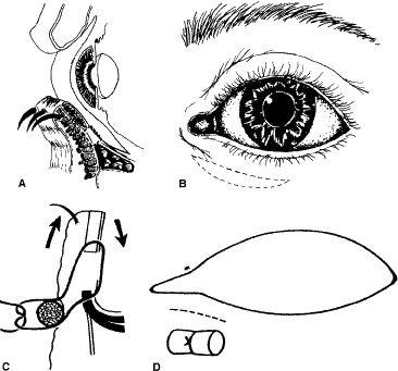
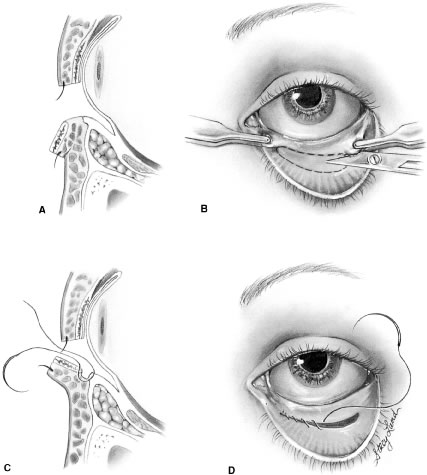
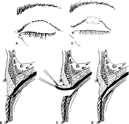
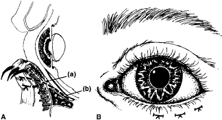
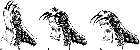
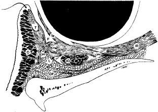
The posterior lamella consists of the eyelid retractor, the tarsus, and the conjunctiva. Hawes and Dortzbach2 reviewed and highlighted lower eyelid anatomy, specifically the lower eyelid retractor (Fig. 1). The lower eyelid is analogous to the upper eyelid; the main variation is the eyelid retractor system. The upper eyelid has a distinct eyelid retractor, the levator muscle, to enhance upper eyelid mobility. The lower eyelid does not have a specialized eyelid retractor. The lower eyelid retractor system originates as a fascial extension of the inferior rectus muscle (capsulopalpebral head). This fascial system splits to encapsulate the inferior oblique muscle and then reunites to form a dense fibrous sheet (capsulopalpebral fascia) to insert onto the inferior tarsal border. The inferior tarsal muscle is a smooth muscle analogous to the superior tarsal muscle (Müller's muscle) of the upper eyelid. This muscle originates in the inferior fornical area and extends toward the inferior tarsal border but does not insert on the tarsal border as its counterpart in the upper eyelid does. The inferior tarsal muscle receives sympathetic innervation, and interruption of its innervation will result in a slightly elevated position of the lower eyelid margin as observed in Horner's syndrome. Otherwise, the inferior tarsal muscle has little pathologic significance.

Fig. 1. Sagittal section of the lower eyelid. The anterior lamella consists of the skin and orbicularis muscle (o). The posterior lamella consists of the conjunctiva, capsulopalpebral fascia (CPF), inferior tarsal muscle (ITM), and tarsus (T). F, fornix; Te, Tenon's capsule; SO, septum orbitale; l, Lockwood's ligament; IO, inferior oblique muscle; CPH, capsulopalpebral head; IR, inferior rectus; OF, orbital fat. (From Hawes JJ, Dortzbach RK: The microscopic anatomy of the eyelid retractors. Arch Ophthalmol 100:1313, 1982.)

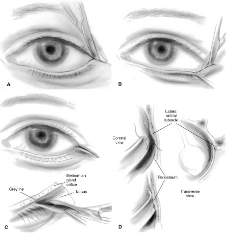
The tarsus provides the primary support or foundation for the eyelids. Although degeneration of the tarsus may promote eyelid laxity,3 the principle focus of weakness of the eyelids is at the lateral and medial canthal tendons.4 Jones and Wobig5 described the medial canthal tendon with a prominent anterior component firmly connecting the medial canthal angle to the maxillary process of the frontal bone. The posterior limb of the medial canthal tendon provides deep support to the posterior lacrimal crest. Anderson6 also emphasized the role of a superior branch of the medial canthal tendon to support the canthal angle. Gioia and coworkers7 have provided a clear description of lateral canthal anatomy. The lateral canthal tendon has contributions from the lateral aspects of the tarsus and the preseptal and pretarsal orbicularis muscle; these insert on the inner aspect of the lateral orbital rim at Whitnall's (lateral orbital) tubercle. The posterior deep insertion of the lateral canthal tendon allows the lateral aspect of the eyelids to approximate the globe.

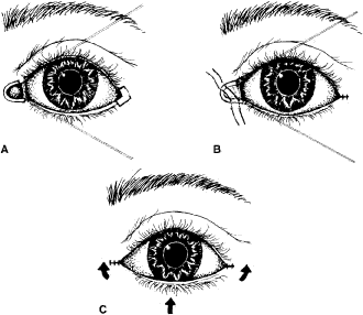
Ectropion and entropion share similar anatomic features. The most common predisposing anatomic factors for lower lid malpositions are horizontal eyelid laxity and weakness or defects of the lower lid retractors. The general weakness of the eyelid is caused by a weakness of the medial and lateral canthal tendons, permitting horizontal eyelid laxity.4 As such, it is best to consider lower eyelid laxity as resulting from laxity of the tarsoligamentous sling or support for the eyelid. The anatomic weakness can be demonstrated by the snap test. Downward retraction is exerted on the lower lid, pulling it away from the globe. On relapse, the lower lid should snap back against the globe spontaneously. Failure of the eyelid to snap back against the globe with a single blink indicates excessive laxity of the tarsoligamentous sling.

The lower eyelid retractors may be weakened or may actually become disinserted from the inferior tarsal border. Weakness or defect of the lower eyelid retractors creates in an instability of the inferior tarsal border that is manifested clinically as ectropion or entropion.

In addition, enophthalmos is associated with both ectropion and entropion. However, the role of enophthalmos as a pathophysiologic feature of lower eyelid malpositions has been grossly overstated.8,9 Enophthalmos may secondarily reduce tarsal stability by exaggerating horizontal lid laxity. It reduces the tension exerted on the eyelids by retraction of the globe and soft tissues. The normal arc or outward bowing of the eyelids is minimized with the enophthalmic globe, and eyelid tension is reduced. Retrobulbar injections stabilize eyelid malpositions by reducing horizontal eyelid laxity. Volume introduced in the retrobulbar area re-establishes the normal outward arch, or bowing, of the eyelid.

The development of ectropion and entropion is a reflection of the relationship between the anterior and the posterior lamellae. The eyelid position is maintained by an appropriate balance of the anterior and posterior lamellae. If tractional forces are unbalanced, eyelid malpositions may manifest as ectropion, entropion, or eyelid retraction. If traction is produced by the anterior lamella, either by chronic cicatricial changes or mechanical aberrations, the eyelid margin is everted away from the globe. This most commonly results from mild solar-induced changes or following surgical procedures or trauma of the eyelid, which produce a shortening or scarring of the skin and underlying orbicularis muscle.

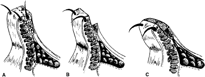
Involutional entropion is more complex because of the multitude of factors producing this abnormality. The tarsal instability of entropic lids is due to horizontal eyelid laxity and loss of support of the lower eyelid retractors.10 The orbicularis muscle dynamics also assume a prominent pathophysiologic role in the production of entropion. In entropic eyelids, the preseptal orbicularis muscle overrides the pretarsal orbicularis muscle, which rolls the superior border of the tarsus internally. This is differentiated from spastic entropion, which is theoretically due to a spasm of Riolan's muscle. However, the primary differentiating feature of spastic entropion is a specific precipitating etiologic factor, such as recent eye surgery, eyelid edema, or ocular inflammation with secondary eyelid edema. An appreciation of the multiple etiologic causes of entropion has resulted in the evolution of combined approaches to correcting the various anatomic abnormalities and stabilizing the tarsus and orbicularis muscle.11–14

This chapter is divided into three sections that discuss eyelid abnormalities. These include ectropion, entropion, and trichiasis. Sections on ectropion and entropion are further divided according to the underlying etiology of the eyelid malposition. Specific surgical approaches are highlighted in each section.

Back to Top
ECTROPION
Ectropion is an eversion of the eyelid away from the globe. It is classified according to its anatomic features as involutional, cicatricial, tarsal, congenital, or paralytic (Fig. 2). Surgical approaches can be directed toward the underlying etiologic factors.

Fig. 2. Clinical presentation of ectropion. A. Medial ectropion. B. Generalized ectropion with retraction of eyelid. C. Tarsal ectropion, with total eversion of tarsus. D. Cicatricial ectropion developing following orbital floor exploration.

INVOLUTIONAL ECTROPION

Involutional ectropion evolves slowly secondary to eyelid laxity and tractional forces of the anterior lamella. This may result from mild cicatricial changes of the skin or orbicularis muscle or a mechanical or gravitational eversion of the eyelid margin. There is generally a progression from eyelid laxity to punctal ectropion, medial ectropion, and then generalized ectropion. If the punctum is slightly everted from the lacus lacrimalis, tears cannot effectively drain into the canalicular system. In addition, the siphoning effect of the lacrimal excretory system, as demonstrated by Doane,15 is not generated if the lacrimal puncta do not occlude on lid closure and do not approximate the lacus lacrimalis. With the puncta approximated, attempted eyelid opening creates negative pressure within the lacrimal excretory system. When the puncta initially separate, the negative pressure siphons tears from the lacus lacrimalis. Also, horizontal eyelid laxity may produce a flaccid canalicular syndrome or poor lacrimal pump, so that tears are not siphoned from the lacus lacrimalis.16 This tearing malfunction is aggravated by the chronic ectropion and eyelid retraction that produce lagophthalmos and secondary exposure keratopathy. With time, the exposed conjunctive thickens and keratinizes, producing further ocular irritation.

Mild medial ectropion is manifested by rotation of the punctum away from the ocular surface causing mild tearing. This can be treated with a one- or two-snip punctal plasty, establishing anatomic continuity between the canalicular system and lacus lacrimalis in the medial canthal angle. This procedure is performed by opening the punctum and vertical canaliculus with a single snip of a sharp iris or Westcott scissors or by excising a very thin “V” wedge (two snips). This procedure may also be helpful after ectropion repairs if a mild eversion of the puncta remains. This can be performed as an office procedure. A cotton-tipped applicator saturated with 4% lidocaine solution held over the punctum and adjacent palpebral conjunctiva usually provides adequate anesthesia. If it does not, a 30-gauge hypodermic needle can be passed through the punctum and vertical canaliculus so that 1% or 2% lidocaine can be infiltrated into the pericanalicular tissue. Medial ectropion, when more severe, may require surgical intervention. Surgery directed at the lateral canthal angle, as discussed later in this chapter, does not fully alleviate medial lid malpositions, and this approach is not universally successful in restoring eyelid position. Removal of a conjunctival and lower lid retractor spindle may be necessary. The spindle is directly beneath the tarsus with its long axis directly parallel to the eyelid margin (Fig. 3). The conjunctival retractor spindle should measure no more than 5 mm in a vertical dimension, to prevent postoperative retraction and entropion resulting from posterior lamellar shortening. In essence, the closure of this internal lamellar resection creates a direct tightening of the medial lower lid retractors. Full-thickness lid sutures tied over a firmly rolled cotton or silicone bolster have been described by Collin.17 Although this technique is a short-term nuisance for the patient, it does result in a superior inward rotation of the punctum and lid margin without retraction.

Fig. 3. Conjunctival lower lid retractor spindle removal. Medial ectropion can be repaired by removal of a conjunctival lower lid retractor spindle from the medial eyelid. (A, cross-sectional view; B, frontal view). A full-thickness suture closure with one or more 5-0 sutures tied over an external bolster of firmly rolled cotton or silicone sponge not only closes the resection of conjunctiva and the lower lid retractors but also advances upwards the anterior lamella increasing the inversion of the punctum. (C, cross-sectional view; D, frontal view). (From Collin JROC: A Manual of Systematic Eyelid Surgery. Edinburgh: Churchhill Livingstone, 1982:31.)

For cases of prominent medial ectropion, the Byron Smith “Lazy T” procedure combines a horizontal lid-tightening procedure with a lower lid retractor resection.18 This combined procedure inverts the eyelid margin to its appropriate anatomic position. Full-thickness eyelid resections and repairs have been poorly demonstrated and reported in the ophthalmology and plastic surgery literature. The concept of a wedge resection or pie-shaped tarsal resection should be totally discarded. In its place, full-thickness modified pentagonal block resections should be used (Fig. 4). Incision through the eyelid should be perpendicular to the lid margin. If the eyelid resection is wedge shaped, the repair or approximation of the lid results in a notch or valley of the eyelid margin at the incision site. The layered repair of the lid margin defect should start with approximation of the tarsus. Adequate tarsal approximation provides the foundation for lid reconstruction and removes all tension from the eyelid margin. As a result, lid notches or trichiasis should be almost nonexistent.

Fig. 4. Pentagonal block eyelid resection. A. Tarsal approximation provides the foundation for eyelid closure. PDS suture is used to approximate the base of the tarsus (suture a). The lid margin is closed by a suture angled from the eyelid margin (suture b). B. Sutures at the eyelid margin are totally without tension and are generally of 6-0 or 7-0 silk.

The initial interrupted sutures should be placed through the base or inferior border of the tarsus to promote tension-free lid approximation (see Fig. 4A, suture a). The first suture for tarsal approximation is a 5-0 or 6-0 polydioxamone (PDS) or polyglactin (Vicryl) with a spatula-style needle. If excessive lid tension is evident, a 5-0 suture is preferable. The PDS is an absorbable suture that retains its tensile strength for 6 weeks and promotes little tissue reaction. Vicryl is also an absorbable suture that retains its tensile strength for 3 to 4 weeks. It may be more prone to cause tissue reaction in some patients. Horizontal approximation of the cut edges of the lower lid retractor with interrupted 6-0 PDS sutures may also help to reduce horizontal lid tension. Continuous running sutures through the lower lid retractors from the inferior aspect of the modified pentagonal lid resection to the base of the tarsus result in a downward traction of the eyelid resulting from vertical shortening of the lid retractors and should be avoided.19 To avoid a suture that is tied at the lid margin, a superior tarsal suture is initiated on the anterior tarsal surface approximately 2 mm from the cut edge of the tarsus and 2 mm inferior to the lid margin (see Fig. 4A). The suture is then angled to exit the cut surface of the tarsus at the posterior lid margin. The opposing suture is then placed from the posterior lid margin to exit the tarsus at 2 mm from its cut edge and 2 mm from the lid margin. Tying this suture approximates the lid margin perfectly and establishes an appropriate foundation for its repair. Additional sutures of 6-0 or 7-0 silk can be placed directly anterior and posterior to the lashes, but these are not essential and merely stabilize the lid margin (see Fig. 4B). Silk sutures tied as a square knot are used at the lid margin because they do not unravel and abrade the cornea. The skin is closed with a nonabsorbable polypropylene (Prolene) or nylon suture or an absorbable 6-0 gut (Ethicon 1916).

The Byron Smith Lazy T procedure creates a horizontal and vertical tightening of the nasal aspect of the lower eyelid. Subtle modifications of the procedure as originally described by Smith are warranted. Smith originally described the removal of a tarsal conjunctival ellipse as the initial step in the procedure. In addition, he later described a wedge-like resection of the tarsus in the second stage of the procedure. We have found the procedure to be much easier to perform by initially resecting the modified full-thickness pentagonal block from the nasal portion of the eyelid. The initial incision with a sharp iris scissors should be perpendicular to the eyelid margin 3 to 4 mm lateral to the punctum extending to the base of the tarsus (Fig. 5A). A relaxing snip is then performed laterally through the full-thickness eyelid. The lid margins are then grasped with forceps and overlapped to determine the amount of horizontal eyelid laxity. After marking the amount of eyelid laxity, the sharp iris scissors are used to complete the modified pentagonal block resection (see Fig. 5B and C). The conjunctiva and adjacent lower eyelid retractors are then incised at the base of the tarsus and extended nasally (see Fig. 5E and F). After adequate undermining, the conjunctiva and lower eyelid retractor is overlapped, and a wedge or triangle of tissue is removed. The cut edge of the lower eyelid retractor and conjunctiva is then sutured to the inferior tarsal border with 6.0 chromic sutures, with the knots buried or directed away from the conjunctival surface to prevent ocular irritation (see Fig. 5G). Alternatively, a full-thickness lid suturing technique tied over an external bolster may be used as previously described. After repair of the conjunctival lower lid retractor resection, the full-thickness eyelid resection is repaired as previously described.

Fig. 5. Byron Smith Lazy “T” procedure. Pentagonal block resection of the medial eyelid (A to C), followed by removal of a conjunctival lower eyelid retractor triangle in the medial eyelid (D to G). Suture knots are buried to prevent conjunctival irritation (G). H. Final schematic demonstrating site of lower lid full-thickness pentagonal resection and conjunctival lower lid retractor removal.

Prominent laxity of the medial canthal tendon presents a challenge in management because of the deep fixation of the tendon and difficulties in obtaining a patent functional canalicular system.20 Fortunately this condition is rarely encountered. When past trauma or extreme involutional changes necessitate recreation of the deep attachment of the medial canthal tendon to the posterior lacrimal crest, microplate fixation may be a helpful technique.21 Attempts to plicate the anterior aspect of the medial canthal tendon at its insertion to the anterior lacrimal crest may result in kinking or folding of the canalicular system. To alleviate canalicular and medial canthal laxity, a block resection can be performed in the medial canthal angle that includes the medial canthal tendon and the canaliculus. The canaliculus is intubated with silicone, and the deep extension or insertion of the medial canthal tendon is reapproximated. In this fashion, the canalicular system remains patent and functional. In patients with a nonfunctioning lacrimal excretory system, Jordan and coworkers22 have described a medial tarsal strip procedure to elevate and tighten the medial aspect of the lower eyelid. If the medial lower lid is lax and has a nonfunctioning canaliculus resulting from an inadequately repaired trauma, the block resection technique with canalicular intubation and reanastomosis offers an excellent means of improving lid position and function, as well as lacrimal drainage.

If ectropion is generalized, lid-tightening procedures are advocated. Traditionally, lid-tightening procedures have consisted of horizontal shortening of the tarsus to permit better eyelid approximation. The original Kuhnt-Szymanowski procedure involved splitting the eyelid margin at the gray line and shortening the posterior lamella of the lid. This avoided a full-thickness lid resection with its associated difficulties in suture techniques and wound approximation. Smith18 later modified the technique to include a full-thickness resection of the eyelid, and this was the preferred procedure for ectropion repairs for many years. Unfortunately, this procedure involves the resection of normal tarsus and results in a narrowed palpebral fissure and possibly trichiasis and notching resulting from inadequate lid margin approximation.

Tenzel and coworkers24 and Anderson and colleagues25,26 suggested directing attention to the lateral canthal tendon, with the lateral canthal sling and lateral tarsal strip procedures, respectively. The ease in performing the tarsal strip procedure has made it the preferred technique directed at the lateral canthal angle. Tightening at the lateral canthus restores the deep fixation of the lateral eyelid to the inner aspect of the orbital rim. In the enhanced tarsal strip procedure,26 reapproximation of the lateral eyelid corrects increasing degrees of cicatricial ectropion. In this procedure a full-thickness lateral aspect of the lid is sutured to the inner aspect of the lateral orbital rim. Advantages of the tarsal strip procedure include direct repair of the anatomic defect, no eyelid margin sutures near the cornea, preservation of the normal tarsus, avoidance of horizontal eyelid tissue shortening, simultaneous repair of canthal malpositions, and maintenance of lateral canthal contour.

The tarsal strip procedure has multiple uses in lower eyelid surgery, including involutional and cicatricial ectropion and entropion (Fig. 6). Appreciation of subtle anatomic features (as previously described) of the lateral canthal area greatly facilitate this procedure.7 The lateral tarsal strip procedure can be confined to the lateral canthal area or an infralash incision can be extended to permit identification of lower eyelid anatomic features and removal of redundant skin and muscle. A lateral canthotomy is performed with a sharp iris scissors (see Fig. 6A). The attached inferior crus of the lateral canthal tendon prevents mobilization of the lateral canthal angle. To incise the inferior extension of the tendon, the scissors should be directed inferior and nasally (see Fig. 6B). Once the inferior crus of the tendon is incised, the lateral aspect of the eyelid is fully mobile. A free tarsal strip is created by denuding the tarsus of the eyelid margin, orbicularis muscle, lower eyelid retractor, and conjunctiva (see Fig. 6C). Reattachment of the tarsal strip to the periosteum is a potential source of complications. Guidelines should be observed to prevent dystopias of the lateral canthal position. A horizontal plane should be established from the medial canthal angle to midpupil of the eye in primary gaze. The tarsal strip should be attached to the periosteum respecting this horizontal plane. In addition, the tarsal strip should be attached to the inner aspect of the lateral orbital rim, simulating attachment to Whitnall's (lateral orbital) tubercle (see Fig. 6D). If the tarsal strip is attached to the anterior aspect of lateral orbital rim, the deep or posterior forces of the lateral aspect of the eyelid are deficient. As a result the lateral eyelid does not approximate the globe. To further promote posterior forces, the tarsal strip should be reapproximated to the inner aspect of the lateral orbital rim by interrupted sutures tied on the anterior tarsal surface. Attempts to bury sutures beneath the tarsus are counterproductive because they promote elevation of the tarsal strip from the lateral canthus. The lateral canthal angle is reformed with a buried 6-0 PDS or Vicryl suture to restore perfect lateral canthal angle architecture (see Fig. 6E).

Fig. 6. Tarsal strip procedure. A. Lateral canthoplasty. B. Lysis of inferior crus of lateral canthal tendon mobilizes the lateral aspect of the eyelid. C. Creation of denuded lateral tarsal strip. D. Approximation of the lateral tarsal strip to the inner aspect of the lateral orbital rim with two interrupted 5-0 Prolene sutures. E. Re-creation of the lateral canthal angle with absorbable suture, preventing lateral canthal angle malpositions.

Complications of the lateral tarsal strip procedure primarily involve reattaching the tarsus to the periosteum at the inner aspect of the lateral orbital rim. Concerns regarding adequate fixation of the tarsal strip have already been addressed. It is not unusual, however, to have persistent tenderness at the lateral canthal angle until the normal inflammatory response gradually resolves. Occasional persistent lower lid edema can complicate the postoperative course.

CICATRICIAL ECTROPION

Cicatricial ectropion is the result of a shortening of the anterior lamella of the eyelid. This can result from excessive sun exposure, burns, or radiation but is often the result of surgical treatment of the lower eyelids.

Surgeons are often alarmed at the precarious status of the lower lid and how it may be altered by surgical interventions such as blepharoplasty, tumor excision, or orbital floor exploration. Minimal or no skin excision may result in eyelid malpositions such as lid retraction or ectropion, because surgical undermining of skin or orbicularis muscle may result in some tightening during the healing phase. Shorr and Seiff27 explained this phenomenon as resulting from contracture of the middle lamella of the eyelid, the orbital septum. The orbital septum is a multilaminated structure28 that is relatively immobile and if scarred may lead to lower eyelid retraction or frank ectropion. According to Shorr, scarring of the orbital septum is a source of eyelid malpositions following explorations of the lid in which no skin is excised or following blepharoplasties with minimal skin excision. These changes are particularly prevalent in horizontally lax eyelids. A snap test as previously described should be performed on all patients undergoing lower eyelid surgery. If excessive laxity exists, eyelid-tightening procedures (tarsal strip) should be coordinated with the primary procedure to minimize postoperative eyelid malpositions.

The concept of internal scarring and horizontal lid laxity as etiologic factors in cicatricial ectropion is of paramount importance in the management of this disorder. Skin grafting should be restricted to those patients with severe cicatricial changes. Most cases of moderate ectropion are managed by a tarsal strip procedure or a Smith modification of the Kunht-Szymanowski procedure, in which a skin muscle flap is created but no skin is removed. Jordan and Anderson26 have advocated the enhanced tarsal strip procedure as an alternative to skin grafting. The lateral aspect of the lid is immobilized in this procedure, but no skin is excised as in the traditional tarsal strip procedure. The full-thickness lateral aspect of the eyelid is then approximated to the inner aspect of the lateral orbital rim. The firm deep attachment of the skin provides a sharp lateral canthal angle and supports tension from the anterior lamellar shortening.

If the procedures described previously prove ineffective in the management of cicatricial ectropion, a full-thickness skin graft is the final alternative (Fig. 7). Split-thickness grafts should be avoided in eyelid reconstruction because of graft scarring and contracture producing a pale immobile eyelid. Split-thickness grafts are effective in extensive tumor resections in which grafts are placed on avascular areas such as bone. The use of full-thickness skin grafting to the lower eyelid often must be accompanied by additional horizontal lid tightening (see Fig. 7C). Suitable skin graft donor sites include the upper eyelid, preauricular or postauricular areas, and the supraclavicular areas. The skin graft should be carefully thinned and debrided of connective tissue to enhance graft survival. Localization and mobilization of the orbicularis muscle to provide a vascular bed also promotes graft survival and produces a more natural-appearing eyelid.29 The eyelid is immobilized by a superior traction suture or temporary suture tarsorrhaphies. The compressive bolster tied over the graft remains in place for 5 to 7 days.

Fig. 7. Cicatricial ectropion: Skin grafting. A. Ectropion secondary to anterior lamella shortening. B. Elevation of the lower eyelid. Note orbicularis muscle provides a vascular bed for the skin graft. C. Lateral tarsal strip may be required to horizontally tighten the lower eyelid before placement of the skin graft (D and E).

In some cases of cicatricial ectropion, the anterior lamellar deficiency may be at least in part the result of a descent (sagging) of the midface. Depending on the goals of the patient, the need for full-thickness skin grafting may be decreased or even eliminated by surgical techniques that advance the midface soft tissue upward.30

TARSAL ECTROPION

Tarsal ectropion is an unusual form of ectropion produced by a disinsertion of the lower lid retractors, the capsulopalpebral fascia, from the inferior tarsal border31–33 (Fig. 8A). It is characterized by a total eversion of the tarsus, with the inferior tarsal border positioned at the level of the inferior limbus. Defects of the lower lid retractors are most often associated with involutional entropion. However, in patients with tarsal ectropion the tractional forces from the anterior lamella will evert the entire tarsus. Indications of a defect in the lower lid retractors in these patients include (1) a deep inferior fornix, (2) a higher than normal resting position of the lower eyelid, (3) a diminished lower lid excursion, and (4) direct visualization of the disinsertion through the conjunctiva. A prominent bulge of orbital fat can sometimes be seen in the area of the disinserted lower lid retractors when the lower lid is pulled down while the patient looks upward.

Fig. 8. Tarsal ectropion. A. Ectropion secondary to disinsertion of the lower lid retractors. B. Everted lower eyelid and the area to be excised. C and D. Resected conjunctival spindle and reapproximation of the lower eyelid retractors. (From Wesley RE: Tarsal ectropion. Am J Ophthalmol 93:491, 1982.)

Putterman31 has described a patient with tarsal ectropion following a lid-tightening procedure and questioned the role of eyelid laxity in the production of this abnormality. Tse and coworkers33 advocated horizontal lid-tightening in most of their reported cases. In cases of a totally everted tarsus, alleviation of the anatomic abnormality by reapproximating the lower eyelid retractor to the inferior tarsal border corrects the condition (see Fig. 8C and D). Surgical repair may be directed from the skin or the conjunctival side, and excision of the redundant conjunctiva is optional. If significant horizontal eyelid laxity exists, a lateral tarsal strip procedure could be used to stabilize eyelid position. However, conjunctival approaches are recommended to minimize potential scarring of the anterior lamella.

CONGENITAL ECTROPION

Congenital ectropion is rare and is generally associated with severe shortening of the anterior lamella and underlying anatomic developmental abnormalities. These cases must be individually evaluated for the underlying cause, and direct repair of the anatomic defects are then undertaken. A full-thickness lid reconstruction is required sometimes because of shortening of the anterior and posterior lamellae, as seen in patients with developmental abnormalities. Surgical repair may require a split-level reconstruction as described by Brown.34 In this procedure both the anterior and posterior lamellae are lengthened, using skin grafts anteriorly and mucosal grafts posteriorly.

PARALYTIC ECTROPION

The characteristic features of facial paralysis are exaggerated in the aged face, in which anatomic features are highlighted by gravitational effects on the denervated musculature. These features include the loss of midforehead furrows, lower set brow, dermatochalasis, upper lid retraction, lower eyelid retraction, ectropion, flattening of the nasolabial fold, and the inability to elevate the mouth on the affected side. Ocular symptoms are the result of lagophthalmos, resulting in exposure keratopathy. The severity of symptoms correlates with the degree of corneal exposure and the ability of reflex tearing to lubricate the cornea. Proximal involvement of the facial nerve (at the level of the geniculate ganglion) and the facial or fallopian canal will result in the interruption of lacrimal gland secretion. This has been traditionally felt to be a poor prognostic indicator in patients with Bell's palsy, but many question its reliability as an indicator of the final outcome of this disorder.35 Controlled studies suggest a possible herpetic etiology of some cases of idiopathic (Bell's) facial palsy.36 Acyclovir 400 mg orally (PO) 5 times per day for 7 days was shown to hasten recovery if instituted soon after the onset of facial weakness.

Orbicularis muscle paralysis may result in upper and lower eyelid retraction. Pre-existing laxity of the lower lid contributes to the production of frank ectropion. With chronicity, a component of cicatricial ectropion may develop secondary to scarring of the retracted eyelid. Lower eyelid tightening by the tarsal strip procedure will effectively elevate and reposition the lower lid (Fig. 9). Additional procedures such as medial canthoplasties or tarsorrhaphies should be used only when additional lid narrowing is required to prevent exposure keratopathy. Primary lateral tarsorrhaphies to prevent exposure keratopathy are cosmetically unacceptable and should be avoided (Fig. 10). If temporary lid closure is required, suture tarsorrhaphies may be used to reduce exposure keratopathy and possible corneal ulceration.

Fig. 9. Paralytic ectropion. A. Prominent lower lid ectropion and brow ptosis. B. Appearance following lateral tarsal strip procedure and direct supra-brow elevation.

Fig. 10. Facial paralysis. A. Narrowed palpebral fissure secondary to lateral tarsorrhaphy. Associated visual field restriction laterally. B. Revision of lateral tarsorrhaphy with a lateral tarsal strip procedure. No visual field restriction postoperatively; cosmetically improved lid appearance.

If medial canthoplasty is necessary to facilitate lid elevation or to narrow the palpebral fissure, a variation of the standard procedure is helpful to minimize horizontal lid narrowing. Traditionally, medial canthoplasties involve direct approximation in the medial canthal angle in which the medial eyelid is zippered closed.37 This results in a horizontally narrowed palpebral fissure, with minimal elevation of the retracted lower eyelid. A medial canthoplasty is more effectively performed by supporting the lower lid with the superior crus of the medial canthal tendon (Fig. 11). This is accomplished by the approximation of a lateral portion of the lower medial canthal angle to a more nasal portion of the upper medial canthal angle (see Fig. 11B). The nasal portion of the eyelid is then supported by the superior crus of the medial canthal tendon. The lower eyelid thereby assumes a higher position with minimal horizontal narrowing of the palpebral fissure.

Fig. 11. Medial canthoplasty and lateral tarsorrhaphy. Modification of medial canthoplasty is performed to minimize narrowing of the palpebral fissure. The inferior crus of the medial canthal tendon is supported by a nasal portion of the superior crus of the tendon. The lateral tarsorrhaphy is performed by simple approximation of denuded tarsus.

Often, the upper eyelids require surgical intervention to minimize lagophthalmos in patients with facial paralysis. In most texts that deal with eyelid malpositions, the upper eyelid is unfortunately neglected in this discussion. Many procedures have been advocated to minimize lagophthalmos. These procedures include temporalis muscle transfer, upper eyelid weights, stainless steel springs, eyelid magnets, and the fil d'Arion procedure. All of these operations have extensive complications. Lid-loading (eyelid weight) procedures had been discouraged in the treatment of lagophthalmos secondary to facial palsy and orbicularis muscle paralysis, primarily because of the high rate of extrusion of implanted devices. Kelly and Sharpe38 reported an extremely high complication rate of 68% in paralytic eyelids following gold weight implantation. Complications included extrusion (43%), migration (18%), and infection (7%). Gold weight implantation has been re-evaluated and is extremely effective in minimizing lagophthalmos.39,40 The high incidence of complications was a result of improper gold weight implantation techniques. The gold weight procedure can be regarded as re-animation by gravitation.

May39 re-evaluated lid-loading procedures and greatly improved surgical techniques, almost eliminating complications. May recognized that the main disadvantage of traditional loading procedures was the superficial placement of the weight. A muscular pocket could not support the gold weight, allowing migration and extrusion. In the eyelid, the orbicularis muscle fibers easily separated, and the thin, delicate eyelid skin then became the sole barrier to extrusion.

Complications have been dramatically reduced by deeper weight fixation within the eyelid. A small pocket is created by elevating the levator aponeurosis from the tarsus and Müller's muscle (Fig. 12). The gold weight is then sutured to the superior tarsal border and undersurface of the levator aponeurosis and migration and extrusion are virtually eliminated. Also, deep fixation of the gold weight minimizes deformity of the upper lid and will only be apparent in those patients with a sunken superior sulcus. The size of the gold weight is determined preoperatively by taping the weight onto the eyelid and observing the lid closure. Lid closure must be ensured without inducing ptosis from an excessive weight. The most frequently used weights are 1 g and 1.2 g. It should be noted that the gold weight will allow prolonged lid closure but will not recreate the spontaneous blink reflex. Spring implantation is a much more difficult procedure with a high rate of extrusion, but this does provide a more spontaneous blink.35

Fig. 12. Gold-weight implantation for lagophthalmos. A. A 10-mm incision is made in the upper eyelid crease. A to D. A pocket is created between the anterior tarsal surface and the under surface of the levator aponeurosis. The gold weight is sutured in place to prevent migration. E. The final location of the gold weight.

Part of the benefit of upper lid gold weights comes from the elimination of upper lid retraction caused by upper lid retractors working in the absence of normal opposing orbicularis oculi muscle protractor tone. The creation of a very mild upper lid ptosis can further protect against the corneal irritation caused by incomplete spontaneous blinks and incomplete voluntary closure. Müller's muscle excision to lower the upper lid position can improve the symptoms and corneal surface integrity of facial palsy patients even without implantation of a gold weight.41

FLOPPY EYELID SYNDROME

Floppy eyelid syndrome was first described by Culbertson and Ostler in 1981.42 This is a condition most often seen in overweight males. There is significant laxity of the eyelid tissues. It is generally bilateral but may be asymmetric. Retraction of each eyelid leads to its eversion. Ophthalmologically, this condition shows some thickening of the tarsal plate along with low-grade chronic inflammation that may be seen in any of the lid tissues. No specific etiologic factor has been determined.43 There is a high correlation of obstructive sleep apnea in patients with the floppy lid syndrome.44 Perhaps there is some type of connective tissue disorder. However, overweight men are also at more risk for developing sleep apnea. Patients with floppy eyelid syndrome may experience spontaneous eyelid eversion. This was the initial presenting symptom that drew attention to the problem. It is more likely to occur on the side on which a patient sleeps. This is typically the most severely involved side in asymmetric cases. There is also eyelid imbrication in which the lax upper lid rides over the margin of the lower lid on eyelid closure.45 The leads to a chronic irritation of the tarsal conjunctiva of the upper lid. Typically, there is a thickening of the upper lid with erythema and chronic papillary conjunctivitis. Ocular lubricating ointments and corticosteroids can help decrease some of the inflammation that occurs and antibiotics can control secondary infections. However, horizontal eyelid shortening of the upper and sometimes also the lower lids can produce a more lasting improvement.46,47 These eyelids are often extremely lax and fairly large horizontal resections are necessary to restore appropriate horizontal tension to the eyelids. There can also be acquired aponeurosis defects causing ptosis along with dermatochalasis that may require surgical repair.

Back to Top
ENTROPION
Entropion is the inward turning of the eyelid. It is most commonly seen as an aging phenomenon produced by attenuation or detachment of the lower eyelid retractor and associated with horizontal eyelid laxity. Forceful eyelid closure causes the preseptal orbicularis muscle to override the tarsus, resulting in the inward turning of the eyelid. Entropion may be classified as involutional (spastic), cicatricial, and congenital (Fig. 13).

Fig. 13. Entropion. A. Involution entropion. B. Cicatricial entropion. Note the obliteration of the inferior fornix.

INVOLUTIONAL ENTROPION

A multitude of surgical procedures have been advocated for the correction of entropion, reflecting the various pathophysiologic features of the disorder (Table 1). Combination procedures are now used to specifically attack each anatomic aberration.8–11 The correction of a single anatomic feature of entropion results in a less than predictable surgical outcome. Combination procedures are now used to specifically attack each of the anatomic aberrations in an individual patient.11–14


TABLE 1. Previously Advocated Procedures for Involutional Entropion


Corrective MeasureProcedure
Vertical skin-muscle shorteningZiegler cautery
Horizontal tightening of lid at lower tarsal borderFox procedure
 Schimek suture
 Bick procedure
Barricading of orbicularis fibersWies procedure
 Jones procedure
Tightening of orbicularis fibersWheeler procedure
Repair or tightening of inferior retractors (CPF)Jones procedure

CPF, capsulopalpebral fascia.


Internal scarring procedures to stabilize the eyelids have been very helpful in minimizing ocular irritations. Quickert and Rathbun48 advocated suture repair for entropion (Fig. 14A, suture a), and Iliff49 later modified this procedure (see Fig. 14A, suture b). The placement of horizontal mattress sutures from the inferior fornix to exit the skin directly beneath the eyelashes (as in the Iliff modification) enhances the everting forces of the eyelid. This procedure is easy to perform and serves as an excellent temporizing procedure until a more definitive procedure is possible. In eyelids with a minimal degree of horizontal lid laxity, such treatment may suffice indefinitely. In eyelids with a greater degree of horizontal laxity, care must be taken not to use too much eversion effect with the Iliff modification, or an immediate ectropion can be created. Also, in eyelids with spastic entropion, such as following ocular surgery or ocular irritations, this procedure may stabilize the lid until the irritating focus has resolved.

Fig. 14. Entropion suture repair. A. Quickert-Rathbun suture placed directly beneath the tarsus (a); Iliff modification in which the double-armed suture is placed from the inferior fornix to the skin in the infralash location (b). B. Location of the three everting sutures.

The type of suture material used for Quickert-Rathbun sutures is not as important as the tension with which the sutures are tied. The suture tension can lead to focal tissue necrosis and scarring, giving Quickert-Rathbun sutures the potential for some permanent correction. Double-armed sutures provide the safety of making all suture passes away from the globe. 4-0 chromic gut sutures generally remain in place for 2 weeks, which is long enough to provide the necessary effect without the need for scheduled suture removal. Silk and polyglactin sutures will last longer but can lead to tissue infections resulting from the duration and their polyfilament structure. Nylon sutures are less likely to cause infection but are sometimes uncomfortable for the patient because of their stiffness.

Patients with entropion usually require a combined procedure to eliminate horizontal lid laxity and repair the lower lid retractors.50 No procedure addressing an isolated anatomic feature has been universally successful in alleviating entropion. Isolated repair of lower lid retractor defects (analogous to repair of the levator aponeurosis in ptosis procedures) often results in lower lid retraction or actual ectropion.51–53 This is due to the preexisting significant horizontal eyelid laxity, which again promotes an unstable eyelid margin. The tendency is further exacerbated if the lower eyelid retractor is inadvertently advanced onto the tarsus. If horizontal eyelid laxity is evident, a tarsal strip procedure should be performed, as discussed for the treatment of ectropion (see Fig. 6). Various suture placements through the lateral tarsal strip as in the correction of ectropion or entropion described by McCord and coworkers54 are not relevant. The goal of the tarsal strip procedure is to firmly affix the tarsus to the inner aspect of the lateral orbital rim. This is best accomplished by using two interrupted 5-0 nonabsorbable monofilament sutures, although some surgeons prefer 4-0 or 5-0 polyglactin. The sutures should be tied on the anterior tarsal surface; attempts to bury the knots beneath the tarsus will prevent adequate tarsal approximation to the inner aspect of the lateral orbital rim.

The preferred procedure for involutional entropion thus consists of a lateral tarsal strip and repair of the lower eyelid retractors. Isolated lower lid retractor repairs are inappropriate—an invitation for additional procedures. A lateral canthal incision is created and extended in an infralash position, and a skin muscle flap is elevated to expose the orbital septum (Fig. 15B and C). The orbital septum is penetrated and orbital fat identified and retracted inferiorly. Directly beneath the orbital fat is the white fibrous appearing lower eyelid retractor. This is directly analogous to the location of the levator aponeurosis in the upper eyelid. The lower eyelid retractor is normally attached to the inferior tarsal border; however, in entropion, a defect is generally apparent between the lower eyelid retractor and the tarsus. The superior edge of lower eyelid retractor system is identified and carefully reapproximated to the inferior tarsal border (see Fig. 15D and E). Care should be taken not to advance the lower eyelid retractor to prevent lower eyelid retraction or actual ectropion. A lateral tarsal strip procedure as described in the section on ectropion should then be performed to tighten the eyelid horizontally (see Fig. 6). Herniated orbital fat is an important etiologic factor in promoting orbicularis muscle overriding and creating tarsal instability. Redundant orbital fat is carefully removed. To finalize the procedure, redundant skin is draped over the lid margin and is excised as in a lower lid blepharoplasty. Skin removal should be minimal to prevent ectropion. It is this combination type of procedure that produces minimal recurrences and has been extremely successful in managing entropion.

Fig. 15. Lower lid retractor repair for entropion. A. Lower lid retractor defect contributes to tarsal instability. Note the failure of attachment of the lower eyelid retractor to the inferior tarsal border. B to C. A skin-muscle flap is elevated and the orbital septum penetrated to identify the lower eyelid retractor. D to E. The lower eyelid retractor is attached to the inferior tarsal border with interrupted nonabsorbable sutures. F. Final skin closure.

The Weis (transverse tarsotomy) procedure has also been advocated for the correction of entropion. This procedure everts the lid margin and produces an anatomic barrier that prevents preseptal orbicularis muscle override and secondary tarsal rotation (Fig. 16). It has been advocated for involutional entropion but is best suited for patients with varying degrees of cicatricial entropion. The Weis procedure has been called the two-snip procedure and is relatively easy to perform. A stab incision is created through the full thickness of the lid at the inferior aspect of the tarsus approximately 3 mm from the eyelid margin (Fig. 16A). A sharp iris scissors is used to extend the full-thickness lid incision nasally and temporally (hence the two-snip procedure) (see Fig. 16B). The short vertical height of the tarsus of the lower eyelid,55 especially in patients with cicatricial entropion, makes the incision of the tarsus challenging. An alternative method of creating this full-thickness horizontal eyelid incision is to make partial thickness lid incisions with a number 15 blade through skin and orbicularis muscle externally and conjunctiva and tarsal plate internally. Each of these incisions is placed 3 mm below the lid margin. Any remaining tissue can be cut with scissors. Ideally, the incision is through the inferior portion of the tarsus; however, this may be difficult and the incision may occur just below the inferior border of the tarsus. As such, the procedure may be better described as a transverse blepharotomy. Rotation of the tarsus is accomplished by placing horizontal mattress sutures (5-0 chromic) originating from the conjunctiva and lower eyelid retractor, then extending to an infra-lash position (see Fig. 16C). Tying these sutures will evert the eyelid margin and also stabilize the orbicularis muscle. Lance and Wilkins14 evaluated patients treated with a Weis procedure alone and found the recurrence rate to be 11%. However, when a lateral canthal tightening procedure was added, no recurrences were encountered. Thus, if the Weis procedure is used to treat involutional or cicatricial entropion with horizontal laxity, it is best coupled with a tarsal strip procedure to further stabilize the eyelid margin.

Fig. 16. Transverse tarsotomy (Weis) procedure. A. A lower eyelid crease incision is made and (B) extended to full thickness of the lower eyelid. C. Double-armed mattress sutures approximate the conjunctiva and lower lid retractor to the orbicularis muscle and skin, effectively everting the eyelid margin. (Soll DB: Entropion and ectropion. In Soll DB [ed]: Management of Complications in Ophthalmic Plastic Surgery. Birmingham, AL: Aesculapius Press, 1976.)

CICATRICIAL ENTROPION

Cicatricial entropion presents a surgical challenge because of underlying pathologic changes of the eyelid and the associated features of trichiasis, distichiasis, and epidermalization of the eyelid margin. This condition results when contraction of the posterior lamella of the eyelid draws the eyelid margin to the globe. It may result from chemical or thermal injury, trachoma, chronic allergies, or ocular medications. In addition, conjunctival cicatricial changes may result in mucous membrane disorders such as Stevens-Johnson syndrome and ocular cicatricial pemphigoid. Conjunctival cicatrization destroys the lacrimal gland ductules and meibomian gland orifices, severely reducing the aqueous and oily constituents of the tear film. Unfortunately, the progressive nature of ocular pemphigoid may present continued medical and surgical challenges. Medical management with immunosuppressive agents such as diaminodiphenylsulfone (Dapsone), azathioprine (Imuran), or cyclophosphamide (Cytoxan) should be attempted before surgical intervention.56–58 Severe side effects of these immunosuppressive agents necessitate cautionary use in older patients, and these patients should be monitored by an oncologist, hematologist, or rheumatologist. Subconjunctival injections of mitomycin C have been reported as a means of arresting ocular pemphigoid,59 but these results are preliminary. Eyelid or ocular surgery may aggravate ocular cicatricial pemphigoid and convert a chronic form to an acute form, worsening conjunctival shrinkage. Mitomycin C has been reported as a useful adjunct in surgery for cicatricial entropion to prevent additional scar tissue formation.60 Standards for its use in these cases have not yet been established.

Mild to moderate degrees of cicatricial entropion are best treated with a Weis (transverse tarsotomy) procedure (see Fig. 16), in which the tractional forces of the posterior lamella are transferred to the anterior lamella to promote eversion of the eyelid margin. Unlike involutional entropion, moderate degrees of cicatricial entropion do not generally require horizontal lid tightening.

Kersten and coworkers61 re-evaluated the use of tarsotomy with rotational sutures, for mild to moderate degrees of cicatricial entropion (Fig. 17). This procedure consists of fracturing the tarsus approximately 2 mm below the eyelid margin. Double-armed sutures are passed from the tarsus of the inferior edge of incision, and then passed through the orbicularis muscle to exit the skin in an infralash position. Tying these sutures everts the eyelid margin. These authors reported a 94% success with a minimum follow-up period of 6 months.

Fig. 17. Transverse tarsotomy and lid margin rotation. A. Full-thickness incision of the tarsus approximately 2 mm inferior to the eyelid margin. B. Placement of horizontal mattress everting sutures from the inferior tarsus to exit the skin in the infralash position. C. Final everted eyelid position. (From Kersten RC, Kleiner FP, Kulwin DR: Tarsotomy for the treatment of cicatricial entropion with trichiasis. Arch Ophthalmol 110:714, 1992.)

Severe posterior lamella contracture with obliteration of the inferior fornix requires lengthening of the posterior lamella by grafting. Donor materials should provide a mucous membrane and a rigid supporting structure; possible materials are (1) autogenous tarsus and conjunctival (2) nasochondral mucosa, (3) hard palate, and (4) full-thickness buccal mucous membrane and ear cartilage. The use of donor sclera from eye banks has been advocated, but it produces a chronic inflammatory response and is not lined by conjunctiva, limiting the utility of scleral grafts.62,63 In the lower lid, the composite graft is placed between the inferior border and recessed eyelid retractor and conjunctiva (Fig. 18). Attempts to place the graft in the midaspect of the tarsus may be difficult secondary to contraction and shortening of the tarsus. Contraction of the tarsus creates technical difficulties in incising the tarsus and placing the graft within the divided tarsal plate. Ear cartilage or scleral grafts can be used, but these are most effective if they are lined with mucous membrane.

Fig. 18. Cicatricial entropion lower eyelid. A. The tarsus is incised and recessed. (If excessive contracture occurs and the lower lid retractors may be recessed from the inferior tarsal border.) B and C. The graft is sutured between the recessed tarsal margins, everting the eyelid to its normal position.

Worldwide, upper eyelid entropion is most often encountered with trachoma; however, this is rare in the United States. Modifications of lower lid procedures are necessary because of gravitational forces and the length of the tarsus. Mild degrees of cicatricial entropion may be treated with a Weis procedure as in the lower lid. The tarsus is fractured across its horizontal dimension about 4 mm from the eyelid margin, and sutures are used to rotate the lid margin away from the globe. A modified Tenzel64 tarsal trough procedure may also be used to rotate the tarsus; this is a variation of the Weis procedure in which a full-thickness tarsal incision is avoided (Fig. 19). Closure of the tarsal trough will evert the lid margin from the globe. Additional everting forces are induced by the formation of a lower eyelid crease about 4 to 5 mm above the lid margin and the removal of skin and muscle, in essence shortening the anterior lamella. A relaxing incision of the eyelid margin at the gray line, approximately 3 mm in depth, will further evert the ciliary portion of the eyelid margin.

Fig. 19. Tenzel tarsal trough procedure. A. A low eyelid crease incision is formed and redundant skin is excised to increase everting forces. A lid margin relaxing incision at the gray line approximately 3 mm in depth will permit eyelid rotation. B. A tarsal wedge is excised and suture approximation (C) will evert the eyelid margin.

Reacher and coworkers65 evaluated the efficacy of various surgical procedures in the management of trachomatous upper eyelid entropion. For mild to moderate degrees of entropion and trichiasis, the transverse tarsotomy (Weis) procedure was the most effective. Electrolysis and cryotherapy were best used to remove individual recurrent lashes. Initial cryotherapy was associated with a 21% incidence of worsening of trichiasis (an increase of five or more lashes). Also, the authors found tarsal advancement and rotation procedures to be associated with a high degree of failure for milder degrees of entropion and trichiasis. Patients with defective lid closure required lengthening of the posterior lamella. This was accomplished by mobilization and advancement of the tarsus, or free grafting of the tarsus (Fig. 20). Both procedures were equally effective in managing this challenging situation. These procedures are generally successful in reducing the degree of entropion or trichiasis; however, local trichiasis management may be required.

Fig. 20. Cicatricial entropion—upper eyelid. A. The tarsus is incised and the posterior lamella is recessed. B and C. A free graft is sutured to the edges of the recessed tarsus.

CONGENITAL ENTROPION

Congenital entropion is extremely rare and should be differentiated from epiblepharon and congenital distichiasis. Congenital entropion consists of a rotated eyelid margin, whereas epiblepharon is characterized by redundant horizontal pretarsal skin fold that rotates the eyelashes toward the globe. In congenital distichiasis, lashes emanate from the meibomian gland orifices. Tse and coworkers66 have identified retractor aponeurosis disinsertion in patients with congenital entropion and emphasize the importance of its repair (see Fig. 15). Other authors67,68 consider the resection of skin and pretarsal orbicularis muscle a successful method of managing these cases (Fig. 21). This procedure is also excellent for treating epiblepharon even when entropion is not present.

Fig. 21. Congenital entropion epiblepharon. A. Creation of an infralash ellipse, consisting of skin and pretarsal orbicularis muscle. B. Closure incorporating tarsal tissue enhances everting effects.

A few cases have been reported in which the tarsus is folded in a horizontal condition creating a severe congenital entropion referred to as tarsal kink.69,70 This is felt to be a congenital developmental anomaly. It can be repaired during infancy by making a horizontal conjunctival and tarsal incision at the point of its folding.

Back to Top
TRICHIASIS
The eyelid margin is best viewed as a transition zone between the eyelid skin and palpebral conjunctiva. The anterior half of the eyelid margin is the ciliary portion, with eyelashes directed anteriorly away from the globe. The slight indentation or groove in the central aspect of the eyelid margin is the sulcus intermarginalis of Graefe, more commonly known as the gray line. The posterior half of the eyelid margin is the tarsal portion; the meibomian gland orifices are visualized in the central aspect of this half of the eyelid margin. The meibomian glands are unique in that they do not have a pilar apparatus associated with the sebaceous unit.

Trichiasis is an acquired condition in which eyelashes are misdirected posteriorly toward the conjunctiva or cornea. If contact is made with the conjunctiva, a foreign body sensation and localized conjunctival vascular injection result. Corneal involvement produces pain and photophobia, generally correlating with the degree of epithelial involvement. The etiology of trichiasis may be obvious following trauma or surgical interruption of the eyelid margin. However, chronic inflammatory conditions, ranging from blepharitis and chalazion to conjunctival cicatricial disorders such as ocular pemphigoid or Stevens-Johnson syndrome, also frequently result in trichiasis.

Scarring conditions of the conjunctiva produce a particularly frustrating combination of features: entropion, trichiasis, distichiasis, and epidermalization of the eyelid margin. Trichiasis is simply a misdirection of lashes toward the globe. In distichiasis, lashes or hairs emanate from the meibomian orifices. Anderson and Harvey71 have histologically evaluated congenital and acquired distichiasis and have identified pilar units associated with the meibomian glands; these authors view this as representing a reversion toward a pilosebaceous apparatus. A chronic inflammatory stimulus such as chronic cicatricial pemphigoid, erythema multiforme, chemical injury, trachoma, chronic blepharitis, and chronic allergies may initiate this regressive metaplasia of the meibomian glands to produce acquired distichiasis. Scarring of the posterior lid lamella can lead to a rotation of the ciliary portion of the lid margin towards the globe. In this situation, it may appear as if lashes are emanating from the posterior lid margin, but close examination with the slit lamp will show that the most posterior lashes are not arising from meibomian gland orifices and are not distichiasis.

The management of trichiasis can be challenging for both the patient and the physician. Recalcitrant or recurrent lashes are not unusual, despite a systematic approach to this condition. Initial consideration determines the etiology of trichiasis. Trichiasis following interruption of the lid margin either from trauma or surgical manipulation will have a corresponding local lash abnormality. These abnormalities are often avoided with careful approximation and tension-free closure of the eyelid margin (see Fig. 4). In addition, removal of tumors from the eyelid margin, such as papillomas and dermal nevi, should employ a chopping-block type shave biopsy parallel to the surrounding surface of the skin or lid margin. A shave biopsy extending beyond this plane would produce a divot at the eyelid margin and would also interrupt the pilosebaceous apparatus at the anterior lid margin. Trichiasis may result from scarring around the lashes, producing some posterior misdirection of the lash. If complete eyelid margin tumor excision is desired, a full-thickness resection of the eyelid (see Fig. 4) should be performed.

Epithelialization and dryness of the caruncle,73 progressive conjunctival inflammation, cicatrization, subepithelial fibrosis, blunting of the fornices, symblepharon formation, trichiasis, distichiasis, and entropion characterize the clinical presentation of ocular pemphigoid. The disease can progress to keratinization or vascularization of the corneal surface, leading to blindness. The relentlessly progressive nature of ocular pemphigoid can be interrupted by the institution of immunosuppressive therapy, after histologic confirmation of the disorder.56–58

The management of trichiasis requires a stepwise approach. The degree of trichiasis and type of lashes are important in planning therapy. Localized abnormal lashes generally respond better to the ablative procedures than does diffuse trichiasis. Electrolysis is best suited for localized normal sized lashes. Finer lanugo hairs are generally less responsive to electrical ablative techniques. Extensive electrolysis may produce scarring of the eyelid margin and secondary cicatricial changes, complicating later management.

Local anesthesia (2% lidocaine with or without epinephrine) is first administered, then the lid is everted from the globe and an electrolysis needle is introduced directly into the lash follicle under slit-lamp visualization. A useful instrument for electrolysis is the Prolectro epilator with a power setting of 4. The orientation of the follicle generally corresponds to the direction of the lash emanating from the follicle. The electrolysis needle should be advanced into the full extent of the lash follicle, which is approximately 2.4 mm in the upper lid and 1.4 mm in the lower lid.72 Current is applied until frothy bubbles emerge from the lash follicle, which generally requires approximately 15 to 20 seconds of electrical stimulus. A circular or rotating motion of the needle around the lash will fully ablate the follicle. After adequate treatment, the lash is easily removed. Another effective instrument for electrolysis is the Ellman radiosurgery unit using the 0.004 wire electrode, which is insulated except for the tip. A power setting of 2 using the partially rectified current works well. Again, it is essential that the electrolysis tip penetrate the full extent of the eyelash follicle so that it can be ablated. The Ellman unit requires only a few seconds compared with the Prolectro instrument. The success rate for electrolysis should be 80% to 90% with a single treatment. Trichiasis recurrences can be retreated with repeat electrolysis.

Fine lanugo hairs present a challenge for electrolysis. They typically do not have a large enough follicle to permit the advancement of an electrolysis needle. It is possible to create a pathway for the electrolysis needle by first introducing a hypodermic needle. However, this may not actually pass along the true path of the hair follicle. It is also possible to use the blended cutting action of the Ellman unit so that the electrode tip can advance through the tissue to the desired depth as electrolysis is being performed. Again, this may not follow the precise path of the hair shaft in its follicle.

In the past, cryotherapy was considered the optimal treatment for treating trichiasis. The authors have abandoned cryotherapy and now rely on electrolysis and surgical techniques, because of cryotherapy's damage to the integrity of adjacent tarsus and lid margin transitional epithelium. Various techniques of lid splitting in which the cryotherapy is limited to the affected anterior lid lamella have been proposed but do not completely eliminate the shortcomings of cryotherapy. For completeness, the following information is provided regarding cryotherapy. Sullivan and colleagues74 reported the use of cryotherapy for trichiasis, and his guidelines are still recognized as surgical standards. A double freeze-thaw cycle is required for cellular destruction. This is best accomplished by a rapid freeze to -20°C followed by a slow thaw, which causes cell destruction by cell membrane rupture and the formation of intracellular crystals. Liquid nitrogen probes are the most effective means of supplying the rapid level of freezing necessary for ciliary destruction.

A local anesthetic with epinephrine is administered, and cryotherapy is delayed until blanching of the eyelids evidences the full vasoconstriction effect of the injection. Lidocaine may be used, but bupivacaine offers the advantage of prolonged anesthesia with less posttreatment pain. The vasoconstriction facilitates adequate eyelid freezing. A thermocouple is used to monitor tissue temperatures and to ensure freezing to the -20°C level. Care is taken to place the thermocouple near the offending lash follicle. A cryoprobe is placed on the tarsal conjunctive, and the lid is elevated from the globe. The freezing is then initiated, while closely observing the globe to ensure that the cryoprobe does not inadvertently touch the cornea or the bulbar conjunctiva. About 30 to 60 seconds are required to obtain a temperature of -20°C. Placement of the cryoprobe on the anterior lid margin, directed posteriorly, would increase the chance of complications.75 This placement makes it difficult to adequately control the posterior extent of the freeze, resulting in potential injury. Depigmentation of the eyelid is often associated with cryotherapy, limiting its use in darkly pigmented individuals. Additional complications include visual loss (secondary to corneal opacification), corneal ulceration, symblepharon formation, and cellulitis.75 In addition, the destructive process is not selective; normal lashes are also destroyed. Anderson and Harvey71 have described a lid-splitting procedure with selective cryotherapy to the posterior lamella.

Laser ablation of lashes has been advocated76,77 but is best used for a few scattered lashes in patients with disorders such as ocular pemphigoid in which production of minimal inflammation is important. The recommended argon laser settings are a power of 1 watt, a spot of 50 mm, and duration of 0.2 second using the blue-green wavelength. The initial laser spot will vaporize a lash; however, approximately 20 to 30 additional burns are required to destroy the lash follicle. Bartley and Lowry76 reported success in ablating 59% of misdirected lashes with a single treatment. The laser beam must ablate the entire length of the eyelash in its follicle to destroy the follicle. Creative positioning of the eyelid is necessary to align the aberrant orientation of trichiasis follicles with an ophthalmic laser beam. Anesthesia is required for laser lash ablation, just as it is for electrolysis.

Buccal membrane grafting can be used as a last resort in the management of recalcitrant trichiasis, distichiasis, and epidermalization of the eyelid margin. The eyelid margin is split at the gray line, and the superior 2 to 3 mm of the tarsal portion of the eyelid is excised (Fig. 22A and B). Contained within this excised tarsus are the offending lashes and keratinized epithelium. Without buccal membrane grafting, the area of lid excision would likely scar to create cicatricial entropion. As such, buccal membrane is grafted onto the eyelid margin to create nonoffensive eyelid surface (see Fig. 22C). The graft may be obtained from either the lower lip or lateral portion of the mouth at the interdental line. If a slight degree of entropion exists following graft fixation, rotation sutures can be used to establish appropriate eyelid position (see Fig. 14).

Fig. 22. Buccal membrane grafting for trichiasis. A. Abnormal posterior eyelid margin with aberrant lashes is marked and excised (B). C. Placement of buccal membrane graft to the posterior lid margin.

Hard palate mucosal grafts have been proposed for internal lamellar grafting.78,79 There is, however, a risk of ocular irritation from the epithelial surface of this grafted tissue. This makes it a risky replacement for grafts placed in the upper lid or close to the lid margin in the lower lid. Split-thickness dermis grafts have also been proposed. Autogenous split-thickness dermis grafts require a donor surgical site and occasionally develop hair growth. Acellular autologous grafts (Alloderm) eliminate the need for a donor site and do not lead to hair growth; however, their use in cicatricial disorders, such as pemphigoid, has not yet been established.80,81

Back to Top
REFERENCES

1. Wulc A, Dryden RM, Khatchaturian T: Where is the grayline? Arch Ophthalmol 105:1092, 1987

2. Hawes MJ, Dortzbach RK: The microscopic anatomy of the lower eyelid retractors. Arch Ophthalmol 100:1313, 1982

3. Stefanyszyn MA, Hidayat AA, Flanagan JC: The histopathology of involutional ectropion. Ophthalmology 92:120, 1985

4. Neuhaus RW: Anatomical basis of “senile” ectropion. Ophthalmic Plast Reconstr Surg 1:87, 1985

5. Jones LT, Wobig JL: Surgery of the Eyelids and Lacrimal System. Birmingham, AL: Aesculapius Publishing, 1976:33–36

6. Anderson RL: Medial canthal tendon branches out. Arch Ophthalmol 95:2051, 1977

7. Gioia VM, Linberg JV, McCormick SA: The anatomy of the lateral canthal tendon. Arch Ophthalmol 105:529, 1987

8. Kersten RC, Hammer BJ, Kulwin DR: The role of enophthalmos in involutional entropion. Ophthalmic Plast Reconstr Surg 13:195, 1997

9. Hosal BM, Tekeli O, Gursel E: The role of enophthalmos in the development of involutional ectropion. Orbit 18:7, 1998

10. Jones LT: The anatomy of the lower eyelid and its relationship to the cause and cure of entropion. Am J Ophthalmol 49:29, 1960

11. Wesley RE, Collins JW: Combined procedure for senile entropion. Ophthalmic Surg 14:401, 1983

12. Nowinski TS: Orbicularis oculi muscle extirpation in a combined procedure for involutional entropion. Ophthalmology 98:1250, 1991

13. Carroll RP, Allen SE: Combined procedure for repair of involutional entropion. Ophthalmol Plast Reconst Surg 7:123, 1991

14. Lance SE, Wilkins RD: Involutional entropion: A retrospective analysis of the Weis procedure alone or in combination with a horizontal shortening procedure. Ophthalmic Plast Reconst Surg 7:273, 1991

15. Doane MG: Blinking and the mechanics of the lacrimal drainage system. Ophthalmology 88:844, 1981

16. Hill JC: Treatment of epiphora owing to flaccid eyelids. Arch Ophthalmol 97:233, 1979

17. Collin JRO: A Manual of Systemic Eyelid Surgery. Edinburgh: Churchill Livingstone, 1982:30

18. Smith B: The “Lazy-T” correction of ectropion of the lower punctum. Arch Ophthalmol 94:1149, 1976

19. Dryden RM, Doxanas MT: Eyelid malpositions, Part II. Ectropion and entropion. In McCord C (ed): Oculoplastic Surgery. New York: Raven Press, 1981:124

20. Edelstein JP, Dryden RM: Medial palpebral tendon repair for medial ectropion of the lower eyelid. Ophthalmol Plast Reconst Surg 6:28, 1990

21. Howard GR, Nerad JA, Kersten RC: Medial canthoplasty with microplate fixation. Arch Ophthalmol 110:1793, 1992

22. Jordan DR, Anderson RL, Thiese SM: The medial tarsal strip. Arch Ophthalmol 108:120, 1990

23. Smith B. Cherubini TD: Oculoplastic Surgery: A Compendium of Principles and Techniques. St Louis: CV Mosby, 1970:92–94

24. Tenzel RR, Buffam FV, Miller GR: The use of the “lateral canthal sling” in ectropion repair. Can J Ophthalmol 12:199, 1977

25. Anderson RL, Gordy DD: The tarsal strip procedure. Arch Ophthalmol 97:2192, 1979

26. Jordan DR, Anderson RL: The lateral tarsal strip revisited. The enhanced tarsal strip. Arch Ophthalmol 107:604, 1989

27. Shorr N. Seiff SR: Cosmetic Blepharoplasty: An Illustrated Surgical Guide. Thorofare, NJ: Slack, 1986:70

28. Meyer DR, Linberg JV, Wobig JL et al: Anatomy of the orbital septum and associated eyelid connective tissues. Implications for ptosis surgery. Ophthalmic Plast Reconstr Surg 7:104,1991

29. Doxanas MT: Orbicularis muscle mobilization in eyelid reconstruction. Arch Ophthalmol 104:910, 1986

30. Hester TR, Codner MA, McCord CD: The “centrofacial” approach for correction of facial aging using transblepharoplasty subperiosteal cheek lift. Aesthetic Surg Q 16:51, 1996

31. Putterman AM: Ectropion of the lower eyelid secondary to Möller's muscle—capsulopalpebral fascia detachment. Am J Ophthalmol 85:814,1978

32. Wesley RE: Tarsal ectropion from detachment of the lower eyelid retractors. Am J Ophthalmol 93:491, 1982

33. Tse DT, Kronish JW, Buus D: Surgical correction of lower-eyelid tarsal ectropion by reinsertion of the retractors. Arch Ophthalmol 109:427, 1991

34. Brown BZ, Beard C: Split level full thickness eyelid graft. Am J Ophthalmol 87:388, 1979

35. May M: Tearing, salivary flow and taste with facial nerve disorders. In May M (ed): The Facial Nerve. New York: Theime, 1986:238

36. Adour KK, Ruboyianes JM, Doersten PG et al: Bell's palsy treatment with acyclovir and prednisone compared with prednisone alone; a double-blind, randomized, controlled trial. Ann Otol Rhinol Laryngol 105:371, 1996

37. Levine RE: Eyelid reanimation surgery. In May M (ed): The Facial Nerve, p 690. New York, Thieme, 1986

38. Kelly SA, Sharpe DT: Gold eyelid weights in patients with facial palsy: A patient review. Plast Reconstr Surg 89:436,

39. May M: Gold weight and wire spring implants as alternatives to tarsorrhaphy. Arch Otolaryngol Head Neck Surg 113:656, 1987

40. Seiff SR, Sullivan JH, Freeman LN et al: Pretarsal fixation of gold weights in facial nerve palsy. Ophthalmic Plast Reconstr Surg 5:104, 1989

41. Elner VM, Ahmad AZ, Fante RG: Mullerectomy as a primary procedure for exposure keratitis secondary to seventh nerve paralysis. ASOPRS Sci Symp 29:77, 1998

42. Culbertson WW, Ostler HB: The floppy eyelid syndrome. Am J Ophthalmol 92:568, 1981

43. Netland PA, Sugrue SP, Albert DM et al: Histopathologic features of the floppy eyelid syndrome: Involvement of tarsal elastin. Ophthalmology 101:174, 1994

44. McNab AA: Floppy eyelid syndrome and obstructive sleep apnea. Ophthalmic Plast Reconstr Surg 13:98, 1997

45. Karesh JW, Nirankari VS, Hameroff SB: Eyelid imbrication: An unrecognized cause of chronic ocular irritation. Ophthalmology 100:883, 1993

46. Dutton JJ: Surgical management of floppy eyelid syndrome. Am J Ophthalmol 99:557, 1985

47. Moore MB, Harrington J, McCulley JP: Floppy eyelid syndrome: Management including surgery. Ophthalmology 93:184, 1986

48. Quickert MH, Rathbun E: Suture repair of entropion. Arch Ophthalmol 85:304, 1971

49. Iliff NT: An easy approach to entropion surgery. Ann Ophthalmol 8:1343, 1976

50. Danks JJ, Rose GE: Involutional lower lid entropion: To shorten or not to shorten? Ophthalmology 105:2065, 1998

51. Jones LT, Reeh MJ, Wobig JL: Senile entropion. A new concept for correction. Am J Ophthalmol 74:327, 1972

52. Dryden RM, Leibsonn J, Wobig J: Senile entropion. Pathogenesis and treatment. Arch Ophthalmol 96:1883, 1978

53. Schaefer AJ: Variation in the pathophysiology of involutional entropion and its treatment. Ophthalmic Surg 14:653, 1983

54. McCord CD, Tanenbaum M, Dryden RM et al: Eyelid malpositions: Entropion, eyelid margin deformity and trichiasis, ectropion, and facial nerve palsy. In McCord CD, Tanenbaum M (eds): Oculoplastic Surgery. 2nd ed. New York: Raven Press, 1987:286

55. Wesley RE, McCord CD, Jones NA: Height of the tarsus of the lower eyelid. Am J Ophthalmol 90:102, 1980

56. Mondino BJ, Brown SI: Immunosuppressive therapy in ocular cicatricial pemphigoid. Am J Ophthalmol 96:453, 1983

57. Maza MSDL, Tauber J, Foster CS: Cataract surgery in ocular cicatricial pemphigoid. Ophthalmology 95:481, 1988

58. Neumann R. Tauber J. Foster CS: Remission and recurrence after withdrawal of therapy for ocular cicatricial pemphigoid. Ophthalmology 98:858, 1992

59. Donnenfeld ED, Perry HD, Wallerstein A et al: Subconjunctival mitomycin C for the treatment of ocular cicatricial pemphigoid. Ophthalmology 106:72, 1999

60. Secch AG, Tognon MS: Intraoperative mitomycin C in the treatment of cicatricial obliterations of conjunctival fornices. Am J Ophthalmol 122:728

61. Kersten RC, Kleiner FP, Kulwin DR: Tarsotomy for the treatment of cicatricial entropion with trichiasis. Arch Ophthalmol 110:714, 1992

62. Flanagan JC: Eye bank sclera in oculoplastic surgery. Ophthalmic Surg 5:45, 1974

63. Dryden RM, Soll DB: The use of scleral transplantation in cicatricial entropion and eyelid retraction. Trans Am Acad Ophthalmol Otolaryngol 83:669, 1977

64. Tenzel RR: Repair of entropion of the upper eyelid. Arch Ophthalmol 77:675, 1967

65. Reacher NH, Munoz B, Alghassany A et al: A controlled trial of surgery for trachomatous trichiasis of the upper eyelid. Arch Ophthalmol 110:667, 1992

66. Tse DT, Anderson RL, Fratkin JD: Aponeurosis disinsertion in congenital entropion. Arch Ophthalmol 101:436, 1983

67. Bartley GB, Nerad JA, Kersten RC et al: Congenital entropion with intact lower eyelid retractor insertion. Am J Ophthalmol 112:437, 1991

68. Quickert MH, Wilkes TDI, Dryden RM: Nonincisional correction of epiblepharon and congenital entropion. Arch Ophthalmol 101:778, 1983

69. Biglan AW, Buerger GF: Congenital horizontal tarsal kink. Am J Ophthalmol 89:522, 1980

70. Sires BS: Congenital horizontal tarsal kink: Clinical characteristics from a large series. Ophthalmic Plast Reconstr Surg 15:355, 1999

71. Anderson RL, Harvey JT: Lid splitting and posterior lamella cryosurgery for congenital and acquired distichiasis. Arch Ophthalmol 99:631, 1981

72. Elder MJ: Anatomy and physiology of eyelash follicles: Relevance to lash ablation procedures. Ophthalmic Plastic Reconstr Surg 13:21, 1997

73. Francis IC, McClusky PJ, Wakefield D et al: Medial canthal keratinization (MCK): A diagnostic sign of ocular cicatricial pemphigoid. Aust N Z J Ophthalmol 2:350, 1992

74. Sullivan JH, Beard C Bullock JD: Cryotherapy for treatment of trichiasis. Am J Ophthalmol 82:117, 1976

75. Wood JR, Anderson RL: Complications of cryotherapy. Arch Ophthalmol 99:460, 1981

76. Bartley GB, Lowry JC: Argon laser treatment of trichiasis. Am J Ophthalmol 113:71, 1992

77. Huneke JW: Argon laser treatment for trichiasis. Ophthalmic Plast Reconstr Surg 8:50, 1992

78. Cohen MS, Shorr N: Eyelid reconstruction with hard palate mucosa grafts. Ophthalmic Plast Reconstr Surg 8:183, 1992

79. Beatty RL, Harris G, Bauman GR et al: Intraoral palatal mucosal graft harvest. Ophthalmic Plast Reconstr Surg 9:120, 1993

80. Rubin PAD, Fay AM, Remulla HD et al: Ophthalmic plastic applications of acellular dermal allografts. Ophthalmology 106:2091, 1999

81. Shorr N, Perry JD, Goldberg, RA et al: The safety and applications of acellular human dermal allograft in ophthalmic plastic and reconstructive surgery. Ophthalmic Plast Reconstr Surg 16:223, 2000

Back to Top