Chapter 74
Blepharoplasty
ROBERT M. DRYDEN, PERRY M. WAGGONER and KATHLEEN M. DUERKSEN
Main Menu   Table Of Contents

Search

PREOPERATIVE CONSIDERATIONS
PREOPERATIVE EVALUATION
TECHNIQUE FOR UPPER LID BLEPHAROPLASTY
TECHNIQUE FOR LOWER LID BLEPHAROPLASTY
RETROBLEPHAROPLASTY
POSTOPERATIVE CARE
COMPLICATIONS OF BLEPHAROPLASTY
REFERENCES

Blepharoplasty is a surgical procedure performed on the upper or lower lids excising skin, muscle, and fat. It can be cosmetic, to afford a more youthful appearance to the eyes, or functional, to improve visual function. It is always advisable to follow general guidelines for preoperative evaluation and surgical technique, individualizing each case to achieve a pleasing aesthetic outcome and to decrease the risk of developing complications such as ectropion, entropion, lid retraction, and exposure keratopathy.
Back to Top
PREOPERATIVE CONSIDERATIONS
The blepharoplasty candidate has certain expectations for the results of surgery. The patient and surgeon must have a common goal and understanding in this regard. The patient must realize that cosmetic blepharoplasty is not a panacea for other social problems and that the final outcome may be subtle or dramatic, depending on how much “excess baggage” there is to begin with. Ideally, an emotionally sound patient desires surgery to enhance an already positive self-esteem and overall healthy body image. On the other hand, a functional blepharoplasty candidate hopes to alleviate dimmed vision or an impaired visual field. In either case, the patient should not consider the surgery to be minor and the surgeon must not deemphasize associated risks and potential complications. A well-documented informed consent written in lay terms should include not only the worse possible, albeit rare outcomes of death, blindness, and severe exposure but also the relatively more common outcomes of residual excess skin, bruising, asymmetry, scarring, transient lagophthalmos, and enhanced scleral show. Before surgery, a complete medical history should be obtained with emphasis on cardiac, pulmonary, renal, metabolic, allergic, and thyroid status. Any abnormal bleeding tendencies are evaluated, and the patient is instructed to stop taking all antiprostaglandin agents 2 weeks before surgery.1 In an effort to enhance the healing process, it is also advisable for patients to stop smoking.2 Preoperative ancillary tests are guided by the patient's age and medical history with medical clearance obtained before surgery if indicated.
Back to Top
PREOPERATIVE EVALUATION
The preoperative assessment should entail an evaluation of all factors that might influence the desired outcome. A visual acuity must be documented regardless of the surgeon's subspecialty. Visual field testing should be used to document a visual field defect in the patient undergoing a functional blepharoplasty. The visual field defect should improve by elevating the excess skin, since similar field defects may result from other causes, such as glaucoma or ischemic optic neuropathy. The patient must relax the brow during testing because a compensating overacting frontalis may artifactually enlarge the field. The method we prefer to use is the tangent screen, which utilizes standardized wands at specified distances, since this can be done quickly and easily in the office.

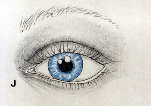
The entire face is examined with subjective quantification of dermatochalasis and steatoblepharon (Fig. 1; see also Fig. 11). Dermatochalasis is baggy eyelid skin, and steatoblepharon is orbital fat that bulges through a thinned orbital septum. There are two primary fat pockets in the upper lid, with the most prominent being the nasal Bourrelet senilis. In the lower lid, the lateral fat pocket is also the smaller and usually less prominent of the two. A coexistent brow ptosis, or blepharoptosis, should be recognized. Additional procedures such as ptosis repair or elevation of the brow may be required at the time of surgery. It may be difficult to convince a patient that a postoperative ptosis was indeed present before surgery unless the patient was evaluated specifically for, and informed preoperatively of, this condition. Likewise, a patient will be disappointed if a functional loss persists after blepharoplasty because a ptotic brow or aponeurotic dehiscence was the true culprit.

Fig. 1. Upper lid showing dermatochalasis and steatoblepharon.

Fig. 11. Lower lid showing dermatochalasis and steatoblepharon.

The examination should then focus on specific findings that might increase the risk of complications, such as ectropion, entropion, or exposure keratopathy. One should evaluate for increased horizontal lid laxity or dehiscence of the lower lid retractors.3 The latter is confirmed when the tarsus remains vertical instead of turning out when the lower lid is pulled down and is associated with an abnormal white line in the fornix. If necessary, a horizontal tightening procedure with or without retractor repair may be incorporated at the time of lower lid blepharoplasty. Any history of dry eyes or clinical finding of lagophthalmos, lid retraction, altered blink, or punctate corneal staining may necessitate further evaluation of the tear breakup time and tear output, since additional exposure may be detrimental to an already compromised cornea. Preoperative and postoperative photographs are an integral part of the patient's medical record. They serve as a learning instrument to the surgeon and are paramount in documentation for medicolegal and insurance purposes. By making certain that the patient's brow is relaxed and the excess skin is illustrated, photographs will reinforce findings on clinical examination and visual field testing. Preoperative 35-mm photographs or slides are taken, and they are repeated 1 to 2 months after surgery. A Polaroid photograph taken before surgery is useful during surgery and for immediate use.

Back to Top
TECHNIQUE FOR UPPER LID BLEPHAROPLASTY
The patient is prepped and draped so that the lids are in their natural position, avoiding undue tension and distortion of the surrounding skin. The patient is asked to look up and down to accentuate the upper lid creases. If the creases are symmetric and in a normal position, they may be used to mark the proposed lower portion of the incision. If they are absent, asymmetric, or high, as may be found with an aponeurotic defect, a new lid crease will need to be created. The height of the new crease may be guided by surgeon or patient preference, a normal contralateral crease, or race. The usual crease in the non-Asian is 10 mm from the lid margin at the midpupillary line. A preincisional marking is then extended toward the medial and lateral canthi as a gently sloping curve matching the configuration of the natural lid crease. A vertical distance of 4 to 6 mm should separate the line from both canthi. At the lateral canthus the line turns slightly upward to continue in a pre-existing crease (Fig. 2). The distance the mark extends laterally is guided by the amount of excess skin and the crease that it forms. The amount of skin to be excised is then determined by placing one tip of a Castroviejo forceps on the lid crease mark in the midpupillary line while the other tip rests more superiorly on the lid. The teeth are gently brought together while another forcep is used to grasp and pull excess skin up between the jaws of the other forceps (Fig. 3). The endpoint is that amount of skin that can be grasped just before the upper lid margin everts. A mark is placed at that point and is extended in both directions to join the lower line, complementing any nasal or lateral upward slope (Fig. 4). The patient is then asked to look up; and, if marked correctly, the lines of the proposed upper and lower incisions should nearly touch. A limiting distance of 1 cm from the lower border of the brow is used for the upper incision to avoid the unpleasant effect of approximating the brow too close to the lid margin. Next, infiltrative anesthesia is given with gentle pressure applied to aid in diffusion. If the patient is receiving monitored anesthesia care, propofol (Diprivan) or methohexitol (Brevital) is given while 0.5% bupivacaine with epinephrine 1:200,000 is injected. However, if the patient is receiving little or no sedation, two injections are given. Buffered 1% lidocaine precedes the bupivacaine and epinephrine mixture. The added bicarbonate not only accentuates onset but also decreases pain, while the extended duration of action of bupivacaine alleviates discomfort in the immediate postoperative period.4–6 While gentle traction is applied, a single continuous skin incision is made along the lower and then the upper marks using a no. 12 Bard-Parker blade placed perpendicular to the skin. The skin is then excised either with a combination of sharp and blunt dissection using appropriate scissors or with the electrocautery unit using a fine-tipped cutting needle (Fig. 5). We prefer the control of the Colorado needle (Colorado Micro Dissection Needle [nos. N103, N104, and N105], Colorado Biomedical, Evergreen, CO), which is insulated except for the tip and provides an excellent combination of precise cutting of delicate tissues and hemostasis. The skin is placed on the instrument stand over the patient's chest, and as fat is excised, it too will be arranged in a manner to signify the pocket from which it came so that symmetry of excision may be compared between sides. Meticulous hemostasis should be achieved during this and every stage of the procedure. A horizontal incision through the orbicularis to the level of submuscular fascia is made just above the lower skin incision (Fig. 6). If fat is to be excised, then the most nasal extent of the incision is deepened through the orbital septum. The fat capsule is incised and the fat is gently grasped and excised (Fig. 7).

Fig. 2. Marking the eyelid crease incision.

Fig. 3. Measurement of the amount of excess skin for excision.

Fig. 4. Eyelid with superior and inferior incisions marked.

Fig. 5. Skin excision utilizing cutting current of the monopolar cautery.

Fig. 6. Incision down to submuscular fascia just above inferior skin incision.

Fig. 7. Fat excision.

We tend to avoid excision from the central fat pocket because of the risk of damage to the underlying aponeurosis. If an aponeurotic dehiscence is to be repaired simultaneously, it may be performed at this time. If excess fat exists centrally, it may be removed under direct visualization.

The orbital septum should not be closed. Formation of the lid crease and skin closure may be achieved using a variety of sutures at the surgeon's discretion (6-0 Vicryl, chromic, catgut, or Prolene). It is important to use proper wound closure technique by alignment and eversion of wound edges (Fig. 8). The lid crease is reformed by taking small bites of submuscular fascia along with bites of the wound edges. The normal lid crease does not extend laterally beyond the canthus, and therefore only skin is closed in this region (Figs. 9 AND 10).

Fig. 8. Eyelid crease formation incorporating deeper submuscular fascia into skin bites.

Fig. 9. Skin closure temporal to lateral canthus.

Fig. 10. Upper lid after excess skin and fat has been removed.

Back to Top
TECHNIQUE FOR LOWER LID BLEPHAROPLASTY
The lower lid incision begins with a small incision placed in a temporal laugh line. Scissors are used to extend this to the punctum medially as an infraciliary incision, preferably camouflaged in the infraciliary groove (Fig. 12). A skin-muscle flap is reflected inferiorly to the orbital rim (Fig. 13). The septum is incised over the nasal and temporal fat pads, and the bulging fat is excised in a fashion similar to that used for upper lids (Figs. 14 AND 15) Before excising excess skin and muscle, the most lateral aspect of the tarsus is exposed and anchored to the inner aspect of the orbital rim using a buried 6-0 Prolene suture (Fig. 16). The patient is then asked to open his or her mouth and look up while the surgeon drapes the skin-muscle flap over the lid margin and marks the proposed line of excision at the level of the anterior ciliary margin (Fig. 17). If in doubt, it is better to undercorrect, since additional skin may be removed at a later time. The skin and muscle is excised and placed aside. The previously placed Prolene is passed through the orbicularis of the flap and tied to provide additional stabilization and superolateral traction (Fig. 18). Closure is achieved with near-far-far-near 6-0 Prolene sutures temporally along with an infraciliary running suture of the same material (Fig. 19).

Fig. 12. Lower lid incision placed in the infraciliary groove.

Fig. 13. Elevation of the myocutaneous flap to the level of the orbital rim exposing the orbital septum and underlying fat pockets.

Fig. 14. Excision of fat, after orbital septum and fat capsule have been incised.

Fig. 15. Appearance after removal of excess fat.

Fig. 16. Exposure of temporal tarsus and internal aspect of orbital rim using cutting current. These structures are apposed with a buried 6-0 Prolene superolateral traction suture. Care is taken not to change the position of the lateral canthal angle.

Fig. 17. Determination of the amount of excess skin and muscle for excision by draping skin in a superior and lateral direction while the patient is looking up with the mouth open.

Fig. 18. After skin and muscle excision, orbicularis is anchored to periosteum of internal aspect of orbital rim utilizing previously placed 6-0 Prolene suture.

Fig. 19. Closure with near-far-far-near sutures temporal to lateral canthus followed by a running infraciliary 6-0 Prolene suture.

LOWER LID BLEPHAROPLASTY COMBINED WITH REPAIR OF HORIZONTAL LID LAXITY

In the event of horizontal lid laxity with the concomitant predisposition toward postoperative ectropion, it is wise to combine a lower lid shortening procedure at the time of the blepharoplasty. We prefer a block excision unless the medial or lateral palpebral tendons are specifically lax.

The infraciliary incision is made, and the skin-muscle flap is reflected as previously described. Straight scissors are used to make a full-thickness marginal incision directed perpendicular to the lid margin approximately 6 mm from the lateral canthal angle. Beneath the full-thickness marginal cut, a tarsal conjunctival incision is extended to just below the inferior tarsal border and is then directed nasally at 100°. The lid margin ends are grasped, overlapped, and marked to determine the amount to be excised. Another incision is directed perpendicular to the lid margin at this point, carried to the lower tarsal border, and then directed temporally at 100° so that a pentagonal block is excised. After hemostasis is obtained, the retractors (inferior tarsal smooth muscle and capsulopalpebral fascia) and tarsus are reapposed with 6-0 Vicryl sutures, including a crucial buried suture placed through the posterior lid margin. The lid margin is approximated externally with 6-0 silk sutures placed through the gray line and the anterior lid margin. The remainder of the procedure is as described previously.

Back to Top
RETROBLEPHAROPLASTY
Occasionally, patients may exhibit bulging lower eyelid fat without excessive skin or orbicularis muscle. These generally younger patients are excellent candidates for retroblepharoplasty with the incision placed through the conjunctiva. An external scar is avoided, and therefore it has an advantage over the traditional approach.

After injection of local anesthesia, an incision is made across the inferior fornix and through the lid retractors, gaining access to the orbital fat. Care should be taken to avoid damage to the inferior oblique muscle. The fat is grasped and excised, again with meticulous attention to hemostasis. The lid retractors need not be closed, and conjunctiva is sutured with interrupted absorbable 6-0 sutures.

Back to Top
POSTOPERATIVE CARE
A small amount of ophthalmic antibiotic ointment is applied to the incisions, but no other dressing is used. Ice packs are begun immediately in the recovery room and are continued at home for the first 48 hours, followed by heat applications four times daily until the swelling is gone. Patients are advised to refrain from taking antiprostaglandin agents for 2 weeks. Wound care, including ophthalmic antibiotic ointment applications twice daily, is explained in a postoperative instruction sheet given to the patient.

Nonabsorbable sutures are removed after 5 to 7 days. Absorbable sutures may be removed if they have failed to dissolve or if social circumstances dictate earlier removal.

Back to Top
COMPLICATIONS OF BLEPHAROPLASTY
The major complications of blepharoplasty include entropion, ectropion, exposure keratopathy, central retinal artery occlusion, and central retinal artery avulsion. Their risk of occurrence may be minimized by a careful preoperative evaluation, judicious excision of excess skin, meticulous hemostasis, and gentle handling of intraorbital fat.

Since there is usually only mild to moderate pain after a blepharoplasty, any complaints of severe pain or decrease in vision warrant immediate evaluation to rule out a retrobulbar hemorrhage. If present, the wound is opened immediately, the blood evacuated, and bleeding vessels identified and coagulated. It may also be prudent to perform a cantholysis of the upper and lower limbs of the lateral canthal tendon since a rapidly rising intraocular pressure may occlude the central retinal artery. The practice of meticulously obtaining hemostasis at the time of surgery cannot be overemphasized, especially while excising orbital fat.

Total blindness may result from avulsion of the central retinal artery. The proposed mechanism is traction transmitted to the central retinal artery while grasping and pulling orbital fat through the septal incisions. Acute unilateral blindness occurs, with an extremely poor prognosis for visual recovery. Therefore, maintaining a gentle approach to orbital fat excision is recommended to decrease the occurrence of this devastating complication.

Postoperative ectropion usually develops due to failure to recognize lid laxity preoperatively but may result from excessive skin and muscle removal in the lower lids. Depending on the etiology, ectropion can be repaired through horizontal tightening procedures with or without retractor plication, a full-thickness skin graft, or a combination of the two. In some cases, mild ectropion may resolve over time with the return of orbicularis tone and with gentle upward massage.

The creation of postoperative “rounded eyes” is common. This appearance results from an increased concavity of the lower lids. This occurs from downward traction on a lid from excessive skin removal, a lax lid, or both. There may be enhanced scleral show and lid retraction inferiorly with or without frank ectropion. We attempt to avoid this by placing the previously mentioned Prolene sutures, giving additional superolateral traction to the lower lid. However, if preoperative ectropion or severe horizontal laxity is present, then a shortening procedure is also indicated.

Exposure keratopathy may be transient, mild, and asymptomatic until swelling resolves and orbicularis tone returns. Patients with coexistent dry eyes, altered blink, and findings of mild corneal punctate staining may benefit from artificial tears during the day and a lubricating ointment at bedtime. However, if exposure becomes more severe, corneal decompensation may progress, resulting in extensive corneal breakdown, ulceration, and perforation. Correction is dictated by the etiology and may include the use of a full-thickness skin graft to correct lagophthalmos or inferior lid retraction resulting from an excessively shortened anterior lamella. If lagophthalmos occurs secondary to inadvertent engagement of the orbital septum during lid crease reformation and wound closure, the involved suture must be identified and released.

Back to Top
REFERENCES

1. Goodman LS, Gilman AB: The Pharmacological Basis of Therapeutics, 8th ed, pp 647–648. New York, Pergamon Press, 1990

2. Moseley LH, Finseth F, Goody M: Nicotine and its effect on wound healing. Plast Reconstr Surg 61:570, 1978

3. Dryden RM, Leibsohn J, Wobig J: Senile entropion--pathogenesis and treatment. Arch Ophthalmol 96:1883, 1978

4. Peterfreund RA, Datla S, Ostheimer GW: pH adjustments of local anesthetic solutions with sodium bicarbonate: Laboratory evaluations of alkalinization and precipitation. Reg Anesth 14:265, 1989

5. Hilgier M: Alkalinization of bupivacaine for brachial plexus block. Reg Anesth 10:59, 1985

6. McKay W, Morris R, Mushlin P: Sodium bicarbonate attenuates pain on skin infiltration with lidocaine, with or without epinephrine. Anesth Analg 66:572, 1987

Back to Top