Chapter 81
Surgical Treatment of Graves' Disease
JOHN S. KENNERDELL
Main Menu   Table Of Contents

Search

TARSORRHAPHY
SURGERY FOR LID RETRACTION
STRABISMUS
ORBITAL DECOMPRESSION
CONCLUSION
REFERENCES

Dysthyroid orbitopathy (DO), also known as thyroid-associated orbitopathy (TAO) or Graves' disease, is the most common orbital disorder in adults.1,2 The diagnosis of this disease is discussed elsewhere in these volumes. Morbidity from this disease occurs from exposure keratopathy, extraocular myopathy, optic neuropathy, and disfigurement (Fig. 1). When medical therapies fail or when they are not expected to benefit the patient sufficiently, surgery may be indicated. As a general rule, surgery during the active phase of Graves' disease is not performed except for vision-threatening disorders (e.g., optic neuropathy or severe exposure keratopathy).

Fig. 1. A 59-year-old woman with severe dysthyroid orbitopathy including bilateral optic neuropathy, left lid retraction with corneal exposure, extraocular myopathy, and disfigurement.

The evaluation of a patient with dysthyroid orbitopathy should begin with a best-corrected visual acuity, including a refraction. The pupils, color vision, and visual fields are tested. The amount of proptosis is measured. The position of the eyelids is documented, including fissure width, the position of the upper and lower lids relative to the limbus, the margin reflex distance, the amount of levator function, and the presence or absence of lagophthalmos and lid lag. When lid surgery is contemplated, a Schirmer's test should be done. The motility is described and the amount of strabismus, if present, is quantified using prisms in the cardinal positions of gaze. In some instances, forced duction testing is performed. Intraocular pressure is measured and a dilated funduscopic examination is performed. Formal visual fields are performed in patients with significant disease. Orbital ultrasonography is often used to evaluate the size of the extraocular muscles. Orbital computed tomography (CT) or magnetic resonance imaging (MRI) is indicated when optic neuropathy is present to look for apical compression of the optic nerve. External photographs are taken to further document proptosis, lid position, and motility deficits.

Surgery to correct functional abnormalities that have developed from the disease is carefully timed and individualized because of the self-limited and variable nature of DO. The procedures used will be discussed individually.

Back to Top
TARSORRHAPHY
A lateral tarsorrhaphy is occasionally used to aid in lid closure and corneal coverage in patients who have significant exposure keratitis due to their DO. This is usually performed in conjunction with orbital decompression or lid retraction surgery. A temporary tarsorrhaphy may be performed after those procedures when there continue to be significant corneal exposure symptoms or signs despite adequate decompression or repair of lid retraction (Fig. 2). It is occasionally used as a procedure to mask mild exophthalmos, but it usually stretches because of lid retraction pulling on the adhesions, which is not cosmetically acceptable.

Fig. 2. A 46-year-old man with persistent left corneal exposure after two-wall orbital decompression for dysthyroid orbitopathy with optic neuropathy and corneal exposure.

The tarsorrhaphy complications are usually failure of the lid adhesion or a stretching of the lid adhesion. Misdirection of the lashes can occur after a tarsorrhaphy. There are seldom major complications such as hemorrhage or infection.

There are many techniques available for tarsorrhaphy. The following approach is effective in creating a permanent tarsorrhaphy that can be reversed later if desired.

First, the lids are infiltrated with local anesthetic containing epinephrine at 1:100,000 dilution. The lateral portions of the upper and lower lids are brought together to assess the amount of horizontal fissure narrowing needed. If the patient has significant lagophthalmos, he can be asked to gently close the eyes while the lids are being held together to determine if the proposed tarsorrhaphy will ensure full closure of the eye. A protective corneal lens may be placed on the eye at this time. The lids are then split into anterior and posterior lamellae at the gray line to a depth of approximately 1 mm. This incision extends from the lateral canthus to the nasal-most portion of the lid that is to be closed. The marginal tissue on the posterior side of this incision is then excised. The posterior lamella is closed with three or four interrupted 5-0 Vicryl sutures, with the knots tied anteriorly. The sutures should be placed so they are not penetrating through to the conjunctival surface. Following this, the skin edges are everted and the lids sutured together with 6-0 suture. If there is significant tension in the lids, a temporary Frost suture may be placed and removed in a week.

Back to Top
SURGERY FOR LID RETRACTION

UPPER LIDS

The most common upper lid disorder requiring surgical therapy is upper lid retraction. Upper lid retraction may cause dry-eye symptoms and exposure keratitis and may even allow for a corneal ulcer due to inadequate lid closure. It also contributes significantly to cosmetic disfigurement. Preoperative evaluation of a patient with lid retraction should include measurement of the lid height, measurement of the distance between the superior limbus and the upper lid margin, examination of the cornea for evidence of exposure keratopathy, and preoperative photographs for documentation. Surgery for isolated lid retraction is usually performed after 1 year of observation because of the tendency for spontaneous improvement. Lid retraction surgery is otherwise performed after decompressive and strabismus surgeries have been completed and the lid position has been stable for 6 months or more. A variety of methods for treating upper lid retraction have been described. These include recession of the levator aponeurosis with or without insertion of a spacer, disinsertion of the levator aponeurosis, recession of Müller's muscle, and excision of Müller's muscle.3,4 The method that we usually employ is a transconjunctival approach to a müllerectomy for 1 to 2 mm of retraction (Fig. 3) together with a graded levator aponeurosis myotomy for 2 to 3 mm of retraction (Fig. 4). A levator recession is performed for patients with over 3 mm of upper lid retraction. This approach allows us to tailor the amount of surgery performed to the severity of the upper eyelid retraction.

Fig. 3. A 40-year-old woman with 1 mm of right upper lid retraction secondary to dysthyroid orbitopathy.

Fig. 4. A 44-year-old woman with 3 mm of right upper lid retraction secondary to dysthyroid orbitopathy.

The müllerectomy and levator marginal myotomy procedure is as follows. Topical anesthetic is placed on the cornea followed by injection of 2% Xylocaine with 1:100,000 epinephrine into the midportion of the upper lid. A 4-0 silk traction suture is placed in the center of the lid margin at the gray line. The lid is then everted over a Desmarres retractor. The same local anesthetic is then injected into the subconjunctival space superior to the tarsus, thus ballooning up the conjunctiva and separating it from Müller's muscle. The conjunctiva is then incised at the upper border of the tarsus and carefully dissected from the underlying Müller's muscle, which is then incised at the superior border of the tarsus and is dissected free of the underlying levator aponeurosis. The dissection of conjunctiva and Müller's muscle is carried up 15 or 16 mm. Care is taken not to carry the dissection too far nasally because this can result in a nasal ptosis. The Müller's muscle is then excised, and hemostasis is obtained by use of cautery. The lid is then placed back into its normal anatomic position. The lid's height and contour are evaluated. If the lid is 1 mm below the level of the upper border of the limbus, then the lid is inverted again and the conjunctiva is reattached to the superior margin of the tarsus with 6-0 Vicryl or 6-0 fast-absorbing catgut suture (Fig. 5). If the lid is still too high, then a marginal myotomy of the levator aponeurosis can be performed laterally. The position of the lid is checked again, and if it is still high, an additional small myotomy of the levator can be performed medially (Fig. 6). The surgeon should avoid a large myotomy of the medial portion of the levator because this may result in a nasal ptosis. After the levator aponeurosis myotomy, the conjunctiva is closed with 6-0 absorbable suture as described earlier. An antibiotic-steroid ointment is placed in the eye, and the patient is given instructions to apply ice to the lid for the next 24 hours. It is common for the patient to have mild to moderate ptosis due to swelling for the first several days postoperatively. This should not be cause for concern.

Fig. 5. Same patient as in Figure 3 , after a right upper lid müllerectomy.

Fig. 6. Same patient as in Figure 4, with a right upper lid müllerectomy and a lateral levator aponeurosis marginal myotomy to correct upper lid retraction.

The levator recession and müllerectomy are performed through an anterior lid crease incision with the same local anesthetic. The skin is incised with a 30° superblade, and the dissection is carried down through the orbicularis with blunt-tipped Stevens scissors to the midtarsus. The levator is dissected from the tarsus and Müller's muscle completely across the tarsus, and the lateral horn is cut. Müller's muscle is then excised. The recessed levator is attached to the conjunctiva 12 to 15 mm from the upper tarsal border after checking the position of the lid in the upright position. The skin is closed with a 6-0 plain catgut suture in a running fashion. Postoperative care similar to that of the first procedure is provided.

The complications of upper lid retraction surgery include hemorrhage, which may be directed posteriorly into the orbit on rare occasions, as well as undercorrection or overcorrection of the lid retraction. Of course, corneal abrasions are sometimes associated with the procedure because of the suture line under the lid and the retrolid procedures. The swelling is significant postoperatively and usually resolves without incident.

Ptosis

The treatment of ptosis in Graves' patients is much the same as that in other patients. For small amounts of ptosis, a tarsal-conjunctival resection or a conjunctivo-müllerectomy may be performed. Patients with larger amounts of ptosis usually have evidence of an aponeurotic defect, so levator aponeurosis surgery is usually indicated. These procedures are discussed elsewhere in these volumes.

LOWER LIDS

Lower eyelid retraction is a common problem in Graves' patients. It is often aggravated by recession of the inferior rectus muscle secondary to restrictive myopathy. Patients with lower eyelid retraction complain of epiphora, dryness, and foreign-body sensation, and they frequently have evidence of exposure keratopathy (Fig. 7). The retraction is primarily due to fibrosis and overactivity of the lower lid retractors.5 The most commonly used method of elevating the lid involves recessing the lower lid retractors, usually by placing a tissue spacer between the inferior margin of the inferior tarsus and the lower lid retractors.6 We usually use tarsus harvested from the patient's own upper lid as a spacer.7 This is adequate for 1 to 3 mm of lower lid retraction. The technique is as follows:

Fig. 7. A 48-year-old woman with left lower lid retraction of 2 to 3 mm after left inferior rectus recession.

The upper and lower lids are anesthetized with 2% Xylocaine with epinephrine. A 4-0 silk suture is placed through the upper lid margin and centered over the pupil; the lid is everted over a Desmarres retractor. The conjunctival surface of the upper lid tarsus is dried with a cotton-tipped applicator. The elliptic area of excision, 20 mm wide and 5 to 7 mm high, is outlined, leaving a margin of 1 mm along the superior margin and 2 mm along the inferior margin. The tarsus is incised with an Alcon 30° blade or a ï15 Bard Parker blade along this outline. The tarsus is then dissected free from the underlying orbicularis muscle with Westcott scissors (Fig. 8). Hemostasis is maintained by use of cautery. The donor tarsal tissue is wrapped in gauze soaked in saline. The upper lid is returned to its normal anatomic position and the silk suture removed.

Fig. 8. A tarsal-conjunctival strip being removed from the upper lid.

Next, a silk traction suture is placed through the lower lid margin and the lid pulled inferiorly over a modified Erhart clamp. The conjunctiva and lower lid retractors are incised at the inferior margin of the lower lid tarsus. The lower lid retractors are dissected bluntly downward toward the orbital rim, ensuring that they are free from the adjacent tissue. The donor tarsus is then attached to the inferior margin of the lower lid tarsus with an absorbable suture such as 6-0 Vicryl or 6-0 mild chromic catgut in a running fashion. The conjunctiva and lower lid retractors are then attached to the inferior margin of the donor tarsus by use of the same suture (Fig. 9). A steroid and antibiotic combination ointment is placed in the inferior fornix. A lower lid traction suture may be taped to the forehead for 1 to 2 days to enhance the effect. About 2 to 3 mm of elevation can be obtained by combining the lower lid retractor recession with the tarsal transplant (Fig. 10). An additional 1 mm can be obtained by adding a lateral tarsal strip procedure.

Fig. 9. A tarsal conjunctival strip implanted in the lower lid. The lower lid is retracted by a modified Weitlawer retractor.

Fig. 10. The left lower lid is elevated into near normal position by a tarsoconjunctival transplant from the left upper lid.

Other spacer tissues may be used in place of autogenous tarsus, such as nasal septal cartilage, auricular cartilage, banked sclera, and hard-palate mucosa. Hard-palate mucosa is particularly useful because it has a thickness and consistency similar to that of tarsus and it regenerates so that it can be reharvested every 6 months.

Undercorrection of the retraction is the most common complication of lower eyelid retraction. Overcorrection is less common. Of course, other complications could include hemorrhage or infection, but they are, fortunately, rare. One complication that we have seen with the tarsal transplant is buckling of the transplant, which occurred in one patient about 4 to 6 weeks after surgery. The implant was removed, but the space that we had created filled in, and the correction of lid retraction was not compromised. We have never seen a patient with upper lid malfunction or disfigurement following removal of the tarsal conjunctival wedge.

Back to Top
STRABISMUS
Strabismus is a common problem in DO. Because it is restrictive in nature, the goal of surgical therapy is not to eliminate diplopia entirely but rather to move the region of single binocular vision into a more functional area. The inferior and medial rectus muscles are the most commonly involved muscles in the strabismus of DO. Therefore, patients present with hypo- and/or eso- deviations in most situations (Fig. 11). Recession procedures are the primary modality of surgical treatment for diplopia in Graves' restrictive myopathy after at least 6 months of stable measurements and lack of congestive signs.8

Fig. 11. A 50-year-old woman with dysthyroid left hypotropia with diplopia on attempted upgaze.

We usually perform the surgery on an adjustable basis because of the large amount of variability in the magnitude of correction. By performing the recessions on adjustable sutures, the alignment of the eyes can be fine-tuned in the postoperative period after the patient is awake and alert.9 In our experience, hypotropia from inferior rectus muscle involvement is the most common strabismus in Graves' disease requiring surgery. A graded recession of the inferior rectus muscle is usually successful in treating this, but occasionally one must resort to inferior rectus disinsertion, or recession of the contralateral superior rectus muscle. We always recess the conjunctiva during recession procedures for restrictive strabismus.

ANESTHESIA

When performing uniocular strabismus surgery, we usually use 2% Xylocaine mixed with 0.75% Marcaine in a retrobulbar block or peribulbar manner combined with intravenous sedation. When bilateral surgery is performed, we use a general anesthetic. A medial rectus recession is described.

After administration of anesthesia, the eye is prepped and draped. We will often leave the contralateral eye exposed in order to roughly assess the adequacy of alignment on the table. A wire lid speculum is placed in the eye, and forced ductions are performed to assess the degree of restriction. A limbal peritomy anterior to the involved muscle is performed with relaxing incisions of approximately 3 to 4 mm at either end. Tenon's fascia is separated from the globe in both quadrants adjacent to the inferior rectus muscle. A Stevens tenotomy hook is used to hook the restricted rectus muscle. A Jameson hook is then placed underneath the muscle and the small hook removed. A second Jameson hook is passed to ensure that the entire tendon has been included (Fig. 12). The conjunctiva is reflected away from the muscle by the use of small hooks, and the fascial attachments are then sharply dissected off of the muscle for a distance of approximately 15 mm. Once the fascial attachments are removed from the muscle, a 6-0 double-armed Vicryl suture with a spatulated needle is placed through the tendon of the muscle just behind the insertion. The suture is doubly locked at both edges, and the muscle tendon is then severed from the globe. When necessary, gentle bipolar cautery is used for hemostasis. The ends of the muscle suture are then passed through the midpoint of the original insertion, adjacent to each other. The muscle is allowed to hang back to achieve the desired amount of recession (usually 6–7 mm), and the suture is tied in a slip-knot fashion (Fig. 13). The ends of the suture are trimmed with enough length to allow the sutures to be tied the next morning after adjustment. A bucket-handle suture is then placed anterior to the original insertion of the inferior rectus muscle, adjacent to the limbus, using 4-0 silk suture. The conjunctiva is then teased back up to the original insertion of the inferior rectus muscle, thus recessing it approximately 5 mm. If necessary, the conjunctiva can be sutured in place with interrupted 6-0 plain gut sutures. The sutures are then tucked into the inferior fornix, antibiotic-steroid ointment is placed in the eye, and a gentle pressure patch is placed.

Fig. 12. A Jameson hook under an exposed medial rectus muscle insertion.

Fig. 13. A slip knot holds a recessed medial rectus muscle temporarily.

The patient is discharged from the surgery unit approximately an hour after surgery and seen in follow-up the next day. Alignment is checked and, if necessary, adjustment made at that time. Topical 4% Xylocaine is used for anesthesia, and a lid speculum is placed in the eye. If adjustment is necessary, the slip knot is untied and the muscle brought up or, if necessary, set back a bit, and the alignment is then re-evaluated. Once the desired alignment has been achieved, the suture is tied down in a permanent fashion and the ends cut close to the knot. The conjunctiva remains recessed. The bucket-handle suture is removed. We find that the patient is most comfortable when given an antibiotic-steroid combination ointment instead of drops after this type of procedure. The patient is seen in follow-up approximately 6 weeks postoperatively (Fig. 14). Recession procedures on all rectus muscles are approached in an identical manner.

Fig. 14. The same woman as in Figure 11 following a successful adjustable left inferior rectus recession with binocularity in the primary position of gaze.

The most common complication of extraocular muscle surgery is undercorrection of the deviation into position. A small percentage of patients are overcorrected, and one can expect limitation of movement of the eye in the direction of the muscle recessed if the recession is maximal. This is particularly true in the case of inferior rectus. Also, lid retraction can occur after inferior rectus recession, even with separation of Lockwood's ligament and the lower lid retractors away from the inferior rectus.

Back to Top
ORBITAL DECOMPRESSION
Patients with dysthyroid orbitopathy may require orbital decompression for a variety of reasons. Optic neuropathy in DO can cause severe visual loss that is usually due to compression of the optic nerve from swelling of the rectus muscles near the apex of the orbit.10 With adequate orbital decompression, this can frequently be reversed. Exposure keratopathy from extreme proptosis can be quite severe, occasionally leading to corneal perforation. Again, orbital decompression can reduce the severity of exposure and therefore benefit patients with extreme proptosis. Orbital decompression may also be performed for cosmesis, particularly in those situations in which there is marked asymmetry in the exophthalmos.

The most common approach for orbital decompression for DO is to remove portions of one or more of the bony walls of the orbit. This accomplishes decompression by expanding the volume available to the orbital fat and extraocular muscles. A variety of procedures have been described, including removal of the lateral wall, the orbital floor, the medial wall, and portions of the roof.11–16 Most surgeons use a variety of these techniques, alone or in combination, depending on the patient's findings, including the amount of proptosis, the presence of optic neuropathy, and pre-existing strabismus.17 Orbital fat excision without bone removal has been advocated for patients with symptoms related to proptosis but without compressive optic neuropathy.18

The surgical approach should be selected after consideration of the patient's findings, including the amount of proptosis, pre-existing strabismus, and presence or absence of optic neuropathy. Lateral orbital wall removal alone usually will produce a reduction in proptosis of 1 to 3 mm and is less often associated with postoperative strabismus, but it does not usually relieve the apical crowding causing optic neuropathy. Removal of the medial wall is more effective in treating optic neuropathy and produces an enophthalmic effect of 2 to 4 mm, but it is more often accompanied by postoperative strabismus. Removal of the floor also produces a larger enophthalmic effect of 3 to 5 mm; this procedure is frequently accompanied by worsening of strabismus and/or numbness or paresthesia in the distribution of the second division of the fifth cranial nerve, the latter of which usually resolves after several months. Combining a lateral wall removal with a medial wall and floor procedure may enable the surgeon to improve the enophthalmic effect to 6 to 10 mm without increasing the risk of induced strabismus. Removal of portions of the orbital roof is reserved for those cases with severe proptosis; it is possible to achieve a reduction in exophthalmos of 10 to 17 mm with a panorbital decompression.16

SURGICAL TECHNIQUE

General anesthesia is used for all orbital decompressions for dysthyroid ophthalmopathy. If a four-wall decompression is being performed, we recommend that a neurosurgeon familiar with cranio-orbital junction surgery be a part of the operating team. Dexamethasone phosphate, 10 to 40 mg, is given intravenously at the beginning of the surgery. We do not routinely use prophylactic antibiotics for orbital decompression.

ONE- AND TWO-WALL DECOMPRESSION

When the degree of proptosis is not severe and optic neuropathy is present, a medial wall and sometimes a medial orbital floor decompression is indicated (Fig. 15). As indicated earlier, a Caldwel-Luc approach may be used to accomplish this, but we prefer the medial approach because of a lesser incidence of diplopia. This has been done by an endoscopic method, which may 
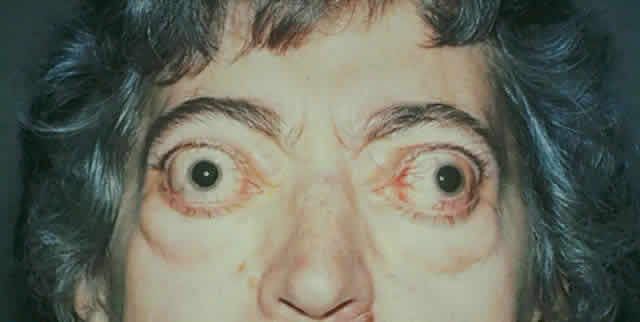
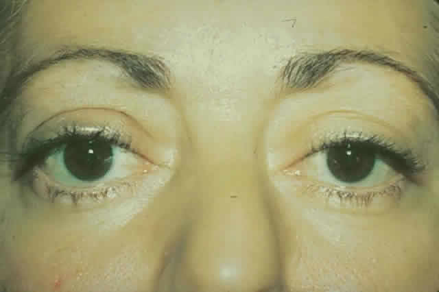
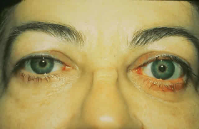
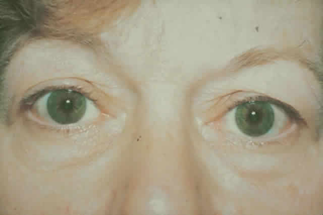
gain favor in the future.

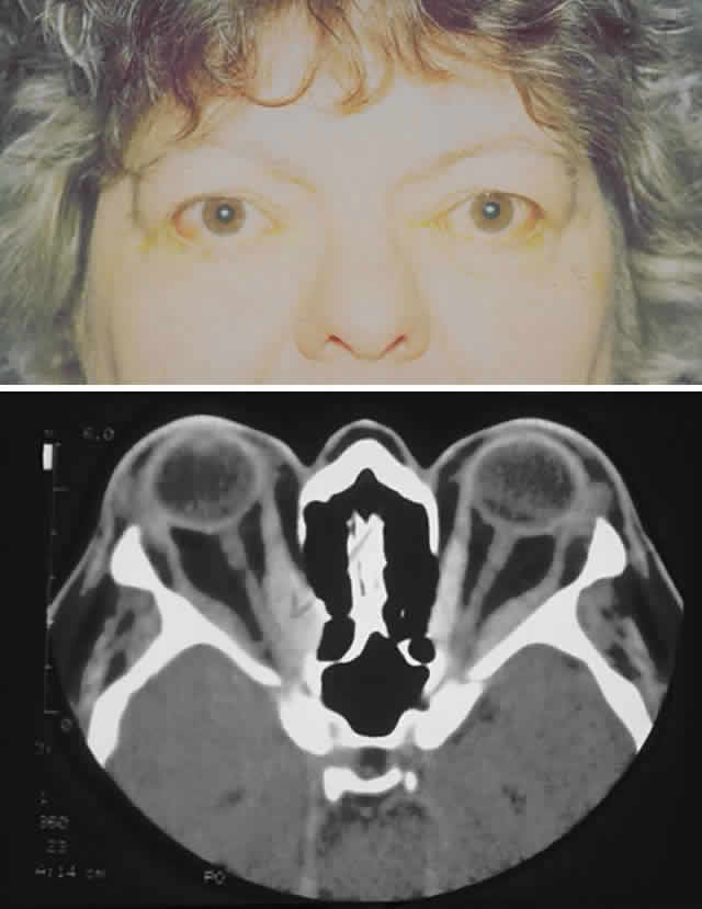
Fig. 15. A. A 48-year-old woman with dysthyroid optic neuropathy. Visual acuity right eye 6/24, left eye 6/9. Color vision right eye 1/12, left eye 10/12--Ishihara plates. Goldmann visual fields showed central scotomas in both eyes. Hertel measurements right 25, left 24, base 102. B. Preoperative axial CT scans showing apical crowding and proptosis.

A medial approach is made through an extended Lynch incision halfway between the medial canthus and the nose, extending from the superior lacrimal crest to 1 cm below the medial brow. The dissection through the skin is done with a No. 15 Bard Parker knife, and the subcutaneous tissue is dissected with cutting cautery down to the periosteum. The periosteum is incised and reflected off the medial orbital wall with a periosteal elevator. Then a Hall drill is used to make a small opening in the anterior ethmoid sinus. A kerasin and a rongeur are then used to remove the medial orbital wall and ablate the ethmoid air cells back to the sphenoid sinus. Care is taken to stay below the anterior and posterior ethmoidal arteries to prevent excessive bleeding and to avoid the cribriform plate. CT scans are displayed in the operating room for study of the anatomy of the cribriform plate prior to surgery. At times, the plate is lower than usual and easily entered if not anticipated. If additional decompression is desired, the medial orbital floor is then removed with rongeurs to the inferior neurovascular bundle. Hemostasis is achieved as well as possible and drainage is obtained through the nostril on the side of the lesion. The deep tissues on the side of the nose are closed with 4-0 Vicryl sutures in an interrupted manner. The skin is closed with a running 6-0 plain catgut suture. A pressure 
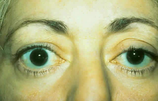
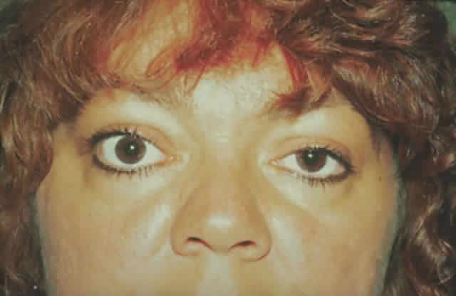
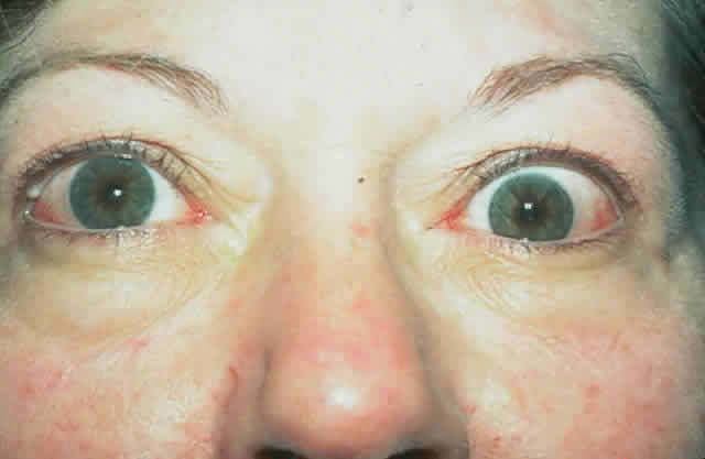
dressing is applied. The patient (see Fig. 15) had a 6 mm decompression (Fig. 16).

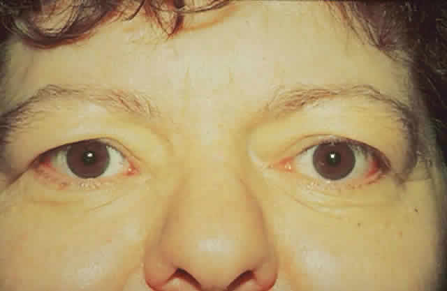
Fig. 16. Postoperative appearance of woman in Figure 15. Visual acuity right eye 6/9, left eye 6/6. Color vision right eye 7/12, left eye 12/12--Ishihara plates. Goldmann visual field showed only enlarged blind spot right eye. Hertel measurements right 19, left 18.

THREE- AND FOUR-WALL DECOMPRESSION

After administration of general anesthesia, with the patient in a supine position, the head is turned approximately 45° to the contralateral side. A 35-mm line is drawn from the lateral canthus posteriorly toward the upper attachment of the ear to the head, and this area is injected with 2% Xylocaine and 1:100,000 epinephrine. The skin is incised along this line to the level of the temporalis fascia and then widely undermined. Anteriorly, the periosteum of the lateral orbital rim is incised 2 mm posterior to the rim for a length of 35 mm with relaxing incisions at the superior and inferior extents. The periosteum is reflected off the underlying bone along the rim, and the temporalis muscle is reflected posteriorly by use of the cutting cautery. The periosteum is elevated anteriorly to the rim and the periorbita is dissected free from the underlying bone. This is firmly adherent only at the lateral orbital tubercle where the lateral canthal structures attach. Cautery may be used on the vascular structures exiting the orbit through the zygomatic bone.

With malleable retractors protecting the periorbita, the superior and inferior lateral orbital rims are cut with a Stryker sagittal saw. The rim is then grasped with a rongeur, outfractured, and removed. The rim of the removed bone is thinned posteriorly with the high-speed air drill and is set aside wrapped in saline-soaked gauze. Bone is removed back to the greater wing of the sphenoid with use of rongeurs and a high-speed air drill.

At this point, if a four-wall decompression is being performed, bone removal continues with the drill, exposing the temporal dura. Bone wax is used to control bone bleeding. With malleable ribbon retractors protecting the periorbita, the diamond-tipped drill removes bone superiorly to expose dura in the roof of the orbit. The dura is dissected from the orbital roof and protected with cottonoids. Small rongeurs are then used to remove bone from the roof anteriorly to the orbital rim, laterally and medially to the inner one third of the roof, avoiding the frontal 
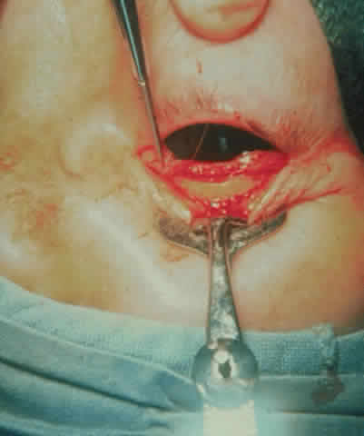
sinus, and posteriorly to the lesser wing of the sphenoid.

The floor of the orbit is then exposed through a canthotomy and fornix incision as follows: The inferior crus of the lateral canthal tendon is severed and the conjunctiva incised with scissors in the fornix along nearly its entire length. This incision is carried to the periosteum of the inferior orbital rim, which is incised just anterior to the rim. A sharp periosteal elevator lifts the periosteum off the rim, exposing the entire floor of the orbit. Small malleable retractors are used to dissect and retract, exposing the ethmoidal and naso-orbital areas.

The floor of the orbit is thin and can usually be fractured with a small, curved hemostat medial and lateral to the inferior neurovascular bundle. Alternatively, a diamond burr may be used to create an opening in the bony floor. Additional bone is removed with rongeurs, and care is taken not to injure the infraorbital nerve and artery. Posteriorly, bone is removed as completely as possible. We leave a strut of bone around the infraorbital nerve anteriorly as a structure to support the globe and prevent globe ptosis. Caution must be used near the orbital rim in the inferomedial area to avoid injury to the nasolacrimal duct. Medially, the ethmoids are removed to the frontoethmoidal suture superiorly and the sphenoid sinus posteriorly. Front-biting rongeurs are used to accomplish this; occasionally, digital pressure can be used to infracture the posterior ethmoid sinuses and attain adequate medial decompressive effect.

Next, the periorbita is opened laterally, inferiorly, and medially to allow orbital fat prolapse into the spaces created by the bone removal. We use a scimitar-shaped blade for this; scissors may be used to enhance or extend the incisions. Frequently, the fat is fibrotic and does not prolapse spontaneously upon opening the periorbita. In this situation, portions of fat may be gently teased through the incisions.

The conjunctiva is closed with a running 6-0 Vicryl suture. The bone of the lateral rim is replaced after the posterior portion is removed, and holes are drilled to allow the rim to be tied into position with 4-0 nylon suture. Some surgeons will secure the rim with titanium miniplates, and others will not fix its position at all but will simply place it in its original bed. The periorbita is closed over the bone with interrupted 4-0 absorbable suture. This same material is used to close the temporalis fascia. A 6-0 running plain catgut suture is used to close the skin. A pressure dressing is placed over the eye and lateral 
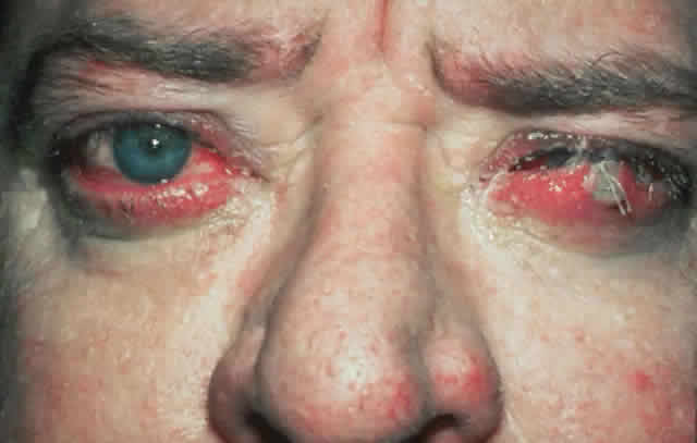
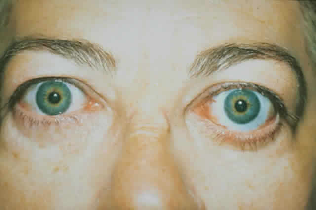
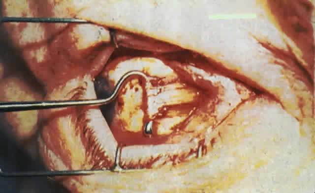
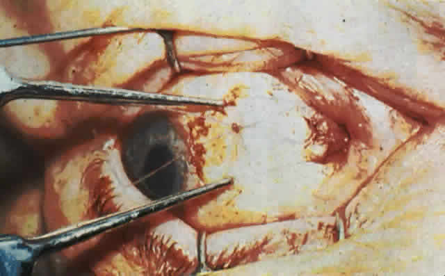
wall of the orbit (Fig. 17).

Fig. 17. A. A 38-year-old woman with marked dysthyroid proptosis and corneal exposure. Hertel measurements right 31, left 30, base 101. B. Postoperative appearance. Hertel right 20, left 18, base 101.

The complications of orbital decompression can be significant and include, most commonly, worsening of eyelid retraction or increased diplopia. Corneal compromise can occur during or after surgery as well and can sometimes be severe. The decompression can be inadequate or too generous, and the ethmoid sinus or maxillary sinus can become infected postoperatively on occasion. There have been injuries to the cribriform plate where spinal fluid leaks from orbital decompression of the medial wall and of the roof. Air can enter the dura directly, causing pneumocrania on rare occasions, and death may ensue from complications of anesthesia or, as in one case, a ruptured aneurysm of an unrelated anterior communicating artery during this major surgery. The optic nerve can be injured or devascularized during decompression and hemorrhage with optic nerve compression, which can result in blindness. Orbital decompression of any type is certainly a major procedure, and all complications should be carefully explained to the patient and family before assuming the risk of this type of surgery.

Back to Top
CONCLUSION
Dysthyroid orbitopathy is a disease that can lead to visual dysfunction and facial disfigurement. A variety of surgical procedures can be used to minimize the sequelae of this disorder. Except under circumstances where vision is threatened if prompt action is not taken, surgical intervention should be delayed until the inflammatory component of the disease is over and the ocular findings have been stable for 6 months or more.
Back to Top
REFERENCES

1. Leone CR: The management of ophthalmic Graves' disease. Ophthalmology 91:770, 1984

2. Kennerdell JS, Maroon JC, Buerger GF: Comprehensive surgical management of proptosis in dysthyroid orbitopathy. Orbit 6:153, 1987

3. Moran RE: The correction of exophthalmos and levator spasm. Plast Reconstr Surg 18:411, 1956

4. Putterman AM, Urist M: Surgical treatment of upper eyelid retraction. Arch Ophthalmol 87:401, 1972

5. Quickert MH, Dryden RM: Lower eyelid advancement. Presented to the American Society of Ophthalmic Plastic & Reconstructive Surgery, Las Vegas, 1971.

6. Baylis HI, Rosen N, Neuhaus RW: Obtaining auricular cartilage for reconstructive surgery. Am J Ophthalmol 93:709, 1982 Ì

7. Gardner TA, Kennerdell JS, Buerger GF: Treatment of dysthyroid lower lid retraction with autogenous tarsus transplants. Ophthal Plast Reconstr Surg 8(1):26, 1992 Ì

8. Evans D, Kennerdell JS: Extraocular muscle surgery for dysthyroid myopathy. Am J Ophthalmol 95:767, 1983

9. Gardner T, Kennerdell JS: Treatment of dysthyroid myopathy with adjustable suture recession. Ophthalmic Surg 21(7):519, 1990

10. Kennerdell JS, Rosenbaum AE, EL-Hoshy MH: Apical optic nerve compression in dysthyroid optic neuropathy. Arch Ophthalmol 99:807, 1981

11. Dollenger J: Die Druckentlastung der Augenhole durch Entefernung der auberen Orbitalwand bei hochgradigem Exophthalmos (Morbus Basedowii) und konsekutiver Hornhauterkrankung. Dtsch Med Wochenschr 37:1888, 1911

12. Hirsch VO, Urbanik AG: Behandlung eines exzessiven exophthalmos (Basedow) durch entefernung von orbitalfett vonder kieferhole aus. Monatschr Ohrenh-Laryngorhinol 64:212, 1930 Ì

13. Nafzinger HC: Progressive exophthalmos. Ann R Coll Surg Engl 22, 1954 Ì

14. McCord CD Jr, Moses JL: Exposure of the inferior orbit with fornix incision and lateral canthotomy. Ophthalmic Surg 10:53, 1979

15. Walsh TE, Ogura JH: Transantral orbital decompression for malignant exophthalmos. Laryngoscope 67:544, 1957

16. Maroon JC, Kennerdell JS: Radical orbital decompression for severe dysthyroid exophthalmos. J Neurosurg 56:260, 1982

17. McCord CD Jr, Current trends in orbital decompression. Ophthalmology 93:476, 1985

18. Trokel S, Kazim M, Moore S: Orbital fat removal decompression for Graves' orbitopathy. Ophthalmology 100(5):674, 1993

Back to Top