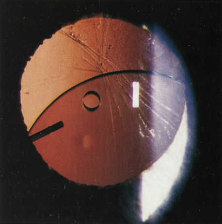
Fig. 3.
Recurrent iritis and hyphema associated with a loose posterior chamber intraocular lens in the ciliary sulcus. Note fine dusting of red blood cells on endothelial surface.