|
|
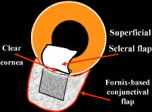
| Fig. 41. Deep sclerectomy with collagen implant: superficial scleral flap. After obtaining adequate limbal exposure, a fornix-based conjunctival flap is prepared. A 5 × 5 mm superficial limbal based 1/3 thickness scleral flap is developed in a uniform fashion. Cautery is used judiciously, but sclerosis of outflow vessels is not as critical as with deep sclerectomy with viscocanalostomy (DSVC). This superficial flap is dissected 1 mm into clear cornea to allow space for dissection of canal area. |