Fig. 5.
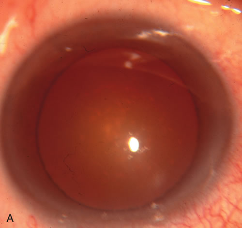
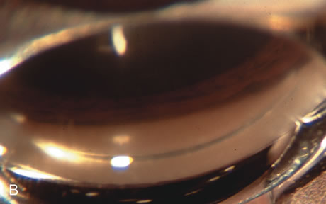
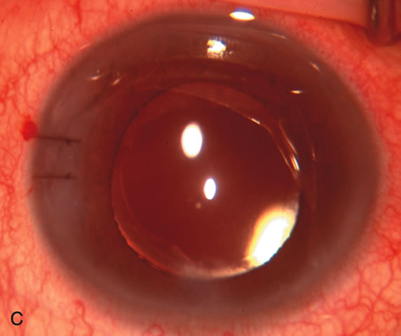
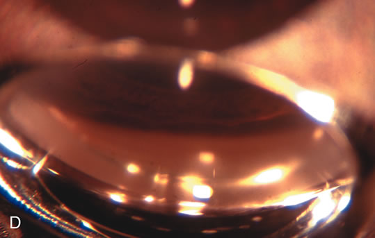
Anterior chamber angle changes associated with lens extraction and PCIOL This 65-year-old Vietnamese woman has a long-standing history of chronic angle-closure glaucoma treated with laser peripheral iridectomy. The optic nerve demonstrated mild glaucomatous damage and IOP was moderately controlled on two antiglaucoma medications. The cataract was removed through temporal clear corneal phacoemulsification with foldable acrylic IOL. A. Symptomatic cataract in narrow-angle glaucoma eye with patent iridectomy. B. Intraoperative goniophotograph showing crowding of angle with increasing narrowness due to phacomorphic component. C. Intraoperative photograph showing temporal clear corneal approach with IOL in the capsular bag. D. Intraoperative goniophotograph demonstrating deepening of chamber angle following lens extraction. Proposed theories for IOP reduction following lens extraction with complete wound closure:
- Anterior chamber deepening with improved access to trabecular meshwork
- Increase in traction on the trabecular meshwork
- Improved outflow facility mediated by an increase in prostaglandin release
- Reduction in aqueous humor production
- Atrophy of ciliary body processes
- Goniosynechialysis due to intraoperative over deepening of AC with viscoelastic
- Relief of undiagnosed pupil block
|


