|
|
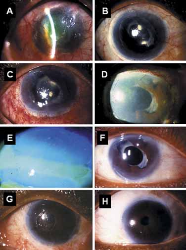
| Fig. 8 Conjuctival limbal autograft (CLAU) with amniotic membrane transplantation. This patient with glaucoma developed drug-induced pseudopemphigoid manifesting total limbal stem cell deficiency (LSCD) with vascularization and epithelial defect in the right eye (A). She received keratolimbal allograft transplant (KLAL) and amniotic membrane transplantation with improved surface that last for 1.5 years with systemic cyclosporin (B), but developed recurrent inflamed ocular surface, LSCD, and epithelial defect (C). Two large strips of CLAU spanning nearly 11 clock hours were removed from the left eye (without visual potential) and transplanted to the right eye over a layer of amniotic membrane (D). Five days later, rapid epithelialization was noted on the amniotic membrane covered cornea from both superior and inferior CLAU (E). This resulted in an improved vision with a stable surface 2.5 years later (F). Because of the large limbal removal, the donor cornea decompensated with surface breakdown 5 days after CLAU (G). Nevertheless, because of the covering of amniotic membrane, a stable surface was recovered 2.5 years post-CLAU. (Modified from Meallet MA, Espana EM, Grueterich M, et al: Amniotic membrane transplantation for recipient and donor eyes undergoing conjunctival limbal autograft for total limbal stem cell deficiency. Ophthalmology 110:1585, 2003, with permission) |