

|
|
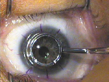
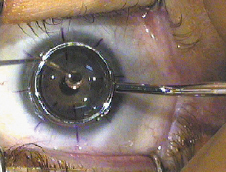
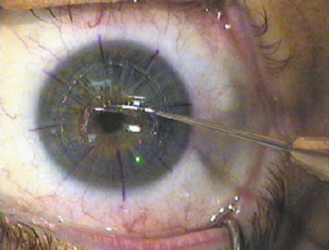
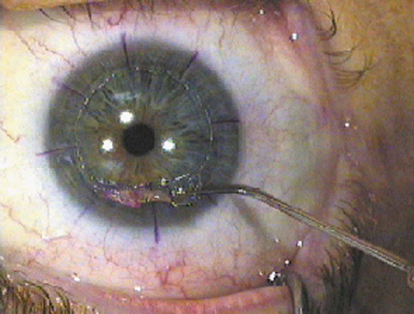
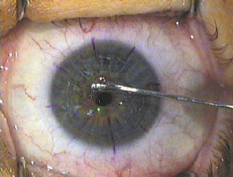
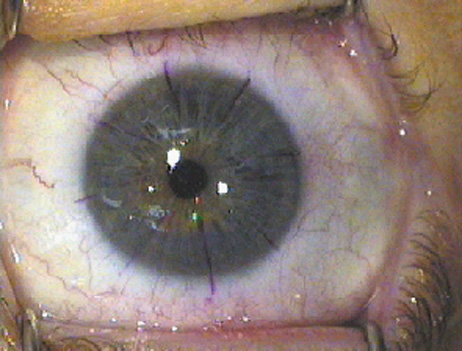
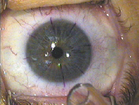
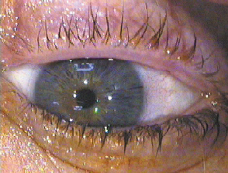
| Fig. 6. Technique for LASEK. A: After the gentian violet radial orientation marks are placed, the epithelial flap is delineated with an optical zone marker or similar instrument. B: Alcohol to loosen the epithelial flap is carefully contained within a trephine-shaped well pressed on the cornea. C: The separation of the flap edge is begun with a “microhoe.” D: The separation of the epithelial flap continues as a “micro hockey stick” strokes the epithelium back, with care taken to not perforate the epithelium. E: The epithelium is fully retracted off the area to be ablated by the laser, leaving a “hinge” of epithelium for about one clock hour in the superior periphery. G: After the laser exposure is completed, the epithelial sheet is gently replaced by floating it and stroking it with a BSS cannula. H: The epithelial flap is realigned using the radial marks. I: The epithelial sheet is briefly dried with filtered air through a microtip to begin adhesion to the stroma. J: After a BSCL is placed, the procedure is complete. (All photographs courtesy of Daniel Durrie, M.D.) |