|
|
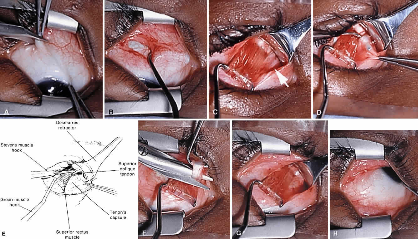
| Fig. 15. Right superior oblique tendon tenotomy performed through a temporal conjunctival approach. A. Conjunctival incision is made lateral to the superior rectus muscle insertion. B. The superior rectus muscle is engaged on a Green hook. C. The conjunctival opening is moved over the muscle hook and the medial border of the superior rectus muscle is exposed. The superior oblique tendon is visible exiting from under the superior rectus muscle (arrow). D and E. The superior oblique tendon is isolated on a muscle hook and Tenon's capsule carefully unloaded. F. Tenon's capsule carefully unloaded. G. The superior oblique tendon is cut adjacent to the medial border of the superior rectus muscle. H. The superior nasal quadrant of the globe is carefully examined for missed tendon fibers. |