Chapter 6
Cataract Surgery
ROBERT J. CIONNI, MICHAEL E. SNYDER and ROBERT H. OSHER
Main Menu   Table Of Contents

Search

THE HISTORY OF CATARACT SURGERY
PREOPERATIVE EVALUATION
SPECIAL PREOPERATIVE TESTING
ANESTHESIA TECHNIQUES
INFECTION PROPHYLAXIS
ANTIBIOTIC COVERAGE
INCISIONS
PHACOEMULSIFICATION
PHACOEMULSIFICATION TECHNIQUES
ALTERNATIVE PHACOEMULSIFICATION MODALITIES
INTRAOCULAR LENS IMPLANTATION
COMPLETION OF CATARACT SURGERY
TO PATCH OR NOT TO PATCH?
POSTOPERATIVE MEDICATIONS
POSTOPERATIVE RESTRICTIONS
POSTOPERATIVE FOLLOW-UP
COMPLICATIONS OF CATARACT SURGERY
CHALLENGING CATARACTS
WHITE CATARACT
BRUNESCENT CATARACT
POSTERIOR POLAR CATARACTS
COMPROMISED ZONULES
THE FUTURE OF CATARACT SURGERY
REFERENCES

THE HISTORY OF CATARACT SURGERY
The unprecedented success of contemporary cataract surgery has been the result of continuing innovation in technique and technology. This section is meant to provide a quick overview of the major events in the history of cataract surgery that are responsible for the current state of the art. For an instructive, entertaining, and detailed review, the reader should peruse “The History of Cataract Surgery” by Norman Jaffe,1 “The History and Development of Phacoemulsification” by Charles Kelman,2 and “Harold Ridley and the Invention of the Intraocular Lens” by David J. Apple.3 Moreover, Stephen Obstabaum and Emmanuel Rosen must be given credit for the rapid dissemination of information in this specialized field, having devoted more than two decades serving as Editors of the American and European Cataract Journals.

It is remotely possible that ophthalmic surgeons would be experts at couching were it not for the French physician, Jacques Daviel, who reported the first series of extracapsular cataract extractions to the Royal Academy of Surgery in 1753.4 However Daviel's operation did not become immediately popular because it required the management of the cortex. At that time, the operating microscope had not yet been invented, which made identification and removal of the cortex virtually impossible. Residual cortex was associated with severe and disastrous inflammatory reactions. Moreover, vitreous loss was a common and serious complication, given that the concept of vitrectomy did not yet exist.

Intracapsular cataract extraction was the standard at the beginning of the 20th century. Leaders of the American Academy of Ophthalmology and Otolaryngology were influenced by the work of Henry Smith in India, who had developed a quick, safe method of delivering the lens within its capsule using external manipulation.5 Intracapsular cataract extraction continued to evolve with the design of capsule forceps, Joaquin Barraquer's discovery of the enzyme α-chymotrypsin to facilitate zonulolysis in 1957,6 and eventually the introduction of cryoextraction by T. Krawicz and Charles Kelman, independently, in 1961.7

Several major events raised the quality of cataract surgery to a new level during the 20th century. Ophthalmic sutures were introduced and anesthesia techniques improved. Intraocular visualization was enhanced by the development of loops and then, the operating microscope. Digital massage was found to decrease the risk of intraoperative complications. Then, two gigantic contributions changed cataract surgery forever.

The first came in 1949, when a British surgeon named Harold Ridley implanted the first intraocular lens (IOL) into the posterior chamber using the posterior capsule to support this heavy discoid implant.8 There were many complications and tremendous controversy concerning his intraocular implants. Even so, a new frontier had been established; superior visual rehabilitation could be achieved with IOL implants.

The next enormous contribution came in 1967, when Charles Kelman introduced the extracapsular small incision technique of phacoemulsification,9 which was at least as controversial as Ridley's historic breakthrough. Both innovators suffered extreme ostracism, yet each was largely responsible for contemporary cataract surgery. Surgeons began to realize that phacoemulsification and IOL implantation resulted in exquisite control of the intraocular environment, greater safety in handling complications or challenging cases, quicker visual rehabilitation, and a reduction in surgically induced astigmatism. Therefore, the pendulum began to swing from intracapsular to extracapsular surgery. The latter enjoyed even greater acceptance as the protective nature of the posterior capsule became better understood when Gass and Norton initiated their fluorescein angiographic study of cystoid macular edema.10 Soon afterward David Kasner declared the “vitreous our enemy,” demonstrating the benefits of manual, and later, automated vitrectomy.11

Intraocular lens pioneers such as Cornelius Binkhorst, Peter Choyce, Jan Worst, Edward Epstein, Steven Shearing, and Svyatoslav Fyodorov among many others modified IOL design and the location for implantation. In England, John Pearce returned the IOL to the posterior chamber preceding the development of the Shearing style lens, which marked the real acceptance of the IOL. The safety and growing popularity of intraocular lenses was intimately linked to the introduction of Healon, a hyaluronic acid derived from Swedish rooster combs. It was Pape and Balaz who pioneered the concept of viscosurgery in 1979.12 Modern implant surgery was revolutionized by Richard Kratz, Robert Sinskey, William Simcoe, John Sheets, Norman Jaffe, Gerald Tennant, Eric Arnott, and Charles Kelman. Outstanding teachers such as David McIntyre, Jared Emery, Henry Hirschman, Herve Byron, Richard Lindstrom, Harry Grabow, Douglas Koch, Roger Steinert, and Samuel Masket spread the word as phacoemulsification techniques, improved instrumentation, and IOL designs proliferated.

Another huge step forward was taken in the early 1980s when French surgeon Danielle Aron-Rosa and her colleagues introduced the neodymium:yttrium-aluminum-garnet (Nd:YAG) laser for performing posterior capsulotomy, which eliminated an additional procedure in the operating room.13 Clifford Terry introduced the surgical keratotomer, which provided us with a new understanding of astigmatism,14 leading to the combination of astigmatic keratotomy with phacoemulsification to reduce pre-existing astigmatism introduced by Robert Osher in 1983.15,16 Then in 1985, Thomas Mazzocco developed and implanted the first foldable IOL17; Australian Graham Barrett and associates ushered us into the materials era with the use of silicone, hydrogels, and acrylics.18 Remarkable insights by David Apple correlated clinical observation with histopathology using the photographic technique developed by Kensaku Miyake of Japan.19

At about the same time, another Japanese surgeon, Kimiya Shimizu, began removing cataracts using topical anesthesia. A Canadian physician, Howard Gimbel and a German surgeon, Thomas Neuhann independently arrived at the concept of capsulorrhexis, although there is mounting evidence that Calvin Fercho20 was the first to perform a continuous curvilinear capsulorrhexis.21,22 Gimbel also popularized the divide and conquer technique,23 John Shepherd disassembled the nucleus into quadrants,24 and Kunihiro Nagahara introduced the concept of chopping in 1993.25 Ken Faust published the technique of hydrodissection26 and Douglas Koch described multilamellar hydrodelineation.27 Robert Osher modified the “phaco” machines by introducing the concept of slow-motion phacoemulsification, which allowed the surgeon to control the intraocular environment more exactly with variable ultrasound, aspiration rate, vacuum, and bottle height.28 John Shepherd introduced the horizontal one-stitch closure,29 eventually leading to the abandonment of sutures altogether, as championed by Michael McFarland.30 Howard Fine returned the smaller phacoemulsification incision back to the cornea using foldable IOLs.31

In the late 1990s, the transition from extracapsular surgery to phacoemulsification was almost complete, as surgeons enjoyed a quicker and safer near-clear or clear corneal incision, capsulorrhexis, hydrodissection, nuclear disassembly, the insertion of a UV-blocking foldable IOL, and a sutureless closure. The incidence of operative and postoperative complications was never lower and the recovery of excellent vision was achieved as technical advances in equipment, lens design, and biometry continued to improve. Intraocular lens formulas were refined by Donald Sanders, Manus Kraff, and Jack Retzlaff; Kenneth Hoffer; and Jack Holladay. Robert Osher challenged ASCRS to adapt a new standard in reporting visual results emphasizing the importance of early-uncorrected vision and promoting the new concept of refractive cataract surgery.32 Industry introduced higher-quality silicone and acrylic IOL materials, viscosurgical tools, and sophisticated phaco machines, which were more versatile and reliable.

At the turn of the century, the incidence of posterior capsular opacification (PCO), the most common untoward event after surgery, was plummeting as the result of a square edge design on the IOL optic. The haptics themselves had evolved from Prolene material to polymethylmethacrylate and then Alcon introduced a soft haptic on a single-piece acrylic platform. Every company developed an injector for improved IOL insertion, and industry was teaming up with innovative surgeons in developing a number of optic modifications. Attempts to achieve multifocality with diffractive optics had been introduced by 3M and their design team of Richard Lindstrom, John Sheets, and Robert Osher. This technology was placed on the back burner until Allergan developed the array multifocal IOL. Alcon purchased and improved the 3M defractive optic and introduced ReStor IOL on a single-piece acrylic platform. The surgical assault on presbyopia accelerated as Eyeonics developed the Crystalens, the first IOL with a pliable optic aimed at generating accommodation, a concept introduced a decade before by Spencer Thornton.33 A toric optic was developed by Staar Surgical; Pharmacia introduced Tecnis, the first spherical aberration-correcting IOL; and Alcon developed the first blue-light–filtering optic to provide macular protection. The once narrowly defined specialty of cataract and IOL surgery was exploding with new ideas and technologies. Smaller-incision IOLs were being designed that could be injected through <2-mm incisions. A host of surgeons, including Amar Agarwal, Jorge Alio, Richard Packard, Hiroshi Tsuneoka, Virgilio Centurion, Howard Fine, and Randy Olson, were exploring microincisional cataract surgery through 1.5-mm incisions by separating the sleeveless ultrasound tip from the irrigating chopper.

Refractive lens replacement and phakic implantation also were gradually earning a rightful place in anterior segment surgery. In the early 1980s, Franco Verzella from Italy initially removed the clear lens for extreme myopia, but surgeons were concerned about the incidence of retinal detachment in this group of high-risk eyes. Robert Osher performed the first clear lensectomy for hyperopia in 1985 and although the risk of retinal detachment was lower, these eyes were challenging both in surgery and in accurately selecting the IOL.34,35 John Gayton's novel approach of using “piggyback” IOLs met with enthusiasm until interlenticular opacification emerged.36 AcriTec engineers from Germany identified a method of manufacturing lens power up to +60 diopters. Phakic implantation with IOLs fixated in the anterior chamber (Baikoff, Kelman, Choyce) and the posterior chamber (Fyodorov, Adatomed, PRL, Staar Surgical) were being developed. The iris-supported Verisyse (Artisan developed by Jan Worst) became the first phakic IOL approved in the United States.

The introduction of innovative adjunctive devices was able to improve the management of challenging cases. The capsular tension ring introduced independently by Tsutomu Hara37 and Toshiyuki Nagamoto38 in Japan was identified as a major weapon in managing zonular weakness by Ulrich Legler and Bernd Witschel of Germany.39 The CTR came to the United States in 1993,40 after which modifications by Robert Cionni,41 Iqbalk Ahmed and Alan Crandall,42 and Burkhard Dick were developed. Iris reconstruction had been primarily limited to suture techniques developed by Malcolm McCannel43 and later Steven Siepser.44 The prosthetic irides were introduced by German surgeons, Ranier Sundmacher45 and Volker Rasch,46 and brought to the United States by Kenneth Rosenthal and then Robert Osher in 1996.47 Although many devices had been developed to mechanically open the small pupil, it was the stretch technique developed by Luther Fry that greatly simplified these difficult cases.48 Improving visibility by staining the anterior capsule of the white cataract was introduced by Masayuki Horiguchi from Japan (ICG)49 and Gerritt RJ Melles from The Netherlands (Trypan Blue).50

It has been said that we can see further because we are standing on the shoulders of those who came before us. Nowhere is this truer than in the field of cataract surgery. We are able to provide our patients with painless surgery, rapid visual recovery, and little chance of complications because of the hard work and innovative thinking of these, and other, dedicated cataract surgeons.

Back to Top
PREOPERATIVE EVALUATION

EXAMINATION

Preoperative evaluation for cataract surgery begins by eliciting the chief complaint, which may be as general as blurred vision or more specific, such as trouble driving at night. In short, it states the reason for the patient's visit and represents the juncture from which future clinical decisions derive. The history of present illness expands on the chief complaint to give a more complete understanding of the nature of the patient's functional problems. Current documentation guidelines suggest that the history of present illness should include certain specific details such as the location, duration, timing, severity, and quality or context of symptoms, associated symptoms, and aggravating or alleviating factors.

The patient's medical history should include all current medications, the reason for their use, and, in addition, information about current illnesses, previous illnesses, and previous surgeries. Any known drug allergies must be documented.

A complete review of systems may reveal information that patients may not have initially recalled about their health history. A form filled out by the patient before the appointment begins can be helpful. The physician must review this information and any uncertainties must be discussed with the patient.

Visual acuity testing is first performed either uncorrected or with the patient's current eyeglasses. A manifest refraction determines the patient's best possible spectacle-corrected vision. If the patient has glare complaints, glare testing can be performed using the patient's manifest refraction. Near vision testing should also be checked because it may help the surgeon better understand the nature of the patient's visual symptoms related to near tasks.

Evaluation of the pupils should not only determine the presence or absence of an afferent pupillary defect, but also pupil shape, size (before and after dilation), and reactivity. Photopic and scotopic pupil size is important when determining IOL size, and it may help the surgeon to select the implant style as well, especially when considering a multifocal IOL. A relative afferent pupillary defect indicates either optic neuropathy or diffuse retinal disease. The origin should be sought before considering cataract surgery, because it will likely have an impact on vision subsequently.

Similarly, evaluation of visual fields may alert the ophthalmologist to pathology unrelated to cataract. Routine confrontational visual field testing can disclose valuable information about the patient's visual system. When confrontational testing results are abnormal, formal visual field testing is indicated.

Extraocular motility and ocular alignment always should be evaluated preoperatively. It is not uncommon to discover a small-angle heterotropia that might indicate amblyopia or alert the ophthalmologist to the possibility of postoperative diplopia. The surgeon thereby can better advise the patient.

Orbital anatomy should be evaluated and the presence of any abnormalities documented. It is best to investigate significant proptosis, lagophthalmus, epiphora, or other external abnormalities that might affect the surgical outcome before surgery. The surgical plan may need modification because of prominent superior orbital rims, marked adipose tissue prolapse, or other significant findings. Additionally, patients often comment on how noticeable wrinkles, drooped lids, or baggy lids appear after cataract surgery. It could be helpful to have documented such findings preoperatively.

Slit-lamp evaluation needs to be thorough, beginning with inspection of the lids and lashes. Blepharitis, trichiasis, ectropion, and other lid abnormalities may increase the risk for infection after cataract surgery and therefore should be addressed before surgery.

The quantity and quality of the tear film should be noted. Patients with significant keratitis sicca often can improve vision by rehabilitating their tear film, occasionally obviating the need for cataract surgery. Conversely, cataract surgery in the setting of an unstable ocular surface risks corneal melting, scarring, or ulceration. Untreated blepharitis may increase the risk of endophthalmitis.

Conjunctival abnormalities should be investigated carefully. Symblepharon formation, for example, may alert the surgeon to the possibility of pemphigoid, which is more likely to reactivate with cataract surgery if appropriate precautions have not been planned in advance. Filtering blebs or scarring from previous surgeries might encourage the surgeon to use a clear corneal approach. An area of limbal thinning might influence the surgeon's decision about incision placement or affect planning for astigmatic reduction by limbal relaxing incisions.

Detailed evaluation of the cornea is essential, with attention given to each layer. Many corneal diseases are possible and many of these may affect the outcome of surgery or the decision to have cataract surgery. Corneal basement membrane dystrophies, including map-dot-fingerprint (MDF) dystrophy, or Salzmann's nodules, for example, may cause irregular astigmatism and therefore could be a contributing factor to the patient's visual complaints. Treating MDF before surgery may improve the patient's vision sufficiently that cataract surgery may be avoided altogether. Additionally, keratometry readings are more accurate after treating significant MDF, reducing the risk of a postoperative refractive surprise. The presence of corneal guttae alerts the surgeon to the increased risk of corneal decompensation with surgery. The knowledge of such increased risk may affect the patient's decision to proceed with surgery or not. The surgeon also may want to change technique to provide the cornea with supplemental protection during surgery.

The depth of the anterior chamber should be noted and any inflammation investigated. If slitlamp examination reveals normal anterior chamber depth, then gonioscopy is not necessary unless indicated for other reasons such as glaucoma, trauma, or for consideration of an anterior chamber IOL (ACIOL).

The iris is inspected for lesions, synechiae, sphincter tears, transillumination defects, abnormal vessels, and other abnormalities. The surgeon should carefully compare the color of the irides; it is easy to miss subtle heterochromia when looking through the slit-lamp at each eye independently. In addition, one must look for iridodonesis and keep in mind that both iridodonesis and phacodonesis often are more evident before dilation.

The crystalline lens, of course, is scrutinized, describing any opacities, their location, and their correlation to the recorded vision. Some opacities are best seen in retroillumination and should be described as such. Careful inspection is necessary to visualize trace amounts of pseudoexfoliative material on the anterior lens capsule. Some patients may have anteriorly placed zonules that might compromise a wide capsulorrhexis.51 Cortical changes typically occur in either spokes, plates, or both. They may invade or cross completely over the entrance pupil from one or more quadrants. Cortical spokes are more commonly anterior but occasionally may involve the posterior aspect of the lens as well.

Nuclear cataractous changes manifest in three general patterns: nuclear color, nuclear opacity, and intralenticular blisters. Color and opacity are best assessed by biomicroscopy with a thin, bright slit beam and should be graded separately.52 Blisters are better visualized by retroillumination. Sometimes nuclear changes are somewhat diffuse, whereas other cataracts display prominent opacity only in the most central fetal nucleus. These findings should be accordingly described.

Posterior subcapsular cataractous changes may be fine or coarse, focal or diffuse, and axial or peripheral. Axial posterior subcapsular cataractous changes are, not surprisingly, more symptomatic in most patients. Even subtle changes on biomicroscopy may induce disabling glare.

The ophthalmologist should take care to evaluate for phacodonesis, zonular dialysis, and lens subluxation, especially with a history of trauma or other factors that make such findings more likely, such as Marfan's syndrome or pseudoexfoliation. These findings indicate that special techniques, devices, and instrumentation may be required for surgery. A subtle gap between the iris and anterior lens capsule may indicate undiagnosed zonular weakness.

The posterior segment is examined by indirect ophthalmoscopy, biomicroscopy, or direct ophthalmoscopy. When evaluating the optic nerve head, the ophthalmologist should direct attention to the cup:disc ratio, health of the neural tissue, and peripapillary anatomy. Stereomicroscopic evaluation of each macula is performed while carefully looking for subtle epiretinal membranes, microaneurysms, or other abnormalities.

The retinal periphery is best examined using the indirect ophthalmoscope. Choroidal nevi, retinal tears, and other abnormalities detected are documented and treated, if necessary. Some retinal abnormalities, such as diabetic macular edema and retinal tears, are best treated and given time to heal before proceeding with cataract surgery.

Back to Top
SPECIAL PREOPERATIVE TESTING
When evaluating a patient before cataract surgery, several special tests may be undertaken, which fall into three broad categories. First, some tests seek to correlate the patient's complaints and functional problems with measurable parameters; other tests help give prognostic information about the potential degree of visual recovery; finally, still further tests guide the ophthalmologist in planning the surgery.

SPECIAL MEASUREMENTS OF VISION

Preoperative measurement of vision is meant to determine the patient's current state of visual function. Snellen acuity is routinely tested on all patients as part of their preoperative evaluation. This usually is performed by having the patient read a standardized chart in a darkened room. Although the Snellen acuity scale is the most ubiquitous measure of vision, it measures only one tiny aspect of visual function. Many patients may be profoundly functionally impaired by their degree of visual disability, yet may test surprisingly well measuring Snellen acuity in a darkened room. In these cases, it is incumbent on the ophthalmologist to seek to better understand and document the patient's problems by performing additional testing.

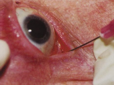
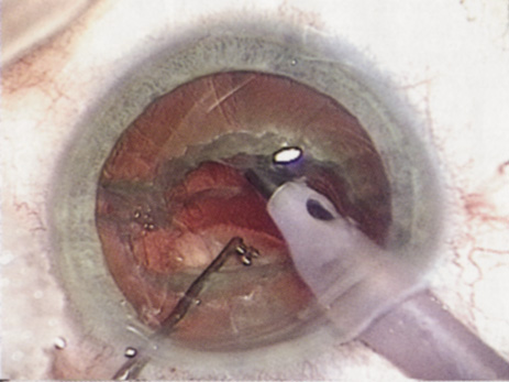
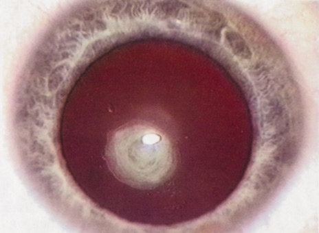
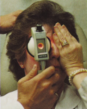
Many patients are most bothered by cataract-induced glare. For these patients, acuity testing under glare situations is indicated. There are several methods to assess visual acuity reduction by glare. The choice of method is often best dictated by the patient's history. If a patient complains of glare problems in the supermarket, or other uniformly illuminated environment, the brightness acuity test can be performed (Mentor Ophthalmics). For this test, the specially illuminated handpiece is held in front of the tested eye using best spectacle correction (Fig. 1). The Snellen acuity is rechecked and can be recorded on each of three light settings.

Fig. 1. Brightness acuity test.

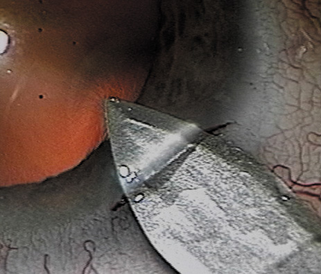
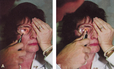
Patients who complain of glare from point sources of light, such as oncoming headlights or bright sunshine, may be best evaluated by a different form of glare testing. To simulate the environment of the patients' symptoms, Snellen acuity is measured while directing a point source of light obliquely toward the eye outside their best spectacle correction or outside of a phoropter dialed in with their best manifest refraction (Fig. 2).

Fig. 2. The correct method for performing a “point light source” glare test. A. Transilluminator is directed toward the eye at an angle. B. Shining the transilluminator from directly in front of the eye is not a valid method for testing glare because it underestimates the glare acuity.

Still other patients' problems may center on difficulty with reading, seeing street signs, or distinguishing fine patterns. In these individuals, the complaints are related more to contrast; therefore, contrast testing is most appropriate. There are a number of ways to assess the effect of contrast on vision. Regan's sine wave gradients have been used frequently for research purposes and are available in some settings. Various commercial devices are now available to measure visual acuity in different contrast settings and each has its relative merits and detractions. The authors have found the Baylor Visual Acuity Tester (BVAT) monitor (Mentor Ophthalmics) testing of contrast to correlate well with patients' complaints and its simplicity is appealing to both patients and technical staff.

In rare instances, patients' complaints may be primarily related to distinguishing colors. Although patients frequently remark about their dramatic improvement in color perception after cataract surgery, there are no convenient methods to document diminished color perception preoperatively. This underscores the importance of correlating patients' complaints with the biomicroscopic examination and the degree of nuclear color change.

PROGNOSTIC TESTS

Physicians often order special tests to help determine a patient's visual potential. Some of these tests are acuity specific. These can be particularly helpful in guiding patients who may have comorbid ocular conditions. Some devices have been designed to project a Snellen chart through the clearest area of the cataractous lens to assess retinal acuity potential such as the potential acuity meter. Studies also have shown a good predictive value by checking vision with a brightly illuminated near card.53 Of course, this can be performed with no additional office equipment. Various other commercial devices, including interferometry and various different pinhole and illumination device combinations, are available.

These approaches are not possible for patients with mature cataracts. Some more general, nonspecific prognostic tests can be performed. If a patient is able to identify the colors of projected lights, this usually indicates that some cone-mediated macular function is present.

Blue field endoscopy also may indicate some macular function. This test is performed by projecting a blue light into the eye. The patient may report seeing small round specks moving around in the vision. These specks correspond to white blood cells passing through the perifoveal capillaries.

The Purkinje phenomenon is tested easily by rapidly wiggling a transilluminator directed toward the globe through the lower lid in a darkened room. If the patient reports a pattern of crooked lines or branches, then he or she is seeing the shadows cast by the retinal blood vessels, indicating that the posterior pole is attached and functioning to at least some degree. Although positive results from the test are encouraging, some patients may still have limited vision after surgery; similarly, some rare patients may test negatively on all these tests and still recover good vision.

Diagnostic Studies

Several diagnostic studies provide information that supplements the historical and clinical data obtained by the surgeon. This information enables proper preoperative patient consultation and surgical planning. This section outlines many preoperative tests used for cataract patients.

A-SCAN BIOMETRY.

Accurate axial length measurement is critical to determine the correct power of the implant lens for the desired refractive result. A-scan biometry is imperative in any patient undergoing cataract surgery. Both contact (applanation) and immersion varieties of A-scan ultrasound units are commercially available. With applanation biometry, a hand-held or slit-lamp mounted probe is gently touched to the corneal surface along the visual axis. Contact A-scans are user dependent and sometimes the authors adjust the surgeon-specific IOL A-constant depending on which ultrasonographer has performed the scan. Nonetheless, outstanding refractive outcomes have been achieved, and the authors have been satisfied with the contact applanation technique.

With an immersion probe, a water bath around the eye acts as the medium to conduct sound waves. Although there is no direct contact of the probe with the globe, the water and water bath must, of course, remain in contact with the ocular surface and periorbita. Immersion scans may reduce interobserver variations but are less comfortable and less convenient for patients.

A-scan biometry is particularly challenging in eyes containing an oil fill. In this instance laser biometry is still able to achieve excellent measures.

LASER PARTIAL COHERENCE OPTICAL BIOMETRY.

Although ultrasound requires continuous contact with media that conduct sound waves, laser light passes easily through any clear media, including air, making this a truly noncontact or “no touch” test. Furthermore, the speed of light is not appreciably different in the clear media of the eye and thus excellent, reliable measures can be achieved in eyes containing intraocular lenses, regardless of type and eyes with oil fills within the vitreous cavity. Although some calculation adjustments can be made depending on the pseudophakic status, the differences among implant material are not appreciable different from a practical clinical perspective. Currently, the only commercially available laser biometry device is the IOLMaster (Zeiss). The measurements obtained by the IOLMaster device are extremely reliable, reproducible, and seem to be relatively technician- and observer-independent.54,55This device also can measure keratometry, optical anterior chamber depth measurements, and “white-to-white” measurements in an automated fashion. Because it relies on the passage of laser light through the ocular media, this instrument is unable to obtain measurements in cases where the media prevent laser light passage; for example, white cataracts, axial posterior subcapsular cataracts, or corneal scarring.

B-SCAN ULTRASOUND.

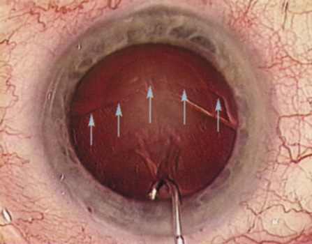
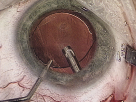
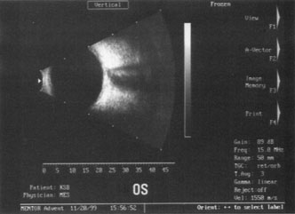
A mature cataract precludes visualization of the fundus. A B-scan ultrasonographic examination provides a real-time, two-dimensional (2D), cross-sectional image of the globe along the marked axis of the probe (Fig. 3). Cataracts are more common in patients with chronic retinal detachment, prior trauma, or intraocular tumors; therefore, a B-scan study is helpful in excluding structural posterior segment pathology before surgery on a mature cataract. Although a negative result to B-scan evaluation is reassuring, the surgeon should remember that it does not predict postoperative visual outcome. The B-scan can be thought of as a picture of Cincinnati from an airplane; the office buildings may all be standing, but you cannot tell whether the people in them are working.

Fig. 3. This B-scan ultrasound shows a normal looking posterior pole, without retinal detachment or intraocular mass. The vitreous cavity is echo lucent.

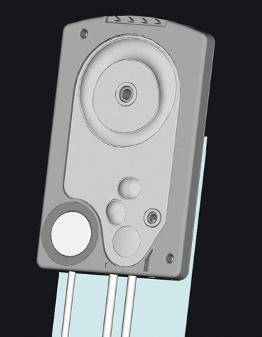
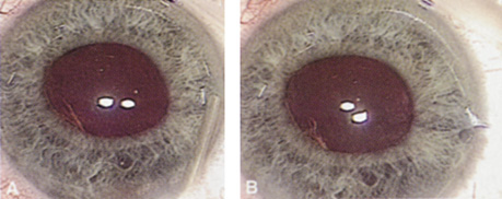
ENDOTHELIAL CELL COUNT.

Although slit-lamp examination can give the ophthalmologist an excellent estimate of endothelial health, sometimes a formal assessment of the corneal endothelial cell density is helpful (Fig. 4). This information is most likely to be helpful in advising patients who may be at greater risk of postoperative corneal decompensation. Specifically, patients with cornea guttata, previous ocular surgery, history of blunt ocular injury,56 exfoliation syndrome,57 iridocorneal-endothelial syndromes,58 or a history of glaucoma59 are known to have reduced endothelial cell counts. Patients with a history of acute angle closure are at particular risk because each episode of elevated intraocular pressure can damage endothelial cells.60

Fig. 4. A noncontact endothelial cell photograph demonstrates a normal cell mosaic and density. The cell count is calculated by identifying those cells within the box and touching two of the four adjacent edges.

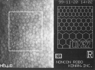
There are qualitative and quantitative methods for endothelial cell evaluation. Cell density can be measured directly with an endothelial cell camera. The surgeon also should view the photograph and qualitatively estimate the regularity of the endothelial cell mosaic. Some instruments calculate a coefficient of variability and percent of hexagonal cells.

When an endothelial cell camera is not available, qualitative assessment of count and cell morphology can be accomplished at the slit-lamp using a technique called specular reflection.61 The ophthalmologist focuses a narrow parallelepiped on the corneal epithelium, directing the beam at the periapical cornea from a 45-degree angle. The slit beam is moved slowly from side to side until the bright corneal reflex strikes the examiner's view from the epithelial surface reflection (first Purkinje-Sanson image). On high magnification, the examiner should focus on the endothelial surface just next to the bright reflex. The image of the endothelial mosaic will come into view. The surgeon can make a qualitative assessment of the cell density and degree of regularity. With practice, these estimates can be surprisingly accurate.

The implications of a reduced endothelial cell count are primarily prognostic and can provide the surgeon with more information to help counsel the patient about the risk of corneal decompensation with cataract surgery. Gentle phacoemulsification without triple procedure is recommended when cornea is clear and compact, given that a significant number of patients may be able to avoid a corneal transplant despite uncountable cell densities. However, these patients should be advised that they may be at an increased risk of requiring a corneal transplant.

PACHYMETRY.

Ultrasonic pachymetry measures central corneal thickness. When endothelial function is tenuous, corneal thickness begins to increase gradually, indicating subclinical stromal edema. Similar to endothelial cell count measurements, pachymetry is primarily of prognostic value.

Surgical planning also may differ for the patient with a compromised endothelium. In this case, use a more highly dispersive viscoelastic and minimal amounts of infusional fluids during surgery. When the degree of compromise is severe, additional dispersive viscoelastic material can be added periodically during the phacoemulsification. Because corneal transplant is a distinct possibility, such patients are not good candidates for a multifocal implant.
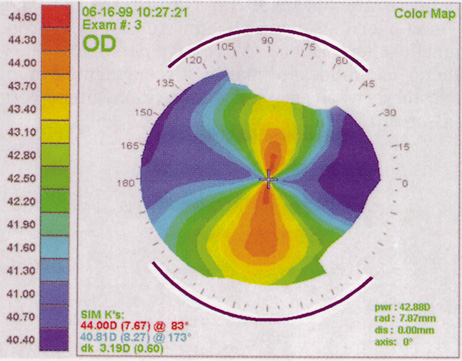
CORNEAL TOPOGRAPHY.

Computed corneal topography may be helpful from both a diagnostic perspective and as an aid to preoperative surgical planning. The diagnostic value of corneal topography is most obvious when keratoconus or irregular astigmatism is suspected. In cases with irregular astigmatism, superficial keratectomy or excimer phototherapeutic keratectomy may be indicated before cataract surgery, particularly when the relevant pathology precludes accurate keratometry.

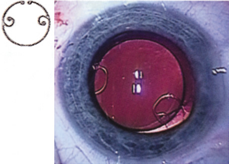
In some cases, corneal topography is an invaluable aid in surgical planning as well. In patients with high astigmatism, the topography can indicate the best meridian for placement of limbal relaxing incisions (Fig. 5). The topography also may be used to confirm the keratometric measurements.

Fig. 5. Corneal topography demonstrates with-the-rule astigmatism. The purple lines drawn suggest the pattern for limbal relaxing incisions.

ULTRASOUND BIOMICROSCOPY.

The ultrasound biomicroscope is a specialized, high-resolution form of 2D ultrasound, similar to the B-scan discussed in the preceding. An ultrasound biomicroscope study may be helpful to the cataract surgeon in cases of mature cataract when retroiridial, anterior segment pathology is suspected. Currently, the ultrasound biomicroscope instrument is expensive and not widely available, and has uncommon indications.

Back to Top
ANESTHESIA TECHNIQUES
Many approaches for ocular anesthesia achieve satisfactory results. Historically, topical anesthesia with cocaine was the first approach. As general anesthetic techniques became more widely available, topical cocaine use for eye surgery was abandoned. Later, orbital blocks became popular to reduce systemic risks associated with general anesthesia. More recently, with the advent of small incision phacoemulsification, many surgeons have returned to topical anesthesia, although with a much higher safety profile than in the days of intracapsular surgery.

GENERAL ENDOTRACHEAL ANESTHESIA

Although general anesthesia is an uncommon choice for today's cataract procedures, in rare instances it may be necessary. Even though improvements in anesthesia techniques and agents have improved the safety of general anesthesia, the risks for patients with other medical conditions may be significant. Although rare, idiosyncratic reactions can still result in dire consequences or death, even in previously healthy patients. This sobering knowledge, combined with the additional patient and physician inconveniences of general anesthesia, makes it an uncommon choice for cataract surgery. Certainly, general anesthesia may be considered when a patient is unable to hold steady or cooperate with regional anesthesia. This is likely to be more commonly required in young children or people with significant cognitive disabilities. Occasionally, general anesthesia is required for patients with neurologic conditions such as high amplitude resting head tremor or spasmodic torticollis.

ORBITAL BLOCKS

The purpose of an orbital block is to provide ocular akinesia and anesthesia. Different anesthetic agents can be administered. Relatively short-acting 1% lidocaine offers a more rapid recovery of vision with less diplopia and lower risk of muscle toxicity, although it may not be appropriate for an anticipated lengthy case. Bupivacaine or lidocaine-bupivacaine combinations induce a longer anesthetic effect, although bupivacaine is reported to have a higher incidence of muscle toxicity with resultant diplopia or ptosis.62–65 The addition of hyaluronidase to the injection mixture improves the anesthetic's ability to spread through the orbital tissues. Agents containing epinephrine increase the longevity of anesthetic action but rarely induce central retinal artery occlusion.66 Varieties of block styles are used with mild variations in effectiveness and complication profiles.

Retrobulbar Block

Retrobulbar blocks were among the earliest of orbital block techniques and have stood the test of time. Although there are many approaches to block administration, each technique intends delivery of the anesthetic medication into the intraconal space. These blocks are highly effective (about 95%) in achieving adequate ocular akinesia and anesthesia. Potential complications include retrobulbar hemorrhage,67 globe penetration,68,69 optic nerve sheath hemorrhage, extraocular muscle toxicity with persistent diplopia, and, rarely, brainstem anesthesia. Visualization of the globe is improved when using a transconjunctival approach (Fig. 6) or when the skin is indented with a cotton-tipped applicator (Fig. 7), making globe perforation less likely.

Fig. 6. Retrobulbar block using a transconjunctival approach.

Fig. 7. The skin is indented with a sterile cotton-tipped applicator until it is past the equator of the globe, thereby reducing the risk of globe perforation when the needle is inserted.

Peribulbar Block

A peribulbar block differs from the retrobulbar approach by delivering the anesthetic dose to the extraconal peribulbar space. The medication then spreads into the muscle cone, aided by a hyaluronidase enzyme, facilitating diffusion through the orbital tissues. The speed of onset is typically a few minutes longer than retrobulbar blocks, and typically a higher volume of anesthetic agent is given. This approach is slightly less effective than the retrobulbar block. Yet, by virtue of staying outside the muscle cone, some potential complications are less likely and others less severe. The sequelae of retrobulbar hemorrhage are less ominous when it occurs outside the muscle cone, as optic nerve compression is less likely. Direct optic nerve or nerve sheath injury is virtually eradicated. As with retrobulbar injections, using a transconjunctival approach or indenting the skin with a cotton-tipped applicator before peribulbar injection, improves visualization of the globe and decreases the likelihood of globe perforation

PARABULBAR ANESTHESIA

Various approaches can instill anesthetic into the subconjunctival or subtenon space using either a needle or a posteriorly directed blunt cannula.70 Parabulbar anesthetics are fairly effective for anesthesia, although they generally achieve less than maximal akinesia. Ballooning of the conjunctiva may cause fluid to build up around the limbus, creating a fluid meniscus lens, markedly limiting surgical visualization (Fig. 8). A posteriorly directed cannula in the parabulbar space could disturb a vortex vein. These injections also may induce unsightly subconjunctival hemorrhage. The authors reserve parabulbar anesthetic techniques primarily for augmentation of anesthesia when an initial anesthetic has either worn off or proved ineffective.

Fig. 8. Chemosis can cause pooling of the irrigation fluid, which creates a negative meniscus lens, inhibits the view, and reduces stereopsis.

LID BLOCKS

Some surgeons combine orbital block with facial nerve block. The main indication for an eyelid block is to limit the patient's forced lid closure against the speculum. Such squeezing can markedly increase posterior pressure and cause anterior chamber collapse. Lid blocks usually are applied only when orbital blocks are given, although occasionally they may be indicated in a case under topical anesthesia when the patient has blepharospasm. A lower lid block can be achieved easily by injecting an additional 1 or 2 ml of anesthetic into the lower lid when withdrawing the needle from an orbital injection. Typically this is satisfactory in most cases, because an upper lid, unopposed by lower lid tone, induces almost negligible effects on the globe.

If more complete facial nerve block is required, an Atkins, Van Lint, modified Van Lint, or Nadbath block may be administered. The surgeon should be reminded that increasing degrees of facial nerve block may have some undesirable effects. Fibers from the lower divisions of cranial nerve VII serving the lower face also may be affected by an eyelid block, producing temporary facial asymmetry and slurred speech, which patients may find frustrating. Transcutaneous facial nerve injections also may cause unappealing facial ecchymosis as well.

TOPICAL ANESTHESIA

With the advent of self-sealing wounds and closed-chamber systems for phacoemulsification, the need for absolute ocular akinesia has diminished. Accordingly, topical anesthesia for cataract surgery has become increasingly more common. In topical anesthesia techniques, the patient receives several drops of a topical anesthetic agent, inducing anesthesia of the ocular surface and allowing the initial incision to occur painlessly. During the surgery, the patients may detect some sensations of pressure or fullness and should be told to expect this at any step in which the globe is pressurized. The patient should be counseled in advance that he or she may feel pressure, but not pain. To explain this to the apprehensive patient, the authors sometimes shake the patient's hand in the office and ask “Can you feel this?” Then, after the patient responds “Yes,” inquire “Does that hurt?” The patient invariably responds, “Of course not.” With topical anesthesia, there is no induced amaurosis and patients may see some steps in the surgery. For example, hydrodissection is associated with a blurring of the distinct image of the microscope light. Continuous patient communication may ease or pre-empt anxiety. Some surgeons instill intracameral preservative-free 1% lidocaine to blunt patients' sensations further. Studies demonstrate different results in assessing safety and efficacy.71–74 Topical anesthesia techniques eliminate the inherent risks of orbital injections and offer rapid visual recovery. Additionally, topical anesthesia is anticoagulant friendly for patients on warfarin (Coumadin) and other blood thinners. However, topical anesthesia is not suitable for all patients or all surgeons.

Selecting the appropriate patient for topical anesthesia is paramount to both the surgeon's and the patient's satisfaction. This determination should be made during the preoperative patient examination. Extremely photosensitive patients or patients who are noted to be “squeezers” in the office might be better suited to regional block. Communication with the patient during surgery is a vital component of the topical anesthetic experience. Accordingly, patients with whom the surgeon cannot easily communicate are not good candidates. For patients who are hard of hearing or patients who do not speak the surgeon's language, an orbital block should be considered. Occasionally, the cataract surgeon encounters patients with disorders of ocular motility that preclude effective topical anesthetic approaches. Certainly, patients with nystagmus require the akinesia of an orbital block. Moreover those patients with large angle strabismus may have difficulty maintaining their gaze while their contralateral eye is unable to fixate.

OPERATIVE MYDRIASIS

One can achieve an adequate pupillary size for routine cataract surgery by a number of different mechanisms. Tradition has favored administration of frequent mydriatic drops on the patient's arrival to the surgical facility. These may include tropicamide, phenylephrine, cyclopentolate, hyoscine, and perhaps a nonsteroidal agent for reducing intraoperative miosis. Although this tried and true approach is still highly effective, other more convenient options have flourished recently. Some centers have patients administer mydriatic drops before arrival, whereas other surgeons have incorporated mydriatic drops into a Xylocaine gel, applied at arrival to the facility in a single dose. Some investigators have instilled miotics into the anterior chamber, while others have demonstrated effective mydriasis by instillation of intraocular, nonpreserved Xylocaine alone.75

Back to Top
INFECTION PROPHYLAXIS
Although infectious endophthalmitis following cataract surgery is now rare, its occurrence can be so devastating that its prevention is of paramount importance.76 Therefore patients should receive appropriate prophylaxis. Patients with blepharitis should be treated with eyelid hygiene measures. In the operating room, a drop of topical povidone-iodine paint instilled at the beginning of surgical preparation has been shown to reduce bacterial flora significantly and decrease the incidence of endophthalmitis.77,78 One survey79 also has shown a correlation between use of preoperative topical povidone-iodine and decreased incidence of endophthalmitis.80 Although there appears to be no benefit to trimming the eyelashes, the authors recommend isolating the lashes with a sterile plastic adhesive drape or a Steri-Strip. Antibiotic use is discussed in detail in the following paragraphs.
Back to Top
ANTIBIOTIC COVERAGE
Despite significant and widespread debates, the use and best method of administration of antibiotics in cataract surgery remains controversial. Traditionally, subconjunctival antibiotic injections were given at the completion of cataract surgery. As topical anesthesia became more popular, surgeons sought alternatives to painful subconjunctival injections. Several regimens of topical, infusional, and intracameral antibiotic agents have since become popular, although there may be a good rationale for avoiding some of these approaches.

It is difficult to determine the best antibiotic coverage choice with existing imperfect and incomplete data. A rational approach should examine the purposes of using an antibiotic, the relative likelihood of achieving those goals, and the possibility of unintended, undesirable effects of each antibiotic choice.

There are essentially two goals of perioperative antibiotic use: prophylaxis of endophthalmitis and avoiding bacterial ulcerative keratitis. Endophthalmitis prophylaxis receives the greatest attention because of the severity of its consequences. Several antibiotic regimens in current practice are reviewed here, stating their strengths and weaknesses.

PREOPERATIVE TOPICAL ANTIBIOTICS

Preoperative topical antibiotic drops certainly can reduce the colony counts on the ocular surface, provided that they are given for a sufficient time in advance of surgery to effect the conjunctival flora. Some believe that preoperative drops create a selection bias for resistant, aggressive organisms, even though there is no evidence of this.81 With sound arguments on either side and data lacking, preoperative drops seem reasonable but not mandated. Many agents address a broad spectrum of gram-positive and -negative organisms. The surgeon should be aware of kill curve data for the selected agent to ensure that the preoperative dosing regimen begins far enough in advance to have a meaningful effect.

PREOPERATIVE ORAL ANTIBIOTICS

There is a wide body of general surgery literature indicating a diminished risk of wound infection with preoperative systemic antibiotics. Some ophthalmic surgeons have theorized that the same philosophies should apply to ophthalmology and the prophylaxis of endophthalmitis. They maintain that a preoperative dose of a broad-spectrum oral antibiotic that produces therapeutic levels in the vitreous cavity should be effective in preventing an errant organism that finds its way into the vitreous gel from colonizing. Although this theory is appealing, no data exist yet on safety or efficacy. Surgeons who consider this method of therapy also should consider the potential for serious idiosyncratic reactions with systemic antibiotics. Remember that some 500 people die in the United States each year from penicillin-induced anaphylaxis,82 1 in 40,000 who receive chloramphenicol can die of aplastic anemia,83,84 and trovafloxacin mesylate, an oral, broad-spectrum fluoroquinolone with a previously presumed high safety profile, has been associated with an infrequent incidence of liver failure.85

ANTIBIOTICS IN THE IRRIGATING SOLUTION

During the past several years, it has become increasingly popular to place antibiotics in the irrigating solution. Although informal surveys seem to report a decreased incidence of endophthalmitis, from the microbiologic perspective, infusional antibiotics are not entirely rational.86 The concentrations of antibiotics that are widely used are not bacteriocidal. Furthermore, such dilute solutions may create a selection bias for resistant organisms on the conjunctival surface and in the operating room. Dilutional errors in preparing the antibiotic in the infusate can have devastating visual sequelae from retinal toxicity.

INTRACAMERAL ANTIBIOTIC INJECTION

In response to the homeopathic dosage criticisms of infusional antibiotics, some surgeons have opted to place a therapeutic antibiotic dose into the anterior chamber at the completion of surgery. Typically, surgeons administer half the dose used for intravitreal injection. This approach makes some theoretical therapeutic sense, given that the antibiotic is present in sufficient concentration to kill introduced organisms. Because the antibiotic-containing fluid does not bathe the ocular surface or access the operating room surfaces, selection of resistant organisms might be less likely. Critics of routine intracameral antibiotics cite the lack of safety and efficacy data and point out that although the administered antibiotic dose may be bacteriocidal, the agent is rapidly diluted by aqueous turnover. Dilutional errors could induce severe retinal or corneal endothelial damage. Insufficient available peer review data preclude widespread recommendations for this mode of endophthalmitis prophylaxis.

SUBCONJUNCTIVAL ANTIBIOTICS

Historically, subconjunctival antibiotic injection has been the mainstay of endophthalmitis prophylaxis. The safety profile is fairly broad, except for aminoglycosides, which have been reported to access the anterior chamber after injection to cause retinal toxicity.87

POSTOPERATIVE TOPICAL ANTIBIOTICS

Topical antibiotic drops are used almost uniformly after cataract surgery. Motivation for their use has been to reduce the risk of endophthalmitis. In one rabbit study, topical 0.5% moxifloxacin was shown to prevent endophthalmitis when an inoculum was placed into the anterior chamber, whereas the control animals treated only with normal saline developed infectious endophthalmitis.88 However, topical agents do have an important role in the prophylaxis of bacterial keratitis, because it may take several days for the corneal epithelial barrier to regain its integrity after even flawless clear corneal surgery. The authors advocate the use of topical antibiotics postoperatively.

Back to Top
INCISIONS
The cataract incision has evolved along with ongoing advances in lens removal techniques and IOL designs. The incisions for intracapsular cataract removal were placed at the anterior limbus or in clear cornea and measured approximately 13 mm in chord length almost 180 degrees. As extracapsular surgery became more popular, the incision size decreased to 11 mm. Next, phacoemulsification was born and the incision size became dependent only on the size required for IOL insertion. At the same time, surgeons began to place their incisions more posteriorly to simplify wound closure and decrease induced astigmatism. As foldable IOLs developed, incision size further decreased from 7 to 3.5 mm and even smaller. During a period of several years, surgeons developed incisional architectures that allowed for one-stitch and eventually sutureless self-sealing incisions. Now most surgeons have returned to clear corneal wounds, taking full advantage of these small self-sealing incisions.

Cataract incisions can be described in terms of size, architecture, and location. Each of these characteristics has a unique effect on the wound. However, these characteristics are all integrated; changing one characteristic often necessitates altering the others to obtain a wound that is structurally sound.

INCISIONAL ARCHITECTURE

A poorly constructed wound can lead to difficulties, such as iris prolapse, throughout the case and a well-constructed wound may help to facilitate a more challenging case. The shape of the incision relative to the limbus is an important factor in wound integrity.89 Incisions that are tangential to the limbus or arched away from the limbus (frown) have been shown to effect better wound closure and induce less astigmatism.90 Likewise, multiplane incisions seem to be more effective in obtaining watertight closure.91

INCISION LOCATION

The location of the incision is described in terms of its meridional axis and placement relative to the limbus. Incisions placed more anteriorly tend to induce more astigmatism. Incisions placed more posteriorly encounter more blood vessels. These factors are considered in more detail in the following sections, which discuss scleral tunnel and clear corneal incisions. The single most important factor when considering axis placement is how it will affect the surgeon's ability to maneuver intraocular instruments. A steep superior orbital rim or an extremely narrow palpebral fissure may limit the surgeon's ability to work superiorly, thus necessitating a temporal approach. A temporal approach may be uncomfortable to a right-handed surgeon when operating on a left eye, especially if the stretcher is not temporal-approach friendly. With this in mind, one can begin to consider other factors that will be affected by the incisional axis. Placement of a sutureless incision at the steep corneal axis can reduce the astigmatism at that axis. A longer incisional length and a more anterior placement will effect a greater astigmatism reduction. Superior incisions have a greater astigmatic effect than incisions placed nearer the horizontal meridian. Supertemporal oblique incisions seem to have little effect on astigmatism, perhaps because of the lack of tractional forces from the rectus muscles.

SCLERAL TUNNEL INCISIONS

A scleral tunnel incision provides the surgeon with a conjunctival covering to the incision that may help to prevent bacteria from entering the eye. Additionally, should a wound leak occur, a bleb may form that may prevent significant chamber collapse. Scleral tunnel incisions induce less astigmatism than more anteriorly placed incisions of similar lengths; therefore, they are more suitable for larger, one-piece IOLs. However, the further posterior the incision is placed the more likely is it that significant bleeding will occur. Additionally, a long scleral tunnel can limit movement of the phacoemulsifier tip, thus impeding maneuverability and perhaps leading to heat build-up and a thermal burn. The internal edge of all cataract incisions should enter the anterior chamber sufficiently anteriorly to prevent iris prolapse. Therefore, the authors do not recommend beginning the portion of the exterior incision where the phacoemulsification tip will be used more than 1.5 to 2 mm behind the limbus.

The early scleral tunnel incisions were fashioned with the external edge following the curve of the limbus. It is now known that this incision style is the least watertight and induces the greatest amount of astigmatism postoperatively. To avoid placing the phacoemulsification handpiece in a long scleral tunnel, surgeons began fashioning the external incision so that only a portion of the incision is near the limbus. These incisions have been termed tangential, frown, and chevron, to name a few. These incisions are all similar in that they move more posteriorly as they are extended away from the limbus where the phacoemulsification handpiece will be inserted. The result is better wound integrity and less surgically induced astigmatism.

Scleral tunnel incisions are typically three-planed. The external incision begins as a groove made perpendicular to the scleral wall approximately half the scleral thickness. A metal or diamond keratome is then tunneled anteriorly until the tip of the blade is just into clear cornea. At that point, the keratome is angled posteriorly to pierce through Descemet's membrane and into the anterior chamber.

CLEAR CORNEAL AND NEAR-CLEAR CORNEAL INCISIONS

When constructing a clear corneal incision, the external aspect of the incision is either at the limbus or slightly anterior to the limbus, offering several advantages. First, a conjunctival flap is not necessary and bleeding is minimal, making a clear corneal approach favorable for patients with bleeding tendencies. Patients undergoing topical anesthesia will be more comfortable during surgery because they do not have to endure the discomfort that cautery can cause. Because a conjunctival flap is not necessary, patients are less likely to have discomfort that can develop from roughness at the conjunctival incision. Patients who have a functional filtering bleb or in whom conjunctiva should be spared for future filtering procedures are excellent candidates for clear corneal cataract surgery. Because these incisions start more anteriorly, iris prolapse is less likely if the surgeon fashions the incision correctly.

There are some potential disadvantages to clear corneal incisions. They may induce more surgical astigmatism because they are nearer the central corneal axis. It has been shown that induced astigmatism is minimized by performing surgery supratemporally or temporally where the corneal diameter is largest.92 If the incision is poorly constructed, and a wound leak occurs, chamber collapse is more likely because the conjunctiva is not covering the incision; therefore, bleb formation is not possible. Finally, some surgeons think that there is a theoretically increased risk of endophthalmitis with clear corneal incisions. This certainly is more likely if the surgeon does not obtain adequate wound closure, because bacteria could more easily gain access to the anterior chamber. Early experiences with clear corneal incisions demonstrated a lack of wound integrity. However, surgeons have improved the architecture of clear corneal and near-clear corneal incisions so that the incisions are much less likely to leak. Indeed, the expected rise of endophthalmitis resulting from clear corneal incisions has not occurred. Hydration of the corneal stroma at the incision site significantly helps obtain a watertight wound. Additionally, when the corneal incision is placed a little more posterior (near-clear), the anterior limbal vessels bleed, forming a fibrin barrier.

There are several ways to fashion a clear corneal or near-clear corneal incision. The incision can be a single-plane stab into the anterior chamber. However, a two- or three-plane incision may create a more secure wound. The external and internal portions of the incision should be straight lines, tangential to the limbus. Shorter tunnels allow for better visualization and facilitate phacoemulsification handpiece maneuverability, whereas longer “squarer” tunnels have been shown to have higher wound rupture pressures.93 The surgeon must weigh these issues when selecting the tunnel length. Special incisional architectures also work well, including hinged incisions, where the external groove is deeper than the tunnel and trapezoidal incisions.94,95 The authors prefer to make a groove perpendicular to the corneal surface at the most peripheral aspect of cornea, where the incision will just engage limbal vessels. A guarded blade set at 300 to 550 μ works well. Then a keratome is used to tunnel the incision forward within the corneal lamellae before angling the keratome slightly more posteriorly to pierce Descemet's membrane (Fig. 9). Regardless of the technique used to fashion the incision, it is important that the surgeon does not over-manipulate the anterior lip of the incision because such trauma may result in a poorly sealing wound at the end of the case. Also, when inserting an instrument or IOL through the incision, be certain that pressure is directed posteriorly and the incision is of adequate size to avoid stripping Descemet's membrane. Clear corneal incisions are best suited for injectable or foldable IOLs.

Fig. 9. A dual beveled metal keratome (Alconlabs, Ft. Worth, TX) is used to fashion a 3.0-mm near-clear corneal incision.

Despite numerous incision choices, one key factor is recommended for all cataract incisions: The incision should be self-sealing. A self-sealing incision decreases the risk of catastrophic expulsive hemorrhage if significant positive pressure develops for any reason. If positive pressure develops at any time during the procedure, simple removal of the instrument should prevent expulsion of the intraocular contents. Additionally, if properly placed and constructed, a self-sealing incision obviates the need for a suture, thereby avoiding suture distortion of the wound and induced astigmatism.

Closure of the incision, regardless of incision type, should ensure that intraocular pressure or external pressure at the posterior lip of the incision, or elsewhere, will not cause wound leak. If leakage occurs, then stromal hydration or suture placement may be necessary. Whenever placing a suture across a cataract incision, the ocular tension should approximate normal pressure before tightening the suture. Doing so helps prevent the surgeon from overtightening the suture, which would induce astigmatism.

ANTERIOR CAPSULOTOMY

One of the most beneficial techniques developed over the last decade is capsulorrhexis. A continuous curvilinear anterior capsular opening helps prevent intraoperative and postoperative complications. With capsulorrhexis, mechanical strength of the capsular opening is superior to that in a can-opener capsulotomy; thus, a tear to the posterior capsule with subsequent vitreous loss is much less likely.96 Capsulorrhexis allows for nuclear manipulation with less risk of posterior capsule rupture. Cortex removal is made easier as well, because it becomes easier to differentiate cortical material from anterior capsule. Should a posterior capsular tear occur, sulcus fixation is more likely attainable with the presence of a clearly visible residual anterior capsular rim. Placement of both IOL haptics into the capsular bag is more certain with capsulorrhexis because the surgeon can more easily visualize the haptics gliding beneath the anterior capsular rim.

Capsulorrhexis can be performed with a cystitome, capsulorrhexis forceps, or combination-type instruments. Regardless of which instrument is used, several principles can help the surgeon successfully complete capsulorrhexis. It is important to maintain the anterior chamber, because making the chamber shallow increases tension on the zonules and causes the tear to run peripherally. The authors recommend the use of a viscoelastic agent for maintaining chamber depth and, of course, for endothelial protection. Therefore, if the tear begins to run peripherally, the surgeon should redeepen the anterior chamber before attempting to redirect the tear. Additionally, folding the capsule margin can aid the surgeon in redirecting the tear more accurately (Fig. 10).

Fig. 10. The capsulorrhexis tear is more easily redirected by folding the capsule over, in advance of the tear.

HYDRODISSECTION

Hydrodissection can be performed after the surgeon has successfully completed capsulorrhexis.97 If the capsulorrhexis is not intact, fluid forced around the interior of the capsule may cause the bag to splay open. With capsulorrhexis, hydrodissection is a safe and extremely useful maneuver. Hydrodissection can be thought of as two maneuvers: hydrodelineation and cortical cleaving hydrodissection. By placing a 27-gauge cannula on a syringe filled with balanced saline solution (BSS), the surgeon can direct fluid beneath the residual anterior capsular rim to create a cleavage plane. Depending on the direction the fluid wave takes, different lamellae of the cataract will be separated. Hydrodelineation is the term used when the cleavage plane separates the adult nucleus from the fetal nucleus or the adult nucleus from the more peripheral epinucleus. Hydrodelineation often results in the characteristic golden ring sign (Fig. 11). Cortical cleavage occurs when the cortex is separated from the capsular bag (Fig. 12). Finding the cortical cleavage plane may be facilitated by gently lifting the capsular margin away from the cortex with the BSS cannula before injecting. Several small bursts of fluid allow the surgeon to monitor progress of the fluid wave. When dealing with a soft nucleus, the authors strive to perform true cortical cleaving hydrodissection. For a hard nucleus, hydrodelineation allows manipulation of less of the nuclear bulk, although the remaining epinuclear shell must be addressed in an additional step. Hydrodelineation is particularly useful if the nucleus is not freely mobile after cortical cleaving hydrodissection.

Fig. 11. A crisp “golden ring” is seen from the fluid cleft between the epinucleus and nucleus with hydrodelineation.

Fig. 12. Hydrodissection, performed subincisionally with a 27-gauge J-cannula, produces a cleavage plane between the capsule and the cortex. The small blue arrows indicate the advancing fluid wave.

Back to Top
PHACOEMULSIFICATION
Phacoemulsification occurs when ultrasound energy resulting from high-frequency vibrations of the metal tip of a phacoemulsification handpiece is delivered to the nucleus of the cataract. This energy results in fragmentation of the nucleus into chips small enough to be aspirated through the hollow center of the vibrating tip.

PHACOEMULSIFICATION EQUIPMENT

The efficiency of phacoemulsification has improved significantly because of improved tip designs, digital control of ultrasonic energy, improved software, and enhanced fluidics. Currently, many different phacoemulsification machines are available with a wide variety of advantages and disadvantages. This chapter describes some of the equipment and modalities currently available but does not attempt to compare or contrast different phacoemulsification machines.

This section discusses fluidics and how it influences phacoemulsification efficiency and safety. Fluidics terminology can be somewhat confusing. To clarify, flow rate refers to the volume of fluid and material drawn through the instrument tip and aspiration line per unit time. It is measured in millimeters per minute and sometimes is referred to as flow, aspiration, or aspiration rate. Flow rate corresponds clinically to the speed with which material comes to the tip of the instrument.

Vacuum refers to the vacuum level created within the cassette of the phacoemulsification machine and is measured in mm Hg. This vacuum is transmitted down the aspiration line, resulting in suction at the aspiration port of the instrument. The vacuum level corresponds to the force with which material is held against the aspiration port of the tip.

Phacoemulsification machines use either flow- or vacuum-based systems. Flow-based systems induce aspiration by directly creating flow within the tubing, whereas vacuum-based systems induce aspiration by first creating a vacuum. Most flow-based systems use peristaltic pumps (Fig. 13). Such pumps employ a series of rollers to pinch compliant tubing serially, thereby creating flow within the aspiration tubing. The vacuum level at the phacoemulsification tip rises when flow toward the pump meets resistance because of partial or full occlusion at the phacoemulsification tip. Computerized pump controls allow the surgeon to set vacuum limits and control flow rates. The surgeon can adjust these independently to balance the degree of hold-ability against the chance of chamber collapse.

Fig. 13. The Infiniti peristaltic pump (Alconlabs, Fort Worth, TX).

Aspiration tubing within a peristaltic pump must be compliant; high vacuum levels can induce an unexpected surge of flow as the compliant tubing springs open after a nuclear chip, occluding the tip is emulsified and aspirated (occlusion break). This phenomenon, known as postocclusion surge, can lead to sudden chamber collapse and thereby result in a posterior capsule rupture, or iris trauma. Some systems have sensors within the tubing to minimize a postocclusion surge by automatically shutting down the pump. Additionally, some systems offer minimally compliant tubing, minimizing postocclusion surge (Fig. 14). Aspiration bypass tips have a small hole within the side of the phacoemulsification needle, allowing continuous low flow, despite tip occlusion, and thereby reducing surge (Fig. 15).

Fig. 14. MaxVax tubing (Alcon Laboratories, Ft. Worth, TX) has thicker walls that limit compression and, thereby, postocclusion surge.

Fig. 15. The aspiration bypass tip (ABS) (Alcon Laboratories, Fort Worth, TX).

Most current vacuum-based systems use a Venturi pump whereby fluid or gas moving across an opening creates a vacuum within the aspiration tubing. The vacuum then draws fluid into the tubing, thus inducing flow. Although the surgeon can set the vacuum level, flow rate depends on the vacuum level and, therefore, cannot be directly controlled. Flow continues at the vacuum-dependent rate until resistance is met owing to partial or complete occlusion of the aspiration port. The flow then decreases while the preset vacuum level is maintained. When an occlusion break occurs, fluid suddenly rushes into the aspiration port as the pump tries to maintain the preset vacuum level. The postocclusion surge is more severe with a larger aspiration port because a larger volume of fluid can move quickly through the port. If the vacuum level is set relatively high, occlusion breaks can lead to dramatic postocclusion surge and chamber volatility. Higher bottle heights are necessary to help prevent such chamber collapses.

Flow is created in a somewhat different manner by the Concentrix scroll pump. Flow is created instead by direct fluid displacement within rigid channels of two rotating discs (Fig. 16). The advantage of such a system or any pump is that the flow rates and vacuum levels are more predictable and consistent because the variability induced by the use of compliant tubing within the pump is eliminated. The same is true for many of the pumps available on newer-generation phacoemulsification machines, which have minimal compliance within the pump itself.

Fig. 16. The Concentrix scroll pump (Storz, St. Louis, MO).

There are advantages and disadvantages to each fluidic system and each phacoemulsification unit. The surgeon must become knowledgeable about the fluidics of the particular machine being used to increase the likelihood of achieving satisfactory surgical results.

PHACOEMULSIFICATION POWER MODULATION

Surgeons and industry have developed innovative methods for ultrasonic power modulation that increase the safety and efficiency of phacoemulsification. The available modalities may differ from machine to machine but all involve software that can induce a duty cycle of the ultrasound power delivery. Duty cycle refers to “on-off” time. The general concept is to limit ultrasound “on” time in order to allow vacuum to build at the phaco tip. This results in lower phaco power and times and, therefore, safer and more efficient emulsification. Linear control of phaco power allows the surgeon to control the ultrasound power level with the foot pedal. The surgeon should depress the foot pedal only far enough to effect lens disruption. Higher-power levels could cause nuclear segments to “chatter” away from the phaco tip. In Pulse mode phaco the surgeon assigns a pulse rate or duty cycle to ultrasound delivery. The power is still controlled in a linear fashion yet there are pauses between ultrasound energy pulses. In some machines, the “on-time” and “off-time” can be set separately, allowing for short “micropulses” of energy delivery. In Burst mode phacoemulsification, the surgeon assigns a burst width that relates only to on-time. The off-time is controlled by the foot pedal so that as the pedal is depressed further, off-time decreases until, as the pedal is fully depressed, ultrasound delivery becomes continuous. The surgeon can use burst mode phacoemulsification with either fixed or linear control of ultrasound power on some of the newer phaco machines. Although the software that controls these power modulation schemes and the terminology differ from machine to machine, the general concept of duty-cycle control works well to improve efficiency and safety during phacoemulsification.

Back to Top
PHACOEMULSIFICATION TECHNIQUES
There are many different approaches to disassembling the lens nucleus; each has advantages and disadvantages. The surgeon should be familiar with all available approaches and select those features that are most appropriate for the equipment used, the particular case, and the surgeon's level of experience.

Slow-Motion Phacoemulsification

Slow-motion phacoemulsification is a technique that seeks to maintain a stable anterior chamber during phacoemulsification while minimizing the volume of fluid that flows through the eye. By minimizing fluid inflow, there should be less endothelial cell loss and, therefore, less early postoperative corneal edema. Additionally, low inflow levels decrease the chance of forcing fluid through any areas of weakened zonules, which could create fluid lacunae within the vitreous gel, leading to positive pressure and possible vitreous prolapse. Chamber collapses are less likely because vacuum levels and aspiration rates are low; therefore, the risk of iris damage or posterior capsule rupture should be diminished. Because the technique involves low vacuum, aspiration, and inflow, it may be difficult to hold onto nuclear fragments for some methods of chopping the nucleus.

Divide Techniques

Divide techniques attempt to emulsify the nucleus by separating it into two or four pieces. Grooves carved into the nucleus facilitate separation of the pieces. The grooves can be achieved using low vacuum levels because complete occlusion of the phacoemulsification tip during is neither needed nor desired at this stage of the procedure. If one makes the groove at least twice the diameter of the phacoemulsification tip, there is better visualization of the walls of the groove, and thus better understanding of the groove's depth. A wide groove also results in the surgeon debulking more of the nucleus in the capsular bag, so that when the pieces are brought forward, less emulsification needs to be performed anteriorly. While fashioning the groove, it is important not to move the phacoemulsification tip faster than it is emulsifying. Advancing the tip too quickly causes the entire nucleus to move with the tip and may lead to subincisional zonular damage. The groove depth required to allow cracking depends on the density of the central and posterior nucleus. A hard nucleus with a dense posterior plate may not crack even if grooved through 90% of its thickness. Likewise, a fetal nuclear cataract may be cracked easily after grooving only halfway through. Regardless of the depth of the groove, the surgeon should place the cracking instruments at the most posterior aspect of the groove before dividing. (Fig. 17). Doing so minimizes stress on the anterior capsulorrhexis and generates forces more likely to result in a successful divide. After the nucleus has been bisected, the surgeon can debulk and emulsify each heminucleus separately. Alternatively, the surgeon can rotate the nucleus 90 degrees and further divide it into quarters in a similar fashion.

Fig. 17. The phacoemulsification tip and an Osher nucleus manipulator (Storz, St. Louis, MO) are placed deep into the groove. By separating the instruments' tips with equal forces, the nucleus is divided.

After the surgeon has completed the divide portion of the procedure, vacuum and aspiration levels can be increased. By increasing vacuum and aspiration levels, the surgeon will find it easier to bring nuclear pieces to the phacoemulsification port. Additionally, higher vacuum levels hold the nuclear segments more tightly to the phacoemulsification port, resulting in more efficient phacoemulsification and better control.

”Chop” Techniques

In 1993, Kunihiro Nagahara introduced a method of chopping the nucleus into pie slice–shaped wedges for emulsification (Fig. 18). Chopping techniques can be defined as vertical or horizontal chopping. Both chopping techniques involve burying the phacoemulsification tip into the center of the nucleus, holding it in place with increased vacuum. When performing a vertical chop, the chopping instrument is inserted through a second incision and placed beneath the anterior capsular rim. The chopper is directed peripherally, and then drawn posteriorly and centrally toward the phacoemulsification tip, causing the nucleus to crack. The horizontal chop differs in that the chopper does not need to be placed under the anterior capsular rim and the chop does not begin at the lens periphery. Instead, the chopping instrument is placed in the nucleus near the anterior capsulorrhexis edge and then directed posteriorly until it meets the buried phaco tip. Both chopping techniques take advantage of the natural cleavage planes between the nuclear lens fibers, similar to chopping a piece of wood with an ax. Each wedge can be brought centrally using a second instrument or the vacuum of the phacoemulsification tip for emulsification away from the posterior capsule, iris, capsulorrhexis edge, and bag periphery. Then the nucleus is rotated and similar maneuvers are performed with each segment until the entire nucleus has been emulsified. Smaller, more manageable wedges can be chopped off the nucleus for dense nuclei and larger pieces sectioned from softer nuclei.

Fig. 18. In “phaco-chop,” small pie-slice segments are serially removed from the larger nuclear fragment.

Chop techniques are especially valuable when the surgeon fails to obtain a complete capsulorrhexis. The forces generated while splitting the nucleus with a divide technique might cause a peripheral extension of an anterior capsular tear. Because forces generated during a chop maneuver are borne by the chopping instrument and the phacoemulsification tip, there is less chance of extending an anterior capsule tear.

Many combinations and variations have been extrapolated from these two basic techniques. It is important to have knowledge of several techniques. What works well for a dense nuclear cataract may not work so well for a softer cortical cataract. Occasionally, the cataract is of such a consistency that it neither divides nor chops well. In these cases, it may be necessary to fall back on earlier techniques that involved sculpting the nucleus to form a nuclear bowl. The nuclear bowl can be brought forward for emulsification using a flip technique or a direct lift maneuver.98,99 Soft nuclei often prolapse anteriorly out of the capsular bag during hydrodissection. Then they can be aspirated easily with little or no phacoemulsification power.

Suprascapular Phacoemulsification

As the fluidics of phacoemulsification machines have improved, it has become possible to increase vacuum levels and aspiration rates. Phacoemulsification times decrease significantly at higher vacuum settings; therefore, it has become possible to emulsify the entire nucleus in the supracapsular space safely.100 A highly retentive viscoelastic agent is injected into the anterior chamber to protect the corneal endothelium. The surgeon fashions a 6-mm or larger capsulorrhexis so that hydrodissection causes the nucleus to tip and prolapse anteriorly. The viscoelastic agent and its cannula are used to completely flip the nucleus upside-down and out of the capsular bag. The nucleus then is completely vulnerable for phacoemulsification. This technique differs from others in that the nucleus is attacked from the outside-in, instead of inside-out. When phacoemulsification is performed anterior to the capsular bag, iatrogenic zonular dialysis may be less likely because the surgeon does not have to struggle to remove the nucleus from the capsular bag during phacoemulsification. However, the surgeon should remember that there are some possible disadvantages to having a large capsulorrhexis, including disruption of the zonular insertion,101 a greater risk for posterior synechiae to the capsule,102 and a higher risk of posterior capsule opacification. Furthermore, supracapsular phacoemulsification may increase the risk of iris damage.

Bowl-Out Techniques

Some surgeons continue to prefer a phacoemulsification approach that essentially nibbles away at the nucleus by carving out layer by layer of lens fibers from the central nucleus, going deeper and more peripheral with each pass of the phacoemulsification tip. Eventually, a bowl is created and the epinuclear shell collapses in on itself. This technique is pleasing in its simplicity, although it requires significant emulsification near the capsular bag periphery, iris margin, and capsulorrhexis. In addition, the peripheral lens material is softer and may be more likely to create a postocclusion surge. Nonetheless, this may be a useful choice if the nucleus is sticky and the pieces cannot be well mobilized without applying significant stress to the zonules.

Back to Top
ALTERNATIVE PHACOEMULSIFICATION MODALITIES
Although ultrasonic excellent results are obtained with ultrasonic phacoemulsification, one should always strive to improve; thus, several alternative lens emulsification modalities have developed. Laser nucleus removal attracted much attention in the last decade. However, interest seems to have waned because of the difficulty of the retaining nuclear material at the tip while delivering laser pulses. Laser nucleus emulsification procedures resulted in a greater degree of lens chatter, did not seem to work well with denser cataracts, and took longer than ultrasonic phacoemulsification.

NeoSonX in its current state is a modality that uses low-frequency, small degree oscillations simultaneous with ultrasound energy delivery. It appears to result in better follow-ability and therefore lower ultrasound times and powers.103 The degree of oscillation can be adjusted to have more or less effect on the nuclear segments.

Mark Andrews theorized that warmed fluid pulses could “melt” the nucleus, making it more amenable for aspiration. This concept has developed into the technique called AquaLase. In its current design, rapid micropulses (4 μl pulses up to 50 pulses/sec) are projected out in front of the AquaLase tip to disrupt the nucleus (Fig. 19). The effect is a delamination of the nucleus with subsequent aspiration of the resultant nuclear debris. This technique seems to work best in softer nuclei. However, as surgeons are gaining experience and this new technology progresses with new tip designs and pulse control, denser cataracts are becoming more amenable to this modality. AquaLase has several desirable advantages over ultrasonic phacoemulsification. Because AquaLase does not create friction at the incision, there is no possibility of developing an incisional burn. In addition, the current AquaLase tip is a soft dull polymer that is much less likely to tear the posterior capsule. Finally, AquaLase also may provide the opportunity to “power-wash” the posterior capsule and capsular bag equator safely to decrease the incidence of posterior capsule opacification.

Fig. 19. Diagram of the AquaLase tip showing fluid pulses disrupting the nucleus.

CORTEX REMOVAL

The improvements made in phacoemulsification fluidics also have improved the ability to remove cortex safely. Smaller incisions now provide surgeons with the opportunity to perform cortical aspiration within a closed system, thereby maintaining a deep anterior chamber. Cortical cleavage hydrodissection may loosen the cortex significantly, which simplifies its aspiration. Sometimes, most or the entire cortex is removed during phacoemulsification of the nucleus. However, most surgeons continue to find residual cortex at the completion of phacoemulsification. If the surgeon has performed capsulorrhexis, cortex removal should be easier to accomplish. One may begin aspiration at the subincisional site so that the remainder of the cortex may help the bag to remain open, thus facilitating subincisional cortex removal. However, if difficulty is encountered aspirating subincisional cortex, the surgeon should temporarily abandon the subincisional site and first remove cortex from the rest of the bag. Doing so may save the surgeon from having to manage an iatrogenic zonular dialysis or posterior capsule rupture while a significant amount of cortex remains. Often, subincisional cortex may just fall out of the capsular bag during aspiration of cortex from other areas, particularly if hydrodissection was initiated subincisionally. For the gentlest cortical stripping, engage the anterior portion of the cortex with the aspiration port just below the capsulorrhexis margin. As vacuum builds, rotate the aspiration port anteriorly and strip the cortex centrally and circumferentially so as to engage the anterior lip of cortex just adjacent to the portion already being stripped. Such a motion decreases zonular stress, maintains an adequate vacuum, and decreases occlusion breaks.

If subincisional cortex remains, it can be aspirated manually with a J-shaped cannula after deepening the chamber with viscoelastic (Fig. 20). This can be accomplished either before or after IOL implantation. Alternatively, subincisional cortex can be aspirated with an automated irrigation/aspiration handpiece after IOL placement. The optic of the IOL provides a protective barrier to prevent posterior capsule rupture during this maneuver. Bimanual irrigation-aspiration systems spilt the infusion aspiration components into separate handpieces that access the anterior chamber through the side-port paracentesis and a second paracentesis on the opposite side of the main wound. This affords a more facile access to the subincisional space, because the location of the aspiration handpiece can be switched but requires a separate incision.

Fig. 20. A J-shaped cannula (Cionni J-cannula, Storz, St. Louis, MO) is used to manually aspirate subincisional cortex after placement of the intraocular lens.

Some irrigation/aspiration handpieces have interchangeable tips and a J-shaped tip can be used to remove subincisional cortex. However, these tips lack a silicone outer sleeve and therefore tend to allow fluid egress from the incision during cortex removal, which necessitates higher bottle heights to maintain the anterior chamber.

Some surgeons prefer to use a bimanual cortex removal system.104 The aspirating and irrigating handpieces can be placed through either of two ports, thereby allowing the surgeon to access subincisional cortex more easily.

A soft, silicone aspiration tip is available now. Because it is soft and smooth, the risk of posterior capsule tear should be markedly reduced compared with a metal aspiration tip, which can develop burs and other defects (Fig. 21).

Fig. 21. Silicone irrigation/aspiration tip (Alconlabs Inc., Ft. Worth, TX).

CAPSULE POLISHING

After the cortex has been removed, the authors routinely vacuum and polish the posterior capsule to remove any debris or lens epithelial cells before implanting the posterior chamber IOL (PCIOL). This is accomplished with the irrigation/aspiration handpiece after checking the tip under the microscope to be certain that there are no sharp burrs. The vacuum level is decreased to 10 mm Hg and the flow rate is set at 5 ml/min. The aspiration port is turned over to contact the posterior capsule while the foot pedal is in position two (vacuum). The tip of the handpiece then is moved left and right while stabilizing the shaft of the handpiece with the forefinger of the opposite hand. It may be best to begin this maneuver centrally so that if a posterior capsule tear occurs, the surgeon can identify the margins of the opening and thereby convert it to a capsulorrhexis, as described later in this chapter. At this low vacuum level, it is unusual for the posterior capsule to become captured in the aspiration port. However, if the posterior capsule is particularly “floppy,” the surgeon may notice capsular striae, which indicate entrapment. Further attempts to vacuum polish the capsule may lead to a tear. Immediately release the foot pedal to position one (irrigation) to discontinue vacuum. If the capsule does not release, it may be necessary to reflux irrigant from the aspiration port. Most systems have reflux switches on the foot pedal.

Occasionally, despite vacuuming, a posterior capsule plaque remains. The surgeon may elect to leave the plaque, planning for a Nd:YAG laser capsulotomy postoperatively. Alternatively, the surgeon may attempt to remove the plaque. If an edge can be raised and grasped with capsulorrhexis forceps, it may be possible to peel the plaque from the posterior capsule. Extreme care must be taken when doing so to avoid rupture of the posterior capsule. If the posterior capsule ruptures, the surgeon should convert the opening to a posterior capsulorrhexis. If a raised edge is not visible, short bursts of BSS can be directed from a 30-gauge cannula to the margins of the plaque. This maneuver often raises an edge and occasionally dissects the entire plaque free. After an edge has been raised, the surgeon can attempt to inject viscoelastic beneath the plaque to dissect it from the posterior capsule or peel the plaque free with capsulorrhexis forceps as just mentioned.

In some instances, a posterior capsular opacity cannot be peeled or otherwise cleared. An additional alternative is for the surgeon to create a primary posterior capsulorrhexis (PCCC). Usually this can be safely accomplished by pressurizing the anterior chamber with a cohesive viscoelastic, then creating a tiny central opening with a cystotome. A highly dispersive agent is then instilled via the paracentesis site with the ostium of the cannula placed at the capsular opening. This expands Berger's space and creates a working space in front of the hyaloid face. A continuous posterior capsular tear then can be created using a capsulorrhexis forceps. Typically, these should be kept fairly small. In the presence of a posterior capsulorrhexis, the surgeon should exercise additional caution in injecting the implant lens to insure that the leading haptic is guided into the capsular fornix and not through the PCCC.

Back to Top
INTRAOCULAR LENS IMPLANTATION
Harold Ridley's introduction of IOLs to ophthalmology more than 50 years ago initially was met with skepticism and, in fact, consternation. Fortunately, albeit slowly, open-mindedness prevailed and implant lenses now are used in virtually all cases of cataract surgery in developed nations. With technologic innovation, many lens styles and choices are widely available. The history of IOLs, including the various designs and materials, is beyond the scope of this chapter. Next there is a discussion of methods for implantation of PCIOLs during cataract surgery.

IMPLANTATION TECHNIQUES

Many implantation techniques are used for IOLs, some because of the surgeon's preference, others for special situations. This section identifies a few approaches. The reader should understand that this section is illustrative but not exhaustive.

Before IOL insertion, filling the capsular bag and deepening the anterior chamber with viscoelastic material maintains a functional working space. The incision should be large enough to allow easy insertion of the IOL or injector tip. Too tight an incision might lead to stripping of Descemet's membrane. The surgeon can further decrease the risk of damaging Descemet's membrane by applying slight posterior pressure during insertion.

Posterior chamber intraocular lenses can be placed with the haptics either within the capsular bag or in the ciliary sulcus. Ideally, intracapsular placement is preferable, although sulcus fixation also can achieve excellent long-term results. The surgeon should avoid asymmetric fixation (one haptic in the bag, the other in the sulcus), because this has a greater chance of lens decentration and tilt.

Rigid Posterior Chamber Intraocular Lenses

Rigid, polymethyl methacrylate (PMMA) PCIOLs are held firmly by the optic with insertion forceps. The leading haptic is guided into the capsular bag while observing the haptic passing posterior to the capsulorrhexis edge. The trailing haptic can be placed directly with a forceps or dialed into position with a manipulating hook while applying slight posterior pressure (Fig. 22). Placing viscoelastic on the anterior surface of the optic after it is in the anterior chamber helps displace the IOL posteriorly, simplifying placement of the trailing haptic. In challenging cases with significant positive pressure or zonular dialysis, it may be helpful to place a second hook through the side-port incision to help guide the trailing haptic into the capsular bag (Fig. 23).

Fig. 22. The trailing haptic is dialed into the capsular bag with a Osher Y-hook (Storz, St. Louis, MO) while applying slight posterior pressure.

Fig. 23. An Osher nucleus manipulator (Storz, St Louis, MO) is used to guide the trailing haptic into the capsular bag while dialing the lens with an Osher Y-hook (Storz).

When trying to place the PCIOL into the ciliary sulcus, the viscoelastic agent should open the potential space between the undersurface of the iris and the residual anterior capsular rim instead of expanding the capsular bag to help the surgeon ensure desired haptic placement.

Foldable Intraocular Lenses

Several commercially available devices can be used to fold a three-piece implant. Most folding forceps have paddles with small notches on the end to hold the edge of the implant during the folding process (Fig. 24). The implant optic is folded in half, then grasped with a slim profile insertion forceps for implantation. The insertion forceps must be clean to avoid adhesion of debris to the IOL optic. This is especially true for acrylic lenses inasmuch as their “sticky” surface makes it nearly impossible to free any debris once attached. The optic can be folded along the long (Fig. 25) or short axis (Fig. 26) of the haptics or, alternatively, along an oblique axis with a “moustache” fold (Fig. 27), according to surgeon preference. When the IOL is folded along the long axis, place the leading haptic through the incision first to avoid crimping the haptic. The leading haptic is directed into the capsular bag while gently nudging the folded optic through the incision with slight posterior pressure. After the haptic is in the bag and the optic is in the anterior chamber, slowly release the forceps while rotating the hand to allow the IOL to unfold into the proper orientation. The direction of the rotation depends on the type of forceps used and the position of the hand while inserting the leading haptic. One can avoid placing the IOL upside-down by observing the leading haptic curling to the left as it moves to the capsular bag periphery (Fig. 28).

Fig. 24. An acrylic posterior chamber intraocular lens is folded before being grasped with Osher insertion forceps (Duckworth and Kent, St. Louis, MO).

Fig. 25. An acrylic intraocular lens, folded along the long axis of the haptics.

Fig. 26. A silicone intraocular lens, folded along the short axis of the haptics.

Fig. 27. An acrylic intraocular lens, folded along an oblique axis (“moustache” fold).

Fig. 28. The leading haptic should always curve to the surgeon's left as it is inserted into the capsular bag.

For a short axis fold, the leading haptic is tucked between the folds of the optic before insertion (Fig. 29). The implant is placed within the capsular bag before unfolding and the haptics open within the capsular bag. This technique is not advisable for acrylic lenses because the haptic may cause an imprint on the optic. Moreover, in the presence of a posterior capsular break or posterior capsulorrhexis, particular caution should be taken when using this technique because the haptics may inadvertently catch the posterior capsular edge and extend a break, causing implant dislocation into the vitreous cavity.

Fig. 29. The end of the leading haptic is tucked into the fold in the optic to prevent the haptic from kinking during insertion through the incision.

When the IOL is folded along the oblique axis, the leading haptic is swept along the limbus until it drops into the wound (Fig. 30). If the tips of the insertion forceps extend a small distance beyond the edge of the optic, they prevent the leading haptic from crimping during insertion. The optic is delivered into the anterior chamber and the haptics are placed into the capsular bag. The implant will open within the capsular bag.

Fig. 30. The elbow of the leading haptic is swept into the wound while the implant is rotated to direct the optic into the tunnel, as indicated by the broad, blue arrow. The small, white arrow shows the tip of the leading haptic.

Silicone PCIOLs tend to unfold rapidly; therefore, they can enlarge a posterior capsule tear if not unfolded carefully. Use of a highly viscous viscoelastic agent dampens the force of unfolding. Some insertion forceps, such as the Fine insertion forceps, allow a more controlled, slower unfolding of silicone implants. Acrylic and hydrogel PCIOLs unfold in a slower, more controlled fashion as well.

INJECTORS.

Several different injector devices (“shooters”) have been designed to place the foldable three- or single-piece implants into the anterior chamber by pushing them through a thin tube and into the anterior chamber or capsular bag. Some of these instruments work with a turn screw mechanism, others with a syringe-style injector. Most injector systems prevent implant contact with the ocular surface by transferring the IOL directly from the injector cartridge into the anterior segment of the eye. Some systems also allow placement through slightly smaller incisions than insertion forceps. Occasionally injectors may tear a lens optic or kink the haptic of a three-piece lens, thus necessitating explantation. Each injector system works differently and improvements are constantly being made in their designs, so this chapter does not attempt to describe specific techniques for using these injectors.

Back to Top
COMPLETION OF CATARACT SURGERY
After the IOL has been safely implanted, the viscoelastic can be removed from the anterior segment. Usually this is performed with an irrigation/aspiration device. Although tedious, manual aspiration of viscoelastic through a paracentesis and exchange for BSS can be achieved with exquisite control when needed in specialized cases. It is helpful to remove the viscoelastic material from behind the implant as well to avoid postoperative pressure spikes or capsular distention.105–107 The irrigation/aspiration tip may be placed directly behind the implant. Alternatively one can use the “rock and roll” maneuver in which the IOL is gently depressed at each edge to push the viscoelastic into the anterior chamber for removal. When highly dispersive agents are used, higher flow may be needed and when highly cohesive agents are used, higher vacuum may be required to ensure complete removal of the viscoelastic material. After the viscoelastic agent has been removed, the anterior chamber is deepened with BSS.

When the scleral or corneal wound has been properly constructed, it is likely to be self-sealing. With clear corneal incisions, the authors usually hydrate the stroma with BSS to augment the water seal (Fig. 31). Occasionally, the wound may continue to ooze slightly. If so, maintaining a gentle pressure over the roof of the tunnel for a few minutes with a cellulose sponge produces a water seal. To test the wound, place moderate posterior pressure a few millimeters behind the incision. If the wound still leaks, suture closure is indicated.

Fig. 31. A 30-gauge cannula is used to hydrate the corneal stroma at the incision site.

Clear corneal, small incision wounds are best closed with a single radial 10-0 nylon suture. Additional sutures may be added if needed. A scleral tunnel wound may be secured with a figure-of-eight 10-0 nylon suture or a horizontal mattress suture.

The conjunctiva can be closed over scleral tunnel wounds by coaptation with electrocautery or suture closure to the limbus. Absorbable suture materials or a releasable suture may be chosen.

Back to Top
TO PATCH OR NOT TO PATCH?
At the completion of small incision cataract surgery, deciding whether to patch depends primarily on the anesthetic technique selected if the wound is essentially secure. When an orbital block is administered in conjunction with cataract extraction, lid function is limited and ocular motility is impaired. If a short-acting agent such as 1% lidocaine is used, recovery of the blink reflex, motility, and ptosis are rapid and a patch becomes optional. When longer-acting agents are employed, corneal anesthesia, levator, orbicularis, and extraocular muscle paralysis are prolonged and a patch is advisable to prevent corneal exposure, inadvertent globe trauma, or distracting diplopia. After routine cataract surgery under topical anesthesia, typically a patch is not required. The blink reflex remains intact and full lid function is present. Occasionally, for patients with severe ocular surface disease, a patch may help allow the epithelium and tear film to recover more completely from the surgical insult before returning to the habitual, perhaps compromised, blink.
Back to Top
POSTOPERATIVE MEDICATIONS
The postoperative medication regimen has two goals: infection prevention and inflammation control. Instillation of antibiotic drops four times daily reduces the flora on the ocular surface, thus reducing the risk of bacterial keratitis or, if the wound is incompetent, endophthalmitis. Although some clinicians have anecdotally reported seeing fluorescein seeping into the anterior chamber when instilled into the cul-de-sac for a postoperative pressure check the first day after the procedure, this should be extremely unlikely with a properly constructed wound. Nonetheless, a broad-spectrum topical antibiotic seems prudent for the first 4 to 7 days after surgery. After the epithelial barrier has been re-established, there appears to be no rationale for continued antibiotic use. Topical antibiotics should be stopped abruptly, not tapered; a subtherapeutic dosing schedule may increase colonization of resistant organisms within the ocular flora.

Postcataract anti-inflammatory therapy remains highly variable. A large number of different steroids and nonsteroidal agents currently are available. Some surgeons advocate the use of steroids, whereas others use only nonsteroidal drops. The authors recommend both, and consider steroids, such as prednisolone, to be superior to nonsteroidal drugs in reducing inflammation. However, nonsteroidal drops may reduce the risk and severity of cystoid macular edema.108 The duration of use and tapering protocols for anti-inflammatory drops vary widely and may be somewhat dependent on the surgeon's preference or technique used. There are no uniformly accepted guidelines, although most surgeons continue treatment for 4 to 6 weeks in routine cases. Even among the authors, routine dosing regimens vary. Furthermore, no rigid schedule is applicable to all patients. The ophthalmologist should titrate the intensity and duration of treatment to the clinical response of patients who develop a more prominent inflammatory reaction from endogenous uveitis, individual constitution, or incomplete cortical cleanup.

Back to Top
POSTOPERATIVE RESTRICTIONS
With the advent of phacoemulsification and small-incision self-sealing wounds, many restrictions and limitations common in the era of intracapsular and extracapsular procedures now are no longer necessary. Restrictions against lifting and bending over initially were instituted to reduce the risk of wound dehiscence, because straining increases intraocular pressure. With a self-sealing tunnel, the wound closes even tighter with higher ocular tensions; therefore, this limitation is no longer necessary. Similarly, routine use of a shield often can be eliminated because a finger inadvertently directed to the eye should not open a properly constructed incision. A well-constructed wound opens only to point pressure.

Although re-epithelialization of the small incision wound probably takes place within 2 days, it is advisable not to get any water in the eye for the first week after surgery. Even normal tap water may harbor bacteria. If water comes in contact with the ocular surface, it spreads by capillary action and potentially inoculates the corneal or scleral wound. Therefore, it is advisable not to splash or rinse tap water into the eye for 1 week and to avoid swimming for 2 or 3 weeks. This is a conservative approach; other surgeons may consider these precautions excessive.

Back to Top
POSTOPERATIVE FOLLOW-UP
Many follow-up regimens in current practice evolved from the eras of intracapsular, then extracapsular, cataract procedures. With phacoemulsification as the overwhelmingly favored technique, the purpose of each follow-up visit should be carefully re-examined to maximize favorable outcomes while minimizing patient inconvenience, and thereby improving efficiency and the overall patient experience.

The primary goal of the first follow-up visit is to determine the intraocular pressure. Within the first day or so after surgery, the primary risk for the patient is from elevated ocular tension, or, perhaps, hypotony from an incompetent wound. Historically, the first visit occurs on the first postoperative day. When retrobulbar blocks are used, this makes perfect sense, given that akinesia usually has resolved by that time and the patch can be removed. In the setting of topical anesthesia, this visit could be scheduled for later in the same day as surgery, in that a pressure spike is equally likely to be found at this time. A first postoperative visit on the same day, a few hours after surgery, may be more convenient for the patient who travels from a long distance or requires the assistance of a third-party driver.

The second postoperative visit is primarily to assess inflammation. The timing of this visit is at the surgeon's discretion in each case but often is scheduled for 3 to 14 days after surgery. At this point, the patient's response to the anti-inflammatory regimen can be assessed and modified as needed. There is a misconception that the second visit is necessary to assess for the presence of infectious endophthalmitis. With the rapid visual recovery typical of phacoemulsification and the essential absence of pain, the patient continuously self-monitors for endophthalmitis. Because vision invariably declines and pain usually is present if infection occurs, patients should contact the physician immediately if there is any decrease in vision or level of comfort. It is probably exceedingly rare to diagnose endophthalmitis at an otherwise routine, scheduled visit.

The third postoperative examination usually is the final visit and typically happens 4 to 8 weeks after surgery. This visit is the best time to assess the final refraction, the presence or absence of cystoid macular edema, inflammation, and the presence or absence of pseudophakic retinal holes or tears. Dilated ophthalmoscopy should be performed at the third visit, although some surgeons perform a dilated funduscopic examination at a 2-week final visit, foregoing a third visit. Several patients have been seen whose spherical and cylindrical refractive status has changed from the second visit. This information can be predictive in selecting an implant and planning astigmatic adjustment for the second eye in the patient with binocular symptomatic cataract. Occasionally, early asymptomatic cystoid macular edema can be detected on funduscopic examination at the third visit and anti-inflammatory therapy with steroids and nonsteroidal anti-inflammatory agents can be augmented, perhaps preventing photoreceptor damage.

Back to Top
COMPLICATIONS OF CATARACT SURGERY
Most cataract surgeries result in happy patients who can see better; however, no surgery is free from risk and occasional complications occur. Cataract surgery complications can be divided into anesthesia-related complications, intraoperative complications, and postoperative complications. This section deals with the most commonly encountered intraoperative problems.

Complications that can occur during surgery include, but are not limited to, scleral or corneal incision burns, Descemet's tears, iris trauma, intraocular hemorrhage, posterior capsule tears, zonular dialysis, dropped nucleus or nuclear fragments, and expulsive hemorrhage. By adhering to the principles outlined in this chapter, the surgeon should be able to decrease the risk of encountering these complications. The management of zonular dialysis is discussed in the section on compromised zonules.

INCISIONAL BURNS

Incisional burns can occur with scleral tunnel or clear corneal incisions. If emulsification power continues while the infusion or aspiration lines are not flowing, the phacoemulsification tip will heat up quickly and cause a burn. There are several ways that one can decrease the risk of this complication. Although a small incision is desirable to achieve a stable anterior chamber for phacoemulsification, the incision should not be so tight that the infusion line could be “choked off.” When using a highly viscous viscoelastic agent in combination with low vacuum and low aspiration settings, it is wise to aspirate a central core of viscoelastic before engaging emulsification power to reduce the risk of the tip becoming clogged and create a space for the cool irrigant to flow around the titanium tip. Phacoemulsification tips designed to allow for continuous aspiration, even when the tip is “occluded,” may reduce this risk (see Fig. 15). Finally, the surgeon should always be alert and discontinue emulsification if the tip remains occluded in the presence of continued ultrasound energy. An early sign of tip occlusion is the appearance of whitish viscoelastic “milk” forming around the tip of the emulsification handpiece.109 Phaco-machines are now available that use microburst technologies in which the ultrasound energy is delivered with a duty cycle that allows cooling of tip between variable microsecond intervals of active ultrasound. These parameters should reduce the risk of wound burn.

Scleral or corneal burns may make incisional closure difficult and may lead to significant corneal astigmatism. The surgeon should be able to minimize the resultant astigmatism by carefully managing wound closure. One should not try to bring the external edges of the incision completely together. Instead, first determine whether corneal stromal hydration will seal the incision. A few different suturing techniques are available and may be appropriate to specific cases. A horizontal mattress suture placed apposing the anterior flap to the posterior stromal bed often results in a sealed wound. If a horizontal mattress suture is chosen, there should be no radial component, because an induced stretch of the anterior flap may cause persistent leakage and astigmatism. Likewise, if radial sutures are used, the goal should be to bring the posterior aspect of the anterior incisional flap in apposition to the anterior aspect of the posterior incisional flap. This should leave an external wound gape. One anecdotal report suggests creating a limbal/scleral groove posterior to the incision to reduce possible induced cylinder effect. The astigmatism caused by the thermal injury often degrades dramatically over several weeks or months.110 Astigmatic relaxing incisions should be strictly avoided until corneal topography has stabilized postoperatively. It is extremely rare to produce a thermal burn that cannot be secured with the techniques just described. If the burn is so severe that the wound cannot be closed to water seal, it may be necessary to use a corneal or scleral patch graft to close the wound. Other materials also may be effective, including preserved, irradiated dura mater or pericardium. In these extreme instances, referral to a surgeon with expertise in this area is appropriate. As a temporizing measure, a conjunctival flap may be used to cover the wound. Epithelium surrounding the wound margins should be débrided before the conjunctival flap in sutured into position. An anterior chamber air bubble and pressure patch are advised.

POSTERIOR CAPSULE TEARS

Posterior capsule tears occur in every cataract surgeon's practice (Fig. 32). If the tear is properly managed, however, the surgeon will likely be able to complete the procedure and place a PCIOL. Proper complication management, including a posterior capsule tear, depends first on the surgeon's ability to maintain composure and operating room decorum. This is particularly important in ophthalmic cases, in which local anesthesia is customary. There is no need to panic! The instrument must not be pulled immediately out of the eye; the chamber will collapse and vitreous is more likely to prolapse into the anterior chamber. If the tear occurs during phacoemulsification, the remaining nuclear pieces often can be manipulated away from the tear with a second instrument, once the tear has been tamponaded with dispersive viscoelastic material. The remaining fragments then may be emulsified cautiously with short bursts of ultrasound power, or alternatively, the fragments can be placed on the iris leaflet into the iridocorneal angle, the remaining cortex can be stripped in a dry aspiration approach, and a three-piece PCIOL can be placed in the sulcus with optic capture through the capsulorrhexis, re-establishing the anterior-posterior barrier. The remaining nucleus can be safely emulsified in the anterior chamber. If the tear is large and most of the nucleus remains, other measures might be needed. First, a viscoelastic agent is injected through the side-port incision before the phacoemulsification handpiece is removed. Continued instillation during handpiece withdrawal prevents the chamber from collapsing, decreasing the risk of vitreous prolapse. Next, a generous amount of a highly retentive viscoelastic agent is placed over the posterior capsule tear to tamponade the hyaloid face. It may be possible now to gently manipulate the nuclear pieces forward with a side instrument or viscoelastic agent into the anterior chamber for emulsification. When emulsifying, it is important to keep the bottle height, vacuum level, and aspiration level low. This minimizes chamber volatility and decreases the risk of vitreous prolapse. If the tear is even more substantial and loss of the nuclear material into the vitreous cavity is imminent, it may be best to convert to extracapsular extraction. The incision is enlarged enough to allow delivery of the remaining nucleus with a lens loop. If lens material already has become entangled with vitreous, the fragments should not be manipulated in any way until the vitreous attachments have been severed.

Fig. 32. A central posterior capsule tear.

Cortical removal is possible without enlarging a posterior capsule tear. Again, a retentive viscoelastic should be used over the tear to tamponade the vitreous. Cortex aspiration begins at a site remote from the tear, and cortex is always stripped toward the tear. Although time-consuming, manual cortex aspiration with a 24- to 27-gauge cannula can further reduce the risk of inducing vitreous prolapse and extending the rent. As much cortex as possible is removed; however, it is not necessary to attempt removal of every small cortical fragment in exchange for enlarging the tear or causing vitreous prolapse.

After cortex is free from the area of the tear, or if the tear occurs during cortical aspiration, it is desirable to convert the tear into a posterior capsulorrhexis.111 This is possible if the edges of the tear are visible. A retentive viscoelastic agent is placed into the tear and anterior chamber, with care taken not to overfill or enlarge the tear. The capsule margin is grabbed with capsulorrhexis forceps, and the tear is directed circumferentially, as one would direct an anterior capsulorrhexis (Fig. 33). The surgeon must finish the capsulorrhexis peripheral to the starting point. After the tear has been converted to a capsulorrhexis, the likelihood of the tear enlarging is greatly diminished. Therefore, cortical aspiration and IOL implantation into the capsular bag will be simplified. If the capsular tear is more peripheral and the edges are not visible, it may not be possible to convert the tear into a posterior capsulorrhexis.

Fig. 33. A posterior capsule tear is redirected circumferentially to fashion a posterior capsulorrhexis.

Since the introduction of capsulorrhexis, the ability to place a PCIOL despite the occurrence of a posterior capsule tear has been improved. If the surgeon has successfully converted the tear into a posterior capsulorrhexis, a PCIOL can be placed into the capsular bag with expectations of long-term stability. The use of silicone plate-haptic style PCIOLs in this setting is discouraged because they may dislocate posteriorly as the capsule contracts. If the surgeon is not able to convert the tear to a posterior capsulorrhexis, a PCIOL usually can be placed in the ciliary sulcus atop an intact anterior capsulorrhexis. If the anterior capsulorrhexis is smaller than the implant optic diameter, the optic may be prolapsed into the capsular bag. The authors prefer to use foldable acrylic PCIOLs or one-piece PMMA PCIOLs when sulcus placement is necessary, and recommend PCIOLs with an overall length of 12.5 mm or larger and an optic size of 6 mm larger. Before a PCIOL is placed into the sulcus, the iris is retracted to be certain that zonular support is adequate. The IOL's power is decreased by 0.5 diopters when adjusting for sulcus placement when posterior optic capture is not possible.112

If at any point during the procedure vitreous is encountered, it must be completely removed from the anterior segment. This can be performed through the primary incision or through a pars plana stab incision 3 mm behind the limbus (Fig. 34).113 The chamber can be maintained by irrigation through the side port incision (Fig. 35). If the amount of vitreous prolapse is minimal, one can perform a dry vitrectomy by maintaining the chamber with intermittent injection of a viscoelastic agent.114 Vitreous can be visualized more easily by injecting purified Kenalog granules.115 The Kenalog granules become entrapped in the vitreous, making the vitreous easily visible and more amenable to complete removal from the anterior chamber (Fig. 36). When a posterior capsule tear occurs, one may consider performing a peripheral iridectomy to decrease the risk of vitreous-induced pupillary block. This can be accomplished easily with the vitrector handpiece if the phacoemulsification machine allows a vacuum-first setting. The cut rate is turned down to 10 or lower and the vacuum to 100 mm Hg. The handpiece is inserted into the incision and the port turned posteriorly to engage the peripheral iris from the front or anteriorly if the vitrector is placed behind the iris. With the foot pedal in position 2, the peripheral iris is drawn into the port. The foot pedal is continued into position 3, and the vitrector is allowed to make one cut. This results in a small, round iridectomy (Fig. 37). This is especially helpful when the surgeon is working through a clear corneal incision.

Fig. 34. A pars plana stab incision is made 3 mm posterior to the limbus with an micro-vitreo-retinal (MVR) blade in preparation for vitrectomy.

Fig. 35. Irrigation is performed through a side-port incision with a 25- to 27-gauge cannula while the vitrector handpiece is inserted through a pars plana stab incision.

Fig. 36. Kenalog granules injected into the anterior chamber improve visualization of the prolapsed vitreous.

Fig. 37. A. The vitrector port is turned posteriorly to engage the iris with 100 mm Hg vacuum. B. Only one cut is required to fashion a small peripheral iridectomy.

Viscoelastic removal should be performed carefully to prevent vitreous prolapse. Attempts to use the automated irrigation/aspiration handpiece behind the PCIOL to remove viscoelastic material are strongly discouraged. The automated handpiece can be used anterior to the PCIOL, but this still risks engaging the vitreous with resulting traction on the retina. When one is removing the handpiece after viscoelastic aspiration, air or BSS should be injected simultaneously to minimize chamber collapse. Alternatively, one can exchange the viscoelastic agent for BSS manually using a 27-gauge cannula through the side port incision. Constricting the pupil with acetylcholine before removal of viscoelastic helps to prevent postoperative vitreous prolapse.

POSTERIOR DISLOCATION OF NUCLEAR FRAGMENTS

Occasionally during phacoemulsification, remaining nuclear fragments can migrate through a posterior capsular tear into the vitreous cavity. This presents a technical and sometimes emotional challenge to the anterior segment surgeon. There is a strong desire to remove the dislocated nuclear fragments, yet this is rarely successful. A few general principles should be kept in mind in attempts to retrieve lost pieces of the nucleus. Fragments that are suspended in the anterior vitreous gel may be addressed by first removing the entangled, surrounding vitreous material with an automated vitrectomy technique. Viscoelastic material then may be placed beneath the fragments to support them. Nuclear remnants then can be gently and slowly elevated into the anterior chamber. Once stabilized, the pieces can be delivered through an enlarged wound with a lens loop or second instrument, or emulsified over a bed of dispersive viscoelastic or Sheets glide. At each step, the surgeon must ensure that no vitreous has become entangled or entered the anterior segment. If any vitreous is detected, it should be promptly severed with an automated vitrector device, although a single discrete strand might be addressed with Vaness scissors. Any manipulation of fragments within the vitreous gel may place traction on the vitreous base, possibly resulting in peripheral retinal holes or tears.

Some lens fragments may migrate through a posterior capsule rent and begin to sink-deep into the vitreous cavity. The anterior segment surgeon should avoid the temptation of trying to grab or retrieve these sinking pieces, given that significant vitreous traction in this setting can result in giant retinal tears.116 Despite the occasional anecdotal reports of “floating the fragment forward” or phacoaspiration and removal of the lens fragments, the patient usually is best served by clean up of any vitreous or remaining cortical material in the anterior segment, implant placement, and timely referral to a vitreoretinal specialist. When the nuclear remnants are large and particularly dense, the surgeon should be familiar with the preference of local vitreoretinal consultants about the placement of an implant. Outcomes typically are excellent when the techniques used minimize vitreous manipulation and traction and the residual lens material is removed in a controlled fashion by three-port pars plana vitrectomy with phacofragmentation.

SUPRACHOROIDAL HEMORRHAGE

Suprachoroidal hemorrhage is among the most dreaded complications in ophthalmology. Its consequences can be devastating. There are several risk factors for suprachoroidal hemorrhage, including axial myopia, advanced age, atherosclerosis, tachycardia, systolic hypertension, glaucoma, ACIOL, uveitis, and prior ocular surgery.117–119 Unfortunately, only two of these risk factors can be modified: intraoperative tachycardia and systolic hypertension. Proper management of suprachoroidal hemorrhage, however, can be critical to a successful visual outcome. First, it is crucial to recognize promptly that suprachoroidal hemorrhage is occurring. The earliest signs include sudden, unexpected positive pressure, chamber collapse, or iris prolapse. When suprachoroidal hemorrhage is suspected, the phacoemulsification handpiece should be removed immediately from the eye while still in foot position 1 (irrigation only). Once the red reflex becomes disturbed and the choroidal mound is visible in the pupillary space, immediate conversion to a closed system is imperative. A venous suprachoroidal bleed may progress a bit more slowly than suprachoroidal hemorrhage from an arteriolar origin. The properly constructed self-sealing corneal or scleral tunnel instantly secures a closed globe. The anterior chamber should be reformed promptly with viscoelastic material, to repressurize the eye and create a tamponade for the hemorrhage. The primary goal is to prevent the disastrous consequences of expulsive suprachoroidal hemorrhage. Once the wound is secured, intravenous hyperosmotics or acetazolamide can be considered if the ocular tension is thought to be high enough to make choroidal infarction imminent. If the location of the suprachoroidal hemorrhage is immediately obvious, the surgeon who is facile in careful scleral cutdown incisions might consider early drainage. Sclerotomy after several minutes probably is fruitless, because the coagulation cascade has already begun. The surgeon should wait at least 15 minutes for the contained hemorrhage to clot before any consideration of repositioning prolapsed iris or continuation of surgery. It may be desirable to delay completion of the phacoemulsification until later that day or the next day, using conservative means for control of the ocular tension. Aqueous release, of course, should be considered with great caution. When suprachoroidal hemorrhage can be contained, the visual outcomes still can be excellent. When retinal tissue has been trapped in the wound, the prognosis for suprachoroidal hemorrhage is miserable.

Postoperative management of suprachoroidal hemorrhage depends on the size and location of the hemorrhage. A small or moderate suprachoroidal hemorrhage may resolve without intervention, whereas a large suprachoroidal hemorrhage may require drainage through a scleral incision. A large hemorrhage may be followed clinically with serial B-scan ultrasonography to determine when the hemorrhagic clot has begun to liquefy, indicating the optimal timing for drainage. Occasionally, suprachoroidal hemorrhage may occur after the posterior capsule has ruptured and vitreous may be present in the anterior segment or attached to the internal sclerocorneal wound. In such cases, it is particularly important to obtain expert vitreoretinal consultation, because retinal detachment may occur as the suprachoroidal hemorrhage is drained or resorbs spontaneously.

Back to Top
CHALLENGING CATARACTS

SMALL PUPIL

Occasionally it may be necessary to enlarge a pupil for better visualization. This decision depends on pupil size, density of the nucleus, anterior chamber depth, corneal health, and the surgeon's level of experience. If a dense nucleus is found in a patient with a shallow anterior chamber, a low cell count, and a small pupil, the pupil probably should be enlarged. If the same pupil reveals a cataract of moderate density and the corneal health is good, the pupil may not need to be mechanically enlarged, depending on the surgeon's level of experience.

Synechiolysis

If posterior synechiae are restricting pupil size, lysis of these synechiae using viscoelastic and the viscoelastic cannula often enlarges the pupil significantly. Additionally, a ring of synechial tissue often can be found at the pupil margin. This synechial ring can be peeled free by grasping it with capsulorrhexis forceps and simply pulling in a tangential motion. After removing the fibrotic ring, the pupil often can be enlarged significantly by injecting a cohesive viscoelastic into the pupillary space. This viscoelastic also tamponades any resultant hemorrhage to maintain visualization for capsulorrhexis.

Pupil Stretch

When synechiae are not present, pupil stretching can be an effective technique for enlarging the pupil. After filling the anterior chamber with viscoelastic, one engages the pupil margin with two dull iris retractors 180 degrees apart. The opposing retractors are moved peripherally, stretching the pupil until they reach the anterior chamber angles.120 This technique usually results in a relatively normal-appearing pupil postoperatively.

Sphincter Incisions

Multiple small incisions can be made in the iris sphincter using Vaness or reverse cutting-style scissors.121 The iris is then gently stretched open further by pushing the iris tissue peripherally with a dull instrument in several different meridians. Alternatively, a single, large-sector incision can be placed through the iris sphincter and tissue in the most advantageous meridian using Vaness scissors. The sphincter incision can be closed at the end of the case with a 10-0 polypropylene (Prolene) suture using the McCannel technique.122 Preplacement of the suture allows for a more reliably round and normal-appearing pupil postoperatively.123

Iris Retractors and Dilators

Disposable or reusable iris retractors are excellent options for improving visualization in small pupil cases. They can be used if the preceding techniques fail to provide a satisfactory pupil or as the initial method of choice.124 Three or four retractors are inserted through strategically placed paracentesis incisions to effect adequate pupil dilation. The incisions should be placed as far posteriorly on the cornea as possible to avoid tenting of the iris and interference with phacoemulsification.125 An S-14 cutting needle provides a sufficient opening for retractor passage and the pathway is invariably self-sealing. The needle tip should enter the anterior chamber just above the iris insertion.

Iris-dilating rings are now available as well and may be easier to insert than iris retractors. These rings are placed through the cataract incision, engaging the pupil margin, where they exert expansile tension to enlarge the pupil for surgery. If the pupil is enlarged enough to tear the sphincter muscle, the pupil may not return to its preoperative size after surgery.

All these pupil-enlarging techniques may cause intraocular bleeding. Use of a cohesive viscoelastic agent usually tamponades bleeding and helps to maintain visualization. However, if significant bleeding continues and the anterior chamber entry is self-sealing, the surgeon can increase intraocular pressure by injecting BSS. A few minutes at an elevated intraocular pressure often achieves hemostasis, although prolonged elevations should be avoided, of course. If bleeding continues, the surgeon can apply diathermy directly to the bleeding iris vessels using a needlepoint cautery tip, with the protection of a viscoelastic agent.

Whenever synechiae are lysed or the iris sphincter is stretched or incised, the iris becomes more flaccid, making iris trauma more likely. A flaccid iris tends to jump into the phacoemulsification aspiration port. Decreasing vacuum and aspiration levels diminishes this risk. Occasionally, the iris may be so flaccid that a second dull instrument or iris retractors must be used to keep it out of the phacoemulsification aspiration port.

Back to Top
WHITE CATARACT
The challenge in phacoemulsification of the white cataract arises from poor visualization during capsulorrhexis. If an intact capsulorrhexis is not obtained, the risk for posterior capsule rupture and vitreous loss is increased and support for a PCIOL is uncertain. The problem with visibility arises from the lack of a red reflex and the lenticular debris that escapes when the anterior capsule is punctured. Additionally, if the lens is intumescent, the elevated intralenticular pressure encourages the tear to extend peripherally. The white cataract has remained challenging for many years and several techniques have evolved to combat poor visualization. These include the use of high magnification, side illumination, lenticular decompression, and the performance of capsulorrhexis under air instead of viscoelastic. Although helpful, none of these suggestions makes capsulorrhexis much easier.

In 1998, Horiguchi reported on the used of indocyanine green (ICG) dye to stain the anterior capsule.126 His technique has dramatically improved the ability to visualize progression of the capsulorrhexis, making the challenge of the white cataract less formidable. Indocyanine green dye is available currently as a powder. Once dissolved, it is reported to be stable for only a few hours, so it must be put into solution just before surgery. Because ICG is relatively insoluble in saline solutions, Horiguchi recommends first dissolving the precipitate in a small amount (0.5 ml) of sterile water. After the ICG is completely dissolved, it can be added to 4.5-ml BSS so that the solution will be tolerated by the corneal endothelium. A filter is used to prevent injection of any undissolved particles. The anterior chamber is filled with air or viscoelastic and one or two drops of the ICG solution are spread directly on the anterior capsular surface. After waiting 20 or 30 seconds, if air was used, it is replaced with viscoelastic. Although the capsule may not appear darkly stained, the increased visibility becomes evident immediately after capsulorrhexis is begun (Fig. 38). Trypan blue is also an efficacious capsular dye and has been approved recently for use in the United States.

Fig. 38. Indocyanine green staining of the anterior capsule markedly improves visualization of the capsulorrhexis in white cataracts.

Although there still may be some increased intralenticular pressure to deal with, the improved anterior capsule visualization afforded by ICG staining has greatly improved the ability to manage these cases, making them almost routine.

Back to Top
BRUNESCENT CATARACT
The brunescent cataract presents a great threat for intraoperative complications. Zonular dialysis and posterior capsule rupture are more likely while trying to manipulate the large dense nucleus or its fragments. Longer phacoemulsification times and higher power levels can lead to corneal endothelial damage and might induce scleral or corneal burns.

A retentive viscoelastic should be used to protect the corneal endothelium during phacoemulsification. The capsulorrhexis should be sufficiently large to allow the surgeon ample room for grooving and sculpting the nucleus without risk of nicking the edge of the capsulorrhexis. A larger anterior capsular opening also makes nucleus manipulation easier. Hydrodelineation should be attempted, because manipulating the smaller fetal portion of the nucleus is far easier than attempting to manipulate the entire nucleus. However, hydrodelineation and hydrosis-section of a large, dense nucleus and its compacted cortex are not always successful.

A curved phacoemulsification tip delivers power more directly to the hard nucleus, thereby decreasing phacoemulsification time. Additionally, retracting the silicone sleeve further back than normal on the titanium tip increases cutting efficiency but risks thermal damage to the iris if the surgeon is not careful. Higher vacuum levels can greatly increase phacoemulsification efficiency. However, higher vacuum levels also can lead to a volatile anterior chamber, increasing the risk for iris trauma and posterior capsule rupture. Phacoemulsification tubing designed to resist collapse at higher vacuum levels decreases chamber volatility, thereby allowing the surgeon to use these higher vacuum levels.

One effective method for emulsification of the brunescent cataract involves nucleus disassembly with a “divide-and-debulk” technique. The initial groove is best made about the width of two to three phacoemulsification tips. This allows the phacoemulsification tip to reach the hard posterior plate of the nucleus without the silicone sleeve hanging up on the edges of the groove. Additionally, a large groove results in carving out more space for nucleus manipulation. Obtaining a deep groove at the superior pole of the nucleus can be difficult. To access the superior pole, rotate the nucleus 180 degrees. After the groove is adequate, place the phacoemulsification tip and a second, dull instrument deep into the groove just on top of the posterior plate. The nucleus is split by applying gentle, opposing forces at the posterior aspect of the groove. Attempts to crack the nucleus more anteriorly usually fail because of the hard posterior plate and require much more displacement. After dividing the nucleus, the surgeon can sculpt the halves, leaving a thin, divided bowl. If the phacoemulsification tip becomes embedded during this process, free it with the help of a second instrument to avoid displacing the entire nucleus and inducing zonular damage. Each half of the remaining bowl then can be manipulated with a second instrument or the vacuum power of the phacoemulsification tip into the central pupillary space for final emulsification.

A second alternative, after dividing the nucleus into halves, is to rotate the divided nucleus 90 degrees to groove and further divide or chop it into quadrants. These quadrants tend to prolapse anteriorly, thus making nucleus manipulation easier, yet often lead to more emulsification near the cornea. By increasing vacuum levels, the surgeon can decrease ultrasound power and time, and thereby decrease the risk of corneal endothelial damage. Small, hard nuclear chips tend to chatter away from the grasp of the phacoemulsification tip. The phacoemulsification power should be kept low to avoid this phenomenon. Given that the time required to emulsify these dense cataracts is longer than average, larger volumes of irrigation fluid often are necessary. To decrease the risk of corneal decompensation, the bottle height should be kept at the minimum level required to maintain the anterior chamber. Additionally, phacoemulsification burns are more likely because of the longer phacoemulsification times at higher power levels. As mentioned, the surgeon should be certain to make the phacoemulsification incision sufficiently large to prevent choking off the irrigation.

Back to Top
POSTERIOR POLAR CATARACTS
Patients with posterior polar cataracts are at high risk for posterior capsule rupture and vitreous loss (Fig. 39).127 The presence of satellite opacities around the central, posterior polar opacity may indicate a posterior capsule opening (Daljit Singh sign). However, one can never be certain whether the posterior capsule is open, or significantly weakened, at the onset of surgery. Therefore, several precautions should be taken when attempting to remove any posterior polar cataract.

Fig. 39. A typical posterior polar cataract.

The capsulorrhexis should be large enough to allow easy manipulation of the cataract thereby decreasing stress on the posterior capsule. Hydrodissection should be avoided and any hydrodelineation should be limited and gentle to avoid pressurizing the bag and rupturing the weak posterior capsule or extending a previous defect.

Phacoemulsification is best performed using low aspiration, low flow, and low bottle height settings. High bottle height leads to higher anterior chamber pressure, especially with smaller, tighter incisions. High pressure in the anterior chamber causes excessive deepening of the anterior chamber and stress on the posterior capsule. By using low vacuum and aspiration settings, chamber volatility is minimized; therefore, the bottle height can be lowered. Low settings also allow phacoemulsification to occur more slowly with better control of the intraocular environment.

Special care should be taken to avoid stretching the bag because this challenges the decreased strength of the posterior capsule. Therefore, avoid the “divide and conquer” technique for posterior polar cataracts. Chop techniques can be used carefully so long as posterior forces are kept to a minimum. The authors prefer to sculpt the nucleus, leaving a thin nuclear and epinuclear shell. The nuclear bowl then can be viscodissected forward for emulsification. The epinuclear bowl may be similarly viscodissected, emulsified, and aspirated.

If direct visualization fails to disclose an underlying posterior capsule defect, automated irrigation and aspiration can be performed. However, if a defect is detected, several steps should be altered to allow successful completion of cortical removal and IOL implantation. First, one can tamponade the hyaloid face with a highly retentive viscoelastic. Intravenous mannitol may shrink the vitreous gel, which reduces the risk of prolapse. If the capsular defect is small, it can be converted to a posterior continuous capsulorrhexis using capsulorrhexis forceps. If the defect is already large, greater care needs to be taken to avoid further enlargement during cortical stripping, vitreous removal (if needed), and IOL placement. In the face of a posterior capsule opening, cortex is best removed manually under the protection of viscoelastic using a 25- or 27-gauge cannula. The cortex is stripped from the periphery toward the opening while redeepening the chamber intermittently with viscoelastic. If vitreous prolapses, it should be removed as described previously in this chapter, taking care not to extend the posterior capsular opening.

Back to Top
COMPROMISED ZONULES
Cataract surgery in the patient with zonular dialysis or generalized weakness can be challenging. Various disorders (e.g., Marfan's syndrome, Weill-Marchesani syndrome, pseudoexfoliation syndrome, homocystinuria, sulfite-oxidase deficiency, trauma) are associated with weakened zonules. Complications such as vitreous loss or posterior lens dislocation are more likely in these cases. Once the cataract has been removed, adequate support for a posterior chamber IOL may be lacking with a greater chance for late IOL decentration. By minimizing zonular stress during surgery and taking advantage of any remaining healthy zonules, the surgeon can increase the likelihood of a successful outcome in these patients.

If the zonular defect is noted preoperatively, the surgeon should determine the degree of zonular loss, the location of the zonular defect, and the presence or absence of vitreous prolapse at the preoperative examination. The surgeon also should look for phacodonesis before and after dilation. It may be wise to perform gonioscopy to assess for angle recession or synechiae in cases of trauma or if the surgeon is considering placement of an ACIOL as an alternative.

The surgeon should select an incision location to limit the amount of stress on the remaining zonules during the procedure. Ideally, the incision should be distant from the area of weakness. Maintenance of a deep, stable anterior chamber helps to prevent or limit vitreous prolapse. Incisions should be just wide enough to provide adequate access for each step of the procedure, which decreases fluid or viscoelastic egress and minimizes chamber volatility. Use of a retentive viscoelastic agent helps maintain the anterior chamber. Generous viscoelastic use over the area of zonular dialysis assists in vitreous tamponade.

Usually it is best to begin the capsulorrhexis by tearing in a direction that uses the countertraction of remaining intact zonules (Fig. 40). For patients with significant generalized weakness, it may be necessary to make the initial tear with a sharp-tipped 15-degree blade or diamond blade. Additional countertraction may be needed as well. If so, the surgeon can secure the capsular margin with a second instrument or iris hook to stabilize the lens for the remainder of the capsulorrhexis (Fig. 41). The capsulotomy should be large enough to simplify nucleus manipulation but not so large as to engage the zonular attachments. Thorough hydrodissection minimizes zonular stress during nucleus rotation and cortical aspiration. Alternatively, one can hydrodissect or viscodissect the entire nucleus anteriorly out of the capsular bag for supracapsular phacoemulsification. This greatly minimizes zonular stress during phacoemulsification.

Fig. 40. Capsulorrhexis is begun in an area that takes advantage of remaining or stronger zonules.

Fig. 41. An Osher nucleus manipulator (Storz, St. Louis, MO) is used to provide countertraction and to hold the lens in view for capsulorrhexis.

Slow-motion phacoemulsification settings allow the surgeon to keep the bottle height low and minimize chamber volatility. A higher bottle height may push fluid boluses through the weakened or missing zonules and cause vitreous prolapse. A supracapsular phacoemulsion, divide, or chop technique is recommended. Equal opposing forces will not displace the nucleus. If the nucleus or epinucleus resists manipulation, placing viscoelastic agent between the peripheral capsular bag and resistant pieces improves fragment mobility. This is especially helpful at the area of the zonular weakness. Cortical viscodissection also helps the surgeon atraumatically remove cortex from a weakened area (Fig. 42).128 A 24- to 27-gauge cannula can be used to manually aspirate cortex while maintaining the chamber with viscoelastic to avoid pressure fluctuations associated with an automated irrigation and aspiration device. Flexible iris retractors engaged on the capsulorrhexis margin can help stabilize the capsular bag during phacoemulsification, cortical aspiration, and IOL implantation.129 The capsulorrhexis edge is engaged by retractors at the site of a zonular dialysis, then the silicone stop is gently adjusted to support the lens, with care taken not to tighten excessively to avoid a capsule tear. For generalized weakness, several hooks may be required (Fig. 43). In some countries, an Ahmed capsular tension segment may be available to support a quadrant of weakened or missing zonules. The segment can be held in place with an iris retractor while the nucleus and cortex are removed.

Fig. 42. Viscoelastic is placed between the peripheral capsular bag and the cortex (viscodissection).

Fig. 43. Disposable iris hooks are used to grasp the capsulorrhexis edge and stabilize the lens.

If a small amount of vitreous migrates into the anterior chamber, a “dry” vitrectomy can be performed using a viscoelastic agent to maintain the anterior chamber. If there is more significant vitreous prolapse, a two-handed vitrectomy should be performed using a side-port incision for irrigation with a 27- or 25-gauge cannula. The vitrectomy handpiece can be inserted through the main incision or through a pars plana sclerotomy (see Fig. 35).

Use of an IOL with a 6-mm or larger optic is recommended to avoid edge glare if a small degree of lens decentration occurs postoperatively. Haptics designed to provide broad contact with the peripheral capsular bag are preferred for long-term centration. If the dialysis is opposite the incision site, the inferior haptic is placed into the bag. If subincisional zonules remain strong, the trailing haptic can be placed routinely. If the dialysis is situated at the incision site, lens placement is more difficult. One can secure the capsulorrhexis edge with an iris hook at the site of zonular dialysis, making IOL placement easier. Alternatively, the entire IOL may be placed into the anterior chamber first. Then, the superior haptic is placed into the capsular bag with a two-handed technique before the inferior haptic is placed in a similar fashion. Foldable IOLs can be inserted so that both haptics unfold into the capsular bag without having to “dial in” the trailing haptic. If the haptics are placed into the area of the dialysis, capsulorrhexis “ovaling” will be diminished. However, only one of the haptics is supported then by intact zonules. Orienting the haptics parallel to the dialysis provides better zonular support, yet induces ovaling of the bag and perhaps a greater chance for decentration. Therefore, the authors prefer to place the IOL into the bag and gently rotate the implant into the axis that results in the best centration. Plate haptic–type silicone IOLs are not recommended because they are more likely to result in lens decentration postoperatively in patients with zonular dialysis or significant zonular weakness. After the IOL is safely within the capsular bag, the viscoelastic material can be removed manually to prevent further risk of vitreous prolapse. A 27-gauge cannula through the side port incision is used to exchange BSS for viscoelastic.

The introduction of the capsular tension ring (CTR) in 1993 by Legler and Witschel has greatly improved the ability to successfully manage the patient with compromised zonules.130 The ring can be placed into the capsular bag at any point after successful capsulorrhexis, using dull forceps or an inserter designed specifically for the CTR (Fig. 44). The CTR expands the capsular bag and transmits stability from any strong, remaining zonules to areas with less support. Because the CTR remains in place after IOL placement, the IOL is more likely to remain stable and centered after surgery. Removal of peripheral cortex trapped under the ring can be difficult. Viscodissection of cortex from the under-surface of the residual anterior capsular rim and peripheral bag before inserting the CTR prevents cortical trapping. Any trapped cortex is best aspirated using a tangential stripping motion.

Fig. 44. A capsular tension ring model 14-A (Morcher, Stuttgart, Germany) is inserted into the capsular bag using a Geuder shooter (Geuder, Hertzstrasse, Germany).

Patients with significant crystalline lens decentration may not achieve adequate capsular bag recentration despite use of the CTR. Several surgeons have devised techniques for suturing the CTR to the scleral wall.131,132 The authors prefer to use a modified CTR (MCTR) that provides for scleral wall suturing without violating capsular bag integrity (Fig. 45). A double-armed, nonabsorbable suture is preloaded through the eyelet of the fixation hook. The ring is inserted into the capsular bag at any time after successful capsulorrhexis. The fixation hook is captured anterior to the capsulorrhexis and can be used to dial the ring so that the hook is oriented at the site of zonular loss or weakness. Then the needles are placed through the incision and pupil, behind the iris and through the scleral wall, always staying anterior to the anterior capsule. A temporary knot is tied to center and secures the capsular bag until the PCIOL is placed in the capsular bag. After the PCIOL is in place, the temporary knot is released and the suture is tightened just enough to achieve IOL centration. A permanent knot is tied and the knot is rotated into the sclera or buried beneath a previously made scleral flap. Three different MCTR models currently are being manufactured, and implantation methods for each vary somewhat (Figs. 45, 46, and 47). Despite its recent introduction, the MCTR has provided unparalleled long-term support and centration in many of the most challenging cases (Fig. 48).133,134

Fig. 45. The diagram illustrates the modified capsular tension ring model 1-L (Morcher, Stuttgart, Germany). The fixation hook can be sutured to the scleral wall without violating capsular bag integrity.

Fig. 46. The photograph demonstrates how the modified capsular tension ring model 2-C (Morcher, Stuttgart, Germany) can be inserted into the capsular bag with the Gueder shooter (Gueder, Hertzstrasse, Germany).

Fig. 47. Modified capsular tension ring model 2-L (Morcher, Stuttgart, Germany) can provide two-point fixation for those patients with the most significant generalized zonular weakness as in this patient with Marfan's syndrome.

Fig. 48. A. Preoperative view of a subluxated crystalline lens in a patient with Marfan's syndrome. B. The modified capsular tension ring model 2-C has been inserted into the capsular bag, showing full expansion of the capsular bag. C. The fixation suture is tightened, resulting in recentration of the capsular bag. D. The posterior chamber intraocular lens is well centered within the capsular bag, along the visual axis, at the completion of surgery.

Use of all CTR and MCTR models is contraindicated if a complete continuous capsulorrhexis is not attained because the forces of the rings may cause the capsular bag to split open. In that situation, and with more than 45 degrees of zonular dialysis, it may be best to secure the IOL with sutures directly to the scleral wall. The entire capsular bag can be removed to prevent future opacification or the IOL sutures can be passed through the capsular bag to stretch it and provide support. Alternatively, an anterior chamber–type IOL can be used. At the time of this writing the MCTR is not yet approved for use in the United States.

Back to Top
THE FUTURE OF CATARACT SURGERY
History makes it crystal clear that innovation will occur as long as surgeons continue to challenge the status quo. Sadly, the US Food and Drug Administration has set up nearly insurmountable hurdles that often keep innovative products from gaining approval in a timely fashion. The capsular tension ring is an excellent example. Eleven years were required from the time that Robert Cionni and Robert Osher implanted the first patients in the United States until it was finally approved. It has been 9 years since Kenneth Rosenthal implanted the first prosthetic device and a compassionate use exemption is still required. Moreover, the drastic cuts in reimbursement from Medicare and private carriers has driven some of the more innovative cataract surgeons to other subspecialties in which their ideas are financially rewarded. The industry also has been handicapped because these severe reductions limit the dollars allocated to research and development.

Yet even with the cost sensitivity that has accompanied year after year of reimbursement cuts, ophthalmologists continue to find more efficient and safer ways to remove the cataract and to implant an IOL through an increasingly smaller incision. Phaco-ersatz was a concept developed by Parel, Gelender, and Norton in the early 1980s whereby the lens content was removed through a tiny opening in the capsule and injectable material was used to reform the lens.135 Okihiro Nishi has continued this intriguing work in Japan, whereas many surgeons continue to explore microincision cataract surgery.136 Phacoemulsification has withstood the test of time, but 40 years is almost forever on the technology timeline. Whatever is in store for cataract removal will need to be fast, cost-efficient, and safe if it is going to be accepted as a replacement for ultrasound.

With respect to the future of IOLs, newer materials will be developed to allow insertion through smaller incisions. Two companies, AcriTec and Thinoptx, already have achieved insertion through a 1.5-mm incision. Moreover, the lens of the future will need to accommodate pharmacologic add-ons that empower the IOL with anti-inflammatory, antibiotic, and posterior capsule opacification–inhibiting characteristics. Gilbert Serpin already has introduced an IOL that leaches out Diclofenac over a 15-day interval.137 This is just the beginning for intracameral drug delivery in cataract surgery.

There will be a time when ophthalmologists will realize the full potential of refractive cataract surgery. Just as there is an increased interest in phakic IOLs, clear lensectomy, toric and aberration correction, power adjustability, and the age-related flaw of presbyopia, more accurate and safer technologies will emerge. Moreover, technology will conquer the concern of IOL accuracy, especially in those patients who have undergone previous refractive surgery. There will also be improved optical systems that will address the severe disability of age-related macular degeneration and it is likely that pressure-sensing devices may be incorporated into the IOL design for continuous monitoring of the glaucoma patient. One prediction is certain: the future for the anterior segment surgeon will continue to be exciting and enormously satisfying.

Back to Top
REFERENCES

1. Jaffe N: History of cataract surgery. Ophthalmology 103(Suppl):S5, 1996

2. Kelman C: The history and development of phacoemulsification. Int Ophthalmol Clin 34:1, 1994

3. Apple D, Sims J: Harold Ridley and the invention of the intraocular lens. Surv Ophthalmol 40:279, 1996

4. Daviel J: Sur une nouvelle methode de guerir la cataracte par l'extraction du crystallin [A method of curing cataract by extracting the crystalline lens]. Mem Acad Roy Chir (Paris) 2:337, 1753

5. Smith H: Please provide article title. Ind Med Gaz 35:241, 1900

6. Barraquer J: Enzymatic zonulysis: Contribution to the surgery of the crystalline lens (preliminary note). Acta Ophthalmol 36:803, 1958

7. Krawicz T: Intracapsular extraction of intumescent cataract by application of low temperature. Br J Ophthalmol 45:279, 1961

8. Ridley H: Further observations on intraocular acrylic lenses in cataract surgery. Trans Am Acad Ophthalmol Otolaryngol 57:98, 1953

9. Kelman C: Phaco-emulsification and aspiration, a new technique of cataract removal: A preliminary report. Am J Ophthalmol 64:23, 1967

10. Gass J, Norton E: Follow-up study of cystoid macular edema following cataract extraction. Trans Am Acad Ophthalmol Otolaryngol 73:665, 1969

11. Kasner D: The technique of radical anterior vitrectomy in vitreous loss. In The New Report on Cataract Surgery: Proceedings of the First-Biennial Cataract Surgical Congress. Miami Beach: Miami Educational Press, 1969:1

12. Pape L, Balazs E: The use of sodium hyaluronate (Healon) in human anterior segment surgery. Ophthalmology 87:699, 1980

13. Aron-Rosa D, Aron J, Griesemann M, et al: Use of the neodymium-YAG laser to open the posterior capsule after lens implant surgery: A preliminary report. Am Intraocular Implant Soc J 6:352, 1980

14. Terry C: Topographical analysis of the cornea with the surgical keratometer. Int Ophthalmol Clin 23:69, 1983

15. Osher R: Paired transverse relaxing keratotomy: A combined technique for reducing astigmatism. J Cataract Refract Surg 15:32, 1989

16. Osher RH: Transverse astigmatic keratotomy combined with cataract surgery. In: Thompson K, Waring G, eds.: Contemporary Refractive Surgery-Ophthalmology Clinics of North America Philadelphia: Saunders, 1992:717

17. Mazzoco T: Soft IOLs: The future. Video J Cataract Refract Surg 1:1, 1985

18. Barrett G, Constable I, Stewart A: Clinical results of hydrogel lens implantation. J Cataract Refract Surg 12:623, 1986

19. Apple D, Lim E, Morgan R, et al: Preparation and study of human eyes obtained postmortem with the Miyake posterior photographic technique. Ophthalmology 97:810, 1990

20. Fercho C: Circular tear capsulotomy. Video J Cataract Refract Surg 3:3, 1987

21. Neuhann T: Theory and surgical technic of capsulorrhexis. Klin Monatsbl Augenheilkd 190:542, 1987

22. Gimbel H, Neuhann T: Development, advantages, and methods of the continuous circular capsulorrhexis technique. J Cataract Refract Surg 16:31, 1990

23. Gimbel H, Evolving techniques of cataract surgery: Continuous curvilinear capsulorrhexis, down-slope sculpting, and nucleofractis. Semin Ophthalmol 7:193, 1992

24. Shepherd J: Cruciate division. Video J Cataract Refract Surg 1:6; 1990

25. Nagahara K: Phaco chop. Video J Cataract Refract Surg 10:4, 1994

26. Faust KJ: Hydrodissection of soft nuclei. Am Intra-Ocular Implant Soc J 10:75, 1984

27. Koch DD, Liu JF: Multilamellar hydrodissection in phacoemulsification and planned extracapsular surgery. J Cataract Refract Surg 16(5):559, 1990

28. Osher R: Slow motion phacoemulsification approach. Video J Cataract Refract Surg 19:667, 1993

29. Shepherd J: Horizontal mattress closure. Video J Cataract Refract Surg 5:3, 1989

30. McFarland M: Sutureless cataract surgery. Video J Cataract Refract Surg 6:1, 1990

31. Fine I: Limitation, logic and language. J Cataract Refract Surg 21:212, 1995

32. Osher RH, Barros MG, Marques DMV, Marques FF, Osher JM: Early uncorrected visual acuity as a measurement of the visual outcomes of contemporary cataract surgery. J Cataract Refract Surg 30:1917, 2004

33. Thornton SP: Lens implantation with restored accommodation. Curr Can Ophthalmol Pract 4:60, 1986

34. Osher RH: The evolution of refractive cataract surgery. In: Wallace RB, ed.: Refractive cataract surgery and multifocal IOLS. 2001

35. Osher RH: Clear lens extraction. J Cataract Refract Surg 20(6):674, 1994

36. Gayton JL, Sanders VN: Implanting two posterior chamber intraocular lenses in a case of microphthalmos. J Cataract Refract Surg 19:776, 1993

37. Hara T, Hara T, Yamad Y: “Equator Ring” for maintenance of the completely circular contour of the capsular bag equator after cataract removal. Ophth Surg 22(6):358, 1991

38. Nagamoto T, Bissen-Miyajima H: A ring to support the capsular bag after continuous curvilinear capsulorrhexis. J Cataract Refract Surg 20:417, 1994

39. Legler U, Witschel B: The capsular tension ring. Video J Cataract Refract Surg 9:4, 1993

40. Osher R: The subluxed lens: An investigational approach. Video J Cataract Refract Surg 9:4, 1993

41. Cionni R, Osher R: Management of profound zonular dialysis or weakness with a new endocapsular ring designed for scleral fixation. J Cataract Refract Surg 24:1299, 1998

42. Ahmed I: Modified CTR combats zonular weakness in cataract surgery. XXI Congress of the ESCRS, 2003

43. McCannel MA: A retrievable suture idea for anterior uveal problems. Ophthalmic Surg 7(2):98, 1976

44. Siepser SB: The closed chamber slipping suture technique for iris repair. Ann Opthalmol 26(3):71, 1994

45. Sundmacher T, Reinhard T, Althaus C: Black diaphragm intraocular lens in congenital aniridia. Ger J Ophthalmol 3(4–5):197, 1994

46. Rasch V: Artificial iris implantation. Video J Cataract Refract Surg 15:4, 1999

47. Osher RH, Burk SE: Cataract surgery combined with implantation of an artificial iris. J Cataract Refract Surg 25:1540, 1999

48. Fry L: Iris stretching. Video J Cataract Refract Surg 11:1, 1995

49. Horiguchi M, Miyake K, Ohta I, Ito Y: Staining of the lens capsule for circular continuous capsulorrhexis in eyes with white cataract. Arch Ophthalmol 116:535, 1998

50. Melles GRJ, Waard PWT, Pameyer JH, Beekhuis WH: Trypan blue capsule staining to visualize the capsulorrhexis in cataract surgery. J Cataract Refract Surg 25:7, 1999

51. Stark W, Streeten B: The anterior capsulotomy of extracapsular cataract extraction. Ophthalmic Surg 15:911, 1984

52. Chylack L Jr, Wolfe J, Singer DM, et al: The Lens Opacities Classification System III. The Longitudinal Study of Cataract Study Group. Arch Ophthalmol 111:831, 1993

53. Hofeldt A, Weiss M: Illuminated near card assessment of potential acuity in eyes with cataract. Ophthalmology 105:1531, 1998

54. Rajan M, Keilhorn I, Bell J: Partial coherence laser interferometry vs conventional ultrasound biometry in intraocular lens power calculations. Eye 16:552, 2002

55. Verhulst E, Vrijghem J: Accuracy of intraocular lens power calculations using the Zeiss IOL master. A prospective study. Bull Soc Belg Ophthalmol 28:61, 2001

56. Fukagawa K, Tsubota K, Kimura C, et al: Corneal endothelial cell loss induced by air bags. Ophthalmology 100:1819, 1993

57. Wirbelauer C, Anders N, Pham D, et al: Corneal endothelial cell changes in pseudoexfoliation syndrome after cataract surgery. Arch Ophthalmol 116:145, 1998

58. Lucas-Glass T, Baratz K, Nelson L, et al: The contralateral corneal endothelium in the iridocorneal endothelial syndrome. Arch Ophthalmol 115:40, 1997

59. Gagnon M, Boisjoly H, Brunette I, et al: Corneal endothelial cell density in glaucoma. Cornea 16:314, 1997

60. Brooks A, Gillies W: Effect of angle closure glaucoma and surgical intervention on the corneal endothelium. Cornea 10:489, 1991

61. Martonyi C, Bahn C, Meyer R: Specular reflection. In Clinical Slit-lamp Biomicroscopy and Photo Slit-lamp Biomicroscopy. Ann Arbor, MI: Time One Ink, 1985:26

62. Hamad L: Strabismus presenting after cataract surgery. Ophthalmology 98:247, 1991

63. Capo H, Roth E, Johnson T, et al: Vertical strabismus after cataract surgery. Ophthalmology 103:918, 1996

64. Singh S, Sekhar G, Gupta S: Etiology of ptosis after cataract surgery. J Cataract Refract Surg 23:1409, 1997

65. Feibel R, Custer P, Gordon M: Postcataract ptosis. Ophthalmology 100:660, 1993

66. Magargal L, Sanborn G, Donoso L, et al: Branch retinal artery occlusion after excessive use of nasal spray. Ann Ophthalmol 17:500, 1985

67. Cionni R, Osher R: Retrobulbar hemorrhage. Ophthalmology 98:1153, 1991

68. Duker J, Belmont J, Benson W, et al: Inadvertent globe perforation during retrobulbar and peribulbar anesthesia: Patient characteristics, surgical management and visual outcome. Ophthalmology 98:519, 1991

69. Edge R, Navon S: Scleral perforation during retrobulbar and peribulbar anesthesia: Risk factors and outcome in 50,000 consecutive injections. J Cataract Refract Surg 25:1237, 1999

70. Friedberg M, Spellman F, Pilkerton A, et al: An alternative technique of local anesthesia for vitreoretinal surgery. Arch Ophthalmol 109:1615, 1991

71. Masket S, Gokmen F: Efficacy and safety of intracameral lidocaine as a supplement to topical anesthesia. J Cataract Refract Surg 24:956, 1998

72. Martin R, Miller J, Cox C, et al: Safety and efficacy of intracameral injections of unpreserved lidocaine to reduce intraocular sensation. J Cataract Refract Surg 24:961, 1998

73. Gills J, Cherchio M, Raanan M: Unpreserved lidocaine to control discomfort during cataract surgery using topical anesthesia. J Cataract Refract Surg 23:545, 1997

74. Gillow T, Scotcher S, Deutsch J, et al: Efficacy of supplementary intracameral lidocaine in routine phacoemulsification under topical anesthesia. Ophthalmology 106:2173, 1999

75. Cionni R, Barros M, Kaufman A, Osher R: Cataract surgery without preoperative eyedrops. J Cataract Refract Surg 29:2281, 2003

76. Hughes D, Hill R: Infectious endophthalmitis after cataract surgery, Br J Ophthalmol 78:227, 1994

77. Boes D, Lindquist T, Fritsche T, et al: Effects of povidone-iodine chemical preparation and saline irrigation on perilimbal flora. Ophthalmology 99:1569, 1992

78. Speaker M, Menikoff J: Prophylaxis of endophthalmitis with topical povidone-iodine. Ophthalmology 98:1769, 1991

79. Masket S: Endophthalmitis: State of the prophylactic art. Eye World 8:42, 1997

80. Schmitz S, Dick H, Krummenauer F, et al: Endophthalmitis in cataract surgery. Ophthalmology 106:1869, 1999

81. Grimes S, Mein C, Trevino S: Preoperative antibiotic and povidone-iodine preparation of the eye. Ann Ophthalmol 23:263, 1991

82. Miles A, Bain B: Penicillin anaphylaxis: A review of sensitization, treatment, and prevention. J Assoc Acad Minor Phys 3:50, 1992

83. Abrams S, Degnan TJ, Vinciguerra V: Marrow aplasia following topical application of chloramphenicol eye ointment. Arch Intern Med 140:576, 1980

84. Chloromycetin. Physicians' Desk Reference. Montvale, NJ: Medical Economics, 1996:1901

85. Nightengale S: From the Food and Drug Administration. JAMA 282:19, 1999

86. Gritz D, Cevallos A, Smolin G, et al: Antibiotic supplementation of intraocular irrigation solutions: An in vitro model of antibacterial action. Ophthalmology 103:1204, 1996

87. Campochiaro P, Conway B: Aminoglycoside toxicity: A survey of retinal specialists. Arch Ophthalmol 109:946, 1991

88. Kowalski R, Romanowski E, Mah F, Yates K, Gordon Y: Topical prophylaxis with moxifloxacin prevents endophthalmitis in a rabbit model. Am J Ophthalmol 138: 33, 2004

89. Mackool R, Russell S: Strength of clear corneal incisions in cadaver eyes. J Cataract Refract Surg 22:721, 1996

90. Singer J: Frown incision for minimizing induced astigmatism after small incision cataract surgery with rigid optic intraocular lens implantation. J Cataract Refract Surg 17(Suppl):677, 1991

91. Ernest P, Kiessling L, Lavery K: Relative strength of cataract incisions in cadaver eyes. J Cataract Refract Surg 17(Suppl):668, 1991

92. Pfleger T, Skorpik C, Menapace R: Long-term course of induced astigmatism after clear corneal incision cataract surgery. J Cataract Refract Surg 22:73, 1996

93. Ernest P, Lavery K, Kiessling L: Relative strength of scleral corneal and clear corneal incisions constructed in cadaver eyes. J Cataract Refract Surg 20:626, 1994

94. Ernest P, Fenzl R, Lavery K, et al: Relative stability of clear corneal incisions in a cadaver eye model. J Cataract Refract Surg 21:39, 1995

95. Langerman D: Architectural design of a self-sealing corneal tunnel, single-hinge incision. J Cataract Refract Surg 20:84, 1994

96. Krag S, Thim K, Corydon L, et al: Biomechanical aspects of the anterior capsulotomy. J Cataract Refract Surg 20:410, 1994

97. Fine I: Cortical cleaving hydrodissection. J Cataract Refract Surg 18:508, 1992

98. Fine I: The chip and flip phacoemulsification technique. J Cataract Refract Surg 17:366, 1991

99. Cionni R, Osher R: Complications of phacoemulsification. In Management and Care of the Cataract Patient. Cambridge, UK: Blackwell 1992:198

100. Maloney W, Dillman D, Nichamin L: Supracapsular phacoemulsification: A capsule-free posterior chamber approach. J Cataract Refract Surg 23:323, 1997

101. Lim SJ, Kang SJ, Kim HB, et al: Analysis of zonular-free zone and lens size in relation to axial length of eye with age. J Cataract Refract Surg 24:390, 1998

102. Holland G, Van Horn S, Margolis T: Cataract surgery with ciliary sulcus fixation of intraocular lenses in patients with uveitis. Am J Ophthalmol 128:21, 1999

103. Vasavada A, Raj S, Lee Y: NeoSoniX ultrasound versus ultrasound alone for phacoemulsification; Randomized clinical trial. J Cataract Refract Surg 30:2332, 2004

104. Reiss G, Dulaney D, Ness J: Bimanual cortex removal. Ophthalmic Surg 25:659, 1994

105. Lane S, Naylor D, Kullerstrand L, et al: Prospective comparison of the effects of Occucoat, Viscoat, and Healon on intraocular pressure and endothelial cell loss. J Cataract Refract Surg 17:21, 1991

106. Masket S: Postoperative complications of capsulorrhexis. J Cataract Refract Surg 19:721, 1993

107. Holtz S: Posterior capsular distention. J Cataract Refract Surg 18:310, 1992

108. Flach A, Stegman R, Kruger L: Prophylaxis of aphakic cystoid macular edema without corticosteroids: A paired-comparison, placebo-controlled double-masked study. Ophthalmology 97:1253, 1990

109. Snyder M, Cionni R, Osher R: Management of intraoperative complication. In Cataract Surgery: The State of the Art. Thorofare, NJ: Slack, 1998:139

110. Sugar A, Schertzer R: Clinical course of phacoemulsification wound burns. J Cataract Refract Surg 25:688, 1999

111. Gimbel H: Posterior capsule tears using phacoemulsification: Causes, prevention and management. Eur J Implant Refract Surg 2:63, 1990

112. Osher R, Corcoran K: Modification of the IOL power: Ciliary sulcus versus capsular bag, presented at Welsh Cataract Congress, Houston, TX, 1986

113. Eller A, Barad R: Miyake analysis of anterior vitrectomy techniques. J Cataract Refract Surg 22:213, 1996

114. Cionni R, Osher R: Complications of phacoemulsification surgery. In Cataract Surgery: Techniques, Complications and Management. Philadelphia: WB Saunders, 1995:327

115. Visualizing vitreous using Kenalog suspension. J Cataract Refract Surg 29:645, 2003

116. Blodi B, Flynn H Jr, Blodi C, et al: Retained nuclei after cataract surgery. Ophthalmology 99:41, 1992

117. Arnold P: Study of acute intraoperative suprachoroidal hemorrhage. J Cataract Refract Surg 18:489, 1992

118. Price F Jr, Whitson W: Suprachoroidal hemorrhage in penetrating keratoplasty. Ophthalmic Surg 25:521, 1994

119. Speaker M, Gueirleio P, Riet J, et al: A case controlled study of risk factors for intraoperative suprachoroidal expulsive hemorrhage. Ophthalmology 98:202, 1991

120. Miller K, Keener JR: Stretch pupilloplasty for small pupil phacoemulsification. Am J Ophthalmol 118:107, 1994

121. Fine I: Pupilloplasty for small pupil phacoemulsification. J Cataract Refract Surg 20:192, 1994

122. McCannel M: A retrievable suture idea for anterior uveal problems. Ophthalmic Surg 7:98, 1976

123. Masket S: Preplaced inferior iris suture method for small pupil phacoemulsification. J Cataract Refract Surg 18:518, 1992

124. Mackool R: Small pupil enlargement during cataract extraction: A new method. J Cataract Refract Surg 18:523, 1992

125. Nichamin L: Enlarging the pupil for cataract extraction using flexible nylon iris retractors. J Cataract Refract Surg 19:793, 1993

126. Horiguchi M, Miyake K, Ohta I, et al: Staining of the lens capsule for circular continuous capsulorrhexis in eyes with white cataract. Arch Ophthalmol 116:535, 1998

127. Osher R, Yu B, Koch D: Posterior polar cataracts: A predisposition to intraoperative posterior capsule rupture. J Cataract Refract Surg 16:157, 1990

128. Cionni R, Osher R: Complications of phacoemulsification. In Management and Care of the Cataract Patient. Cambridge, UK: Blackwell, 1992:198

129. Novak J: Flexible iris hooks for phacoemulsification. J Cataract Refract Surg 23:828, 1997

130. Cionni R, Osher R: Management of zonular dialysis with the endocapsular ring. J Cataract Refract Surg 21:245, 1995

131. Osher RH: New approach: Synthetic zonules. Video J Cataract Refract Surg 8(1):1997

132. Pfeifer V: Video presentation at the American Society of Cataract: Refractive Surgery, San Diego, April, 1998

133. Cionni R, Osher R: Management of profound zonular dialysis or weakness with a new endocapsular ring designed for scleral fixation. J Cataract Refract Surg 24:1299, 1998

134. Cionni R, Osher R, Marques D, et al: Modified capsular tension ring for patients with congenital loss of zonular support. J Cataract Refract Surg 29:1668, 2003

135. Parel J, Gelender H, Trefers W, et al: Phaco-Ersatz: Cataract surgery designed to preserve accommodation. Graefes Arch Clin Exp Ophthalmol 224:165, 1986

136. Nishi O, Nishi K, Mano C, Ichihara M, Honda T: Lens refilling with injectable silicone in rabbit eyes. J Cataract Refract Surg 24(7):975, 1998

137. Serpin G: Loaded IOL: A new anti-inflammatory delivery system. Video J Cataract Refract Surg Vol. XX: Issue 4, 2004

Back to Top