Chapter 28
Superficial Keratectomy
DASA V. GANGADHAR, KENNETH R. KENYON and MICHAEL D. WAGONER
Main Menu   Table Of Contents

Search

INDICATIONS
SURGICAL TECHNIQUE
CONDITIONS APPROPRIATE FOR SUPERFICIAL KERATECTOMY
AUTHORS' EXPERIENCE
THERAPEUTIC ALTERNATIVES
CONCLUSIONS
REFERENCES

Superficial keratectomy involves the surgical removal of subepithelial fibrous membranes and/or anterior corneal opacities that occur in a variety of corneal degenerative and dystrophic conditions. In this chapter the indications, surgical technique, postoperative management, conditions appropriate for treatment, and therapeutic alternatives to this noninvasive minor operative procedure are discussed.
Back to Top
INDICATIONS
A variety of corneal dystrophies, degenerations, and injuries have pathologic changes limited to the anterior cornea. More specifically (and as described in the following sections), those conditions affecting the epithelium, subepithelium, and basement membrane regions are particularly amenable to superficial keratectomy. The most common general indications for performing superficial keratectomy include decreased vision from either corneal opacification or irregular astigmatism and/or recurrent erosions not amenable to more conservative therapies.
Back to Top
SURGICAL TECHNIQUE
Surgery is performed with the aid of an operating microscope in the operating room designated for minor procedures. Local anesthetic consisting of topical proparacaine or tetracaine is usually sufficient. Retrobulbar or peribulbar anesthesia is very rarely necessary. Treatment is limited to the area of superficial corneal abnormality and is not carried out broadly over the entire corneal surface unless the entire cornea is involved. Since these abnormal areas can be subtle in appearance, they must be carefully identified and delineated preoperatively by slit lamp biomicroscopy. An abnormal fluorescein staining or break-up pattern is useful to disclose irregularity of the overlying tear film, as is corneal topography to demonstrate irregular astigmatism.

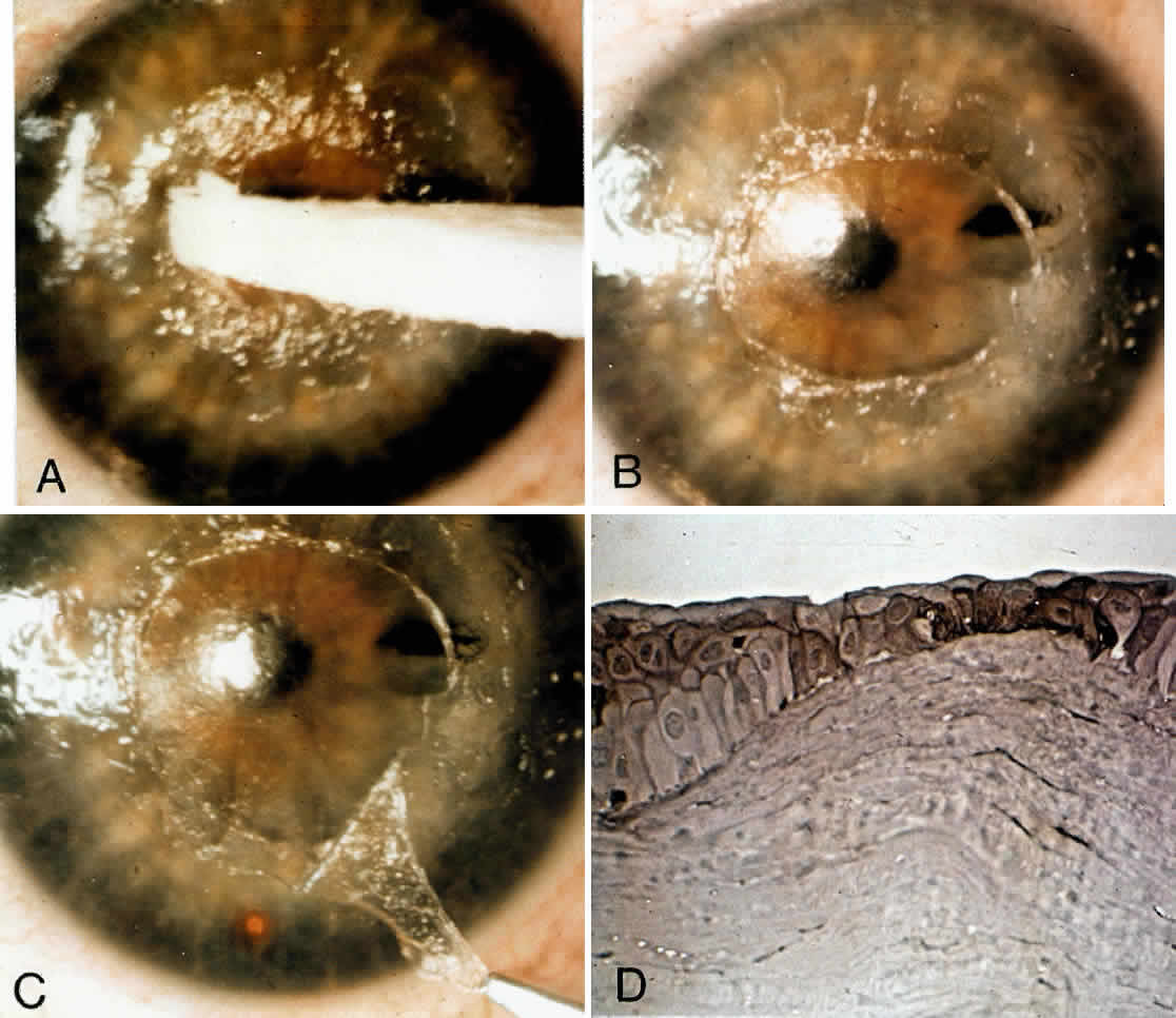
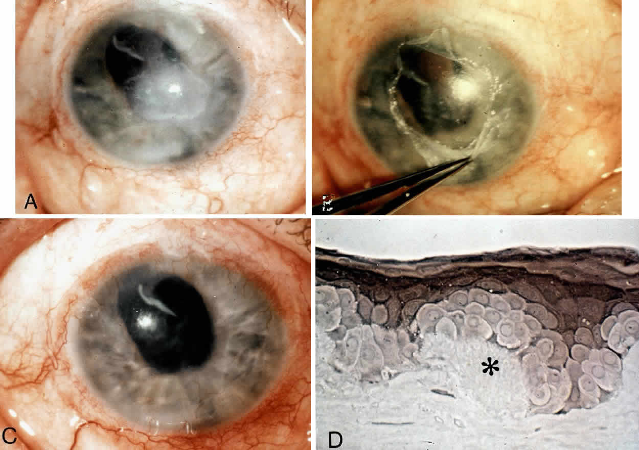
The corneal epithelium is removed over the involved area by gently scraping with a dry cellulose sponge (Weck-cel) or a disposable scarifier blade (Grieshaber 681.01 or Beaver 59). After epithelial removal, the surface should be kept dry to facilitate visualization of subepithelial fibrous plaques or focal accumulations of aberrant basement membrane material. With the aid of a dry cellulose sponge, cleavage planes between the subepithelial fibrous tissue and Bowman's layer can often be located and exploited. After an edge of the subepithelial fibrous membrane is identified, the tissue can be stripped and peeled with the aid of jeweler's forceps, frequently as a continuous sheet, thereby leaving Bowman's layer unharmed. A scarifier may be used to gently scrape or dissect more firmly adherent tissue. Sharp incision or dissection should be avoided to prevent damage to the underlying Bowman's layer or stroma. The cleared corneal surface can be polished with a cellulose sponge or lightly scraped with a scarifier (Fig. 1).1

Fig. 1. Technique of superficial keratectomy. A. A dry cellulose sponge is used to identify a cleavage plane beneath fibrovascular tissue. B. The pannus is stripped using dry sponge and tissue forceps. C. A scarifier may be used for firmly adherent tissue, remaining parallel to the cleavage plane to avoid stromal damage. (Shingleton BJ, Hersh PS, Kenyon KR [eds]: Eye Trauma. St. Louis, CV Mosby, 1991)

Postoperatively, antibiotic ointment is applied and the eye is pressure-patched for 24 hours. Thereafter, antibiotic ointment is initially applied four times a day and reduced in frequency over 2 weeks. Re-epithelialization is usually complete in 5 to 7 days. Contact lens wear can be resumed after 2 weeks.

Back to Top
CONDITIONS APPROPRIATE FOR SUPERFICIAL KERATECTOMY

EPITHELIAL BASEMENT MEMBRANE DYSTROPHY (MAP DOT-FINGERPRINT DYSTROPHY)

Epithelial basement membrane dystrophy is the most common corneal dystrophy, affecting approximately 2% of the population.2 Although most patients remain asymptomatic, about 10% experience recurrent corneal erosions as a consequence of faulty attachment complexes. These attachment complexes consist of hemidesmosomes of the basal epithelial cells, the underlying basement membrane, and the subadjacent anchoring fibrils of Bowman's layer.3 After an erosion, persistence of devitalized epithelium and fragments of basement membrane may inhibit normal re-epithelialization and formation of secure attachment complexes. Simple superficial debridement for removal of abnormal epithelium and basement membrane, thereby leaving a smooth substrate of Bowman's layer, can be performed at the slit lamp. The adjacent normal epithelium can resurface this area, allowing formation of competent attachment complexes and resulting in prompt cessation of erosive symptoms with much reduced frequency of recurrences.4–6 This procedure can be employed if more conservative measures (i.e., lubricants, patching, bandage contact lenses) fail in halting the erosions.

A smaller subset of patients with epithelial basement membrane dystrophy experience reduction of vision and/or recurrent erosions from the extreme deposition of an abnormal basement membrane and fibrillar collagenous material between the epithelium and Bowman's layer.2 This material may lead to irregular astigmatism and abnormal tear breakup. Patients typically complain of monocular visual distortion, diplopia, or “ghost images.” By performing superficial keratectomy, this abnormal material can be readily removed, leaving behind a smooth substrate of intact Bowman's layer. After re-epithelialization a smooth surface is re-established with the elimination of irregular astigmatism.

REIS-BÜCKLERS DYSTROPHY

Reis-Bücklers' dystrophy is an autosomal dominant condition characterized by the extensive accumulation of a fibrous subepithelial tissue that largely replaces Bowman's layer.2 Secondarily defective epithelial attachment complexes result in severe recurrent erosions. The accumulations of fibrous tissue also cause irregular astigmatism and a reticular anterior corneal opacification with diminished vision. The anterior location of this dystrophic process makes it amenable to treatment by superficial keratectomy.7,8 A dissection plane can frequently be developed between the fibrous tissue and the underlying normal stroma. Blunt dissection can then be used to peel the fibrous tissue until it is entirely removed (Color Plate 1: Color Fig. 1A-D). Our experience and that of others7,8 have been very favorable with this technique in achieving cessation of the erosions and improvement of vision. The fibrous tissue may recur after some years, but the procedure can be easily repeated.

Color Plate 1 . Technique of superficial keratectomy in a case of Reis-Bücklers' dystrophy. A. Central epithelium is removed with a dry cellulose sponge. B. Central cornea with epithelium removed. C. Cellulose sponge is used to identify a plane in the fibrous membrane. Fibrous membrane is then peeled as continuous cellophane-like sheets with jeweler's forceps. D. Phase-contrast photomicrograph of keratectomy specimen. Note irregular epithelium, aberrant basement membrane zone, and dense subepithelial fibrous tissue that has replaced Bowman's layer. (Paraphenylenediamine, X 350)

GRANULAR DYSTROPHY

Granular dystrophy is an autosomal dominant condition characterized by the subepithelial and stromal accumulation of an eosinophilic granular material. In advanced cases corneal transplantation is necessary for visual rehabilitation. A superficial variant of granular dystrophy has been described that primarily involves Bowman's layer and clinically and histologically resembles Reis-Bücklers' dystrophy. Transmission electron microscopy is necessary to identify the typical lesions of granular dystrophy in these cases. These superficial variants tend to have an earlier onset and higher frequency of erosive symptoms than typical granular dystrophy.9,10 Fortunately, the predominantly anterior location of the lesions may allow for treatment by superficial keratectomy alone.

Granular dystrophy can recur in corneal grafts. The recurrences in grafted corneas frequently take the form of an avascular fibrous connective tissue between the normal epithelium and Bowman's layer.2 The anterior location of recurrent fibrous tissue allows for easy removal from underlying Bowman's layer by superficial keratectomy.11 Thus, repeat penetrating keratoplasty with its attendant risks can be avoided (Color Plate 2: Color Figs. 2A AND B; 3A and B).

Color Plate 2 . Reis-Bücklers' dystrophy. A. At beginning of superficial keratectomy, markedly irregular reticulum of superficial fibrous tissue is readily evident following removal of corneal epithelium. B. At the completion of superficial keratectomy, major improvement in the smooth contour of the anterior corneal surface is evident.

Color Plate 3 . Granular dystrophy. A. Preoperative appearance. Axial, anterior stromal opacities are responsible for a preoperative decrease in visual acuity to 20/70 (6/21). B. Postoperative appearance. The axial cornea is free of opacities with visual improvement to 20/30 (6/9). Note persistence of solitary stromal deposit.

KERATOCONUS

Keratoconus is an ectactic dystrophy characterized by progressive thinning of the central or paracentral cornea. Indications for corneal transplantation include corneal scarring in the visual axis causing visual reduction or intolerance of contact lenses necessary to correct irregular astigmatism. Some patients become intolerant to contact lenses owing to the formation of a raised, localized area of superficial cornea that is chronically abraded by the contact lenses. Moodaley and co-workers12 have referred to this raised area as a “proud nebula,” and we have termed such a focal nodular scar as a “corneal callus.”13 Superficial keratectomy can be used to remove these focal fibrotic nodules and allow for resumption of contact lens wear. Penetrating keratoplasty can thereby be avoided in many appropriately selected cases.

Histopathologic evaluation of the excised fibrotic nodules reveals fibrillar collagen in random array with few interspersed fibrocytes. The overlying epithelium is fragmented with discontinuous, multilaminar basement membrane and diminished hemidesmosomes. The nodules are anterior to Bowman's layer, and hence, a plane of dissection can be created between the fibrous plaque and the underlying stroma, leaving stroma undisturbed (Fig. 2; Color Plate 3: Color Fig. 4A-C).

Fig. 2. Diagrammatic distinction between keratoconus corneas with focal, elevated nodule (A) versus nonelevated, anterior stromal scarring (B) . Only the former configuration (A) is appropriate for superficial keratectomy.

Color Plate 4 . Keratoconus. A and B. Clinical photographs demonstrate features of focal, elevated, fibrotic nodule with overlying epithelial erosion. This patient is intolerant of contact lenses and has poor spectacle acuity. C. Postoperative appearance. The fibrotic nodule has been removed with resumption of contact lens wear.

VERNAL KERATOPATHY

Vernal disease is a seasonally recurring, bilateral inflammation of the conjunctiva that occurs in children and young adults who have a history of atopy. Vernal keratopathy presents as punctate epithelial keratitis, subepithelial scarring, and large epithelial erosions with plaque formation. This plaque consists of a fibrin-like exudate that is resistant to medical therapy and can become chronic. The vernal plaque can be successfully removed by superficial keratectomy.14

SQUAMOUS CELL CARCINOMA

Corneal intraepithelial neoplasia refers to a spectrum of conditions ranging from a mild dysplastic process to carcinoma in situ. Invasive squamous cell carcinoma of the cornea is diagnosed when the dysplastic process breaks through the epithelial basement membrane. Squamous cell carcinoma of the cornea that is noninvasive can be successfully removed with superficial keratectomy. Supplemental cryotherapy has been advocated.15

OTHER CORNEAL DISORDERS

In our experience, other anterior corneal disorders that we have successfully managed with superficial keratectomy include the following: anterior corneal pannus of aniridia, calcific band keratopathy, bullous keratopathy with secondary amyloidosis (Color Plate 4: Color Fig. 5A-D), superficial pannus following chemical injury, residual scarring following pterygium removal, and Salzmann's nodular degeneration.

Color Plate 5. Post-traumatic bullous keratopathy with secondary amyloidosis. A. Preoperative appearance with visual acuity of counting fingers at 2 feet and symptomatic recurrent erosions. B. Intraoperative photograph. Thick fibrous tissue layer is easily peeled away with jeweler's forceps. C. Postoperative appearance. The amyloid deposits are absent. The corneal epithelium is more regular in appearance, and recurrent erosive episodes are diminished. Visual acuity has improved to 20/400 (6/120). D. Phase-contrast photomicrograph of keratectomy specimen. Marked epithelial distortion due to amyloid deposits (*); Subepithelial pannus is apparent. Bowman's layer is absent.(Paraphenylenediamine; X 350)

Back to Top
AUTHORS' EXPERIENCE
In our more than 10-year clinical experience comprising over 200 cases, we have experienced remarkably few complications with this procedure. Specifically, in two patients with calcific band keratopathy, postoperative persistent epithelial defects (probably related to decreased corneal sensation) resulted in corneal scarring. In one patient with keratoconus, microbial keratitis followed superficial keratectomy performed elsewhere. In our own patients with keratoconus, recurrence of focal scarring developed in less than 10% of approximately 50 cases, and only two of these patients have required penetrating keratoplasty.
Back to Top
THERAPEUTIC ALTERNATIVES
The 193-nm excimer laser has been used to perform anterior keratectomy on patients with superficial corneal opacities. This procedure has been termed phototherapeutic keratectomy.16,17 The excimer laser allows for the removal of precise amounts of superficial corneal tissue and provides an optically smooth surface that is conducive to stable epithelial recovery and basement membrane complex reformation.17

With this approach, anterior corneal dystrophies and scars have been successfully removed, thereby eliminating the need for penetrating keratoplasty in some patients. The excimer laser has also been used to treat intractable recurrent erosions16 as well as to smooth rough irregular corneal surfaces.17,18 The main advantage of the excimer laser lies in its ability to remove deeper corneal opacities than are readily accessible by conventional superficial keratectomy. Its principal disadvantages relative to superficial keratectomy include induced hyperopia, high cost of instrumentation and operation, plus limited accessibility.

Although phototherapeutic keratectomy remains investigational and has shown promise in Food and Drug Administration-approved clinical trials as a comparable or potentially preferable alternative for the treatment of various conditions limited to the anterior cornea, we continue to prefer superficial keratectomy as the initial therapy of choice in these situations.

Back to Top
CONCLUSIONS
Superficial keratectomy or stripping of anterior fibrous membranes can be highly effective in treating anterior corneal dystrophic and degenerative conditions. Disabling epithelial erosions can frequently be eliminated by restoring a substrate suitable for stable epithelial adhesion. Visual improvement can be achieved by eliminating irregular astigmatism and removing anterior corneal opacities.

The simplicity of the procedure is most attractive. Once a dissection plane is established, the entire sheet of aberrant fibrous tissue can be peeled with forceps, thereby obviating the use of sharp instruments. Since the pathologic abnormality is frequently anterior to Bowman's layer, the difficulties with stromal scarring that can result from standard lamellar keratectomy can be avoided.

Recurrence of the pathologic process can certainly occur, and we have rarely observed this phenomenon in Reis-Bücklers' dystrophy, granular dystrophy, and keratoconus. Still, in patients who would otherwise be subjected to penetrating keratoplasty, the improvement in both vision and symptoms with less invasive procedures seems far preferable.

In summary, superficial keratectomy is a simple, extraocular, outpatient procedure with minimal risk of complications. Postoperative recovery is rapid, and the costs, patient restrictions, and potential complications of penetrating keratoplasty are avoided. There is a swift improvement of vision and decrease in erosive symptoms. Although the primary disease process may rarely recur, this minor surgical procedure can be repeated easily. Thus, we continue to recommend its use in these several degenerative and dystrophic disorders of the anterior cornea.

Back to Top
REFERENCES

1. Hersh PS, Kenyon KR: Anterior segment reconstruction following ocular trauma. In Shingleton BJ, Hersh PS, Kenyon KR (eds): Eye Trauma, p 176. St. Louis, CV Mosby, 1991

2. Waring GO, Rodrigues MM, Laibson PR: Corneal dystrophies: I. Dystrophies of the epithelium, Bowman's layer, and stroma. Surv Ophthalmol 23:71, 1978

3. Galbavy EJ, Mobilia EF, Kenyon KR: Recurrent corneal erosions. Int Ophthalmol Clin 24:107, 1984

4. Buxton JN, Fox ML: Superficial epithelial keratectomy in the treatment of epithelial basement membrane dystrophy: A preliminary report. Arch Ophthalmol 101:392, 1983

5. Buxton JN, Constad WH: Superficial epithelial keratectomy in the treatment of epithelial basement membrane dystrophy. Cornea 6:292, 1987

6. Buxton JN, Constad WH: Superficial epithelial keratectomy in the treatment of epithelial basement membrane dystrophy. Ann Ophthalmol 19:92, 1987

7. Wood TO, Fleming JC, Dotson RS, Cotten MS: Treatment of Reis-Bücklers' corneal dystrophy by removal of subepithelial fibrous tissue. Am J Ophthalmol 85:360, 1978

8. Schwartz MF, Taylor HR: Surgical management of Reis-Bücklers' corneal dystrophy. Cornea 4:100, 1985

9. Haddad R, Font RL, Fine BS: Unusual superficial variant of granular dystrophy of the cornea. Am J Ophthalmol 83:213, 1977

10. Kenyon KR, Hersh PS, Starck T, Fogle JA: Corneal dysgeneses, dystrophies and degenerations. In Tasman W, Jaeger EA (eds): Duane's Clinical Ophthalmology, vol 4, chap 16. Philadelphia, JB Lippincott, 1992

11. Lempert SL, Jenkins MS, Johnson BL, Brown SI: A simple technique for removal of recurring granular dystrophy in corneal grafts. Am J Ophthalmol 86:89, 1978

12. Moodaley L, Buckley RJ, Woodward EG: Surgery to improve contact lens wear in keratoconus. CLAO J 17:129, 1991

13. Kenyon BM, Kenyon KR, Korb D: Simplified superficial keratectomy for the management of keratoconus. Ophthalmology, submitted for publication

14. Buckley RJ: Vernal keratopathy and its management. Trans Ophthalmol Soc UK 101:234, 1981

15. Cameron JA, Hidayat AA: Squamous cell carcinoma of the cornea. Am J Ophthalmol 111:571, 1991

16. Sher NA, Bowers RA, Zabel RW et al: Clinical use of the 193-nm excimer laser in the treatment of corneal scars. Arch Ophthalmol 109:491, 1991

17. Stark WJ, Chamon W, Kamp MT et al: Clinical follow-up of 193-nm ArF excimer laser photokeratectomy. Ophthalmology 99:805, 1992

18. Fitzsimmons TD, Fagerholm P: Superficial keratectomy with the 193-nm excimer laser: A reproducible model of corneal surface irregularities. Acta Ophthalmol 69:641, 1991

Back to Top