Chapter 56
Vitrectomy
FRANK J. MCCABE, CARL D. REGILLO and WILLIAM E. BENSON
Main Menu   Table Of Contents

Search

PREOPERATIVE EVALUATION HISTORY TAKING
BASIC VITRECTOMY TECHNIQUE
SPECIAL VITRECTOMY TECHNIQUES LENSECTOMY
VITREOUS SUBSTITUTES PERFLUOROCARBON LIQUIDS
INDICATIONS AND RESULTS GLAUCOMA
COMPLICATIONS
REFERENCES

In 1968, Kasner introduced the “open-sky” technique for removal of formed vitreous. This technique required a large limbal incision, lensectomy, and mechanical removal of the vitreous with scissors.1 Kasner proved that the eye could tolerate removal of the vitreous without severe complications. Nevertheless, few elective vitrectomies were done until 1971, when Machemer performed the first closed, pars plana, vitrectomy.2–4 The instrument for performing the vitrectomy was a single probe called VISC. VISC stood for vitreous-infusion-suction-cutting because this large, bulkyprobe performed all of these functions. Machemer pioneered this technique and later introduced the intraocular fiberoptic light pipe.3,5,6 The next giant step was the separation of the various functions of the single probe into separate probes for aspiration-cutting, illumination, and infusion.7 This ingenious use of a separate infusion line allowed miniaturization and standardization of the size of the various vitrectomy instruments needed for surgery.

Over the years, considerable advancement has been made in vitreoretinal instrumentation, including high-speed vitrectomy cutters, illuminated forceps and picks,8 and the diamond-dusted silicone tip cannula.9,10 Other improvements include less-toxic irrigating solutions,11–13 improved contact lenses, safe intraocular diathermy, flexible iris retractors,14 and endolaser photocoagulation. Motorized scissors have dramatically decreased the incidence of iatrogenic retinal tears because membranes that formerly had to be peeled from the retina can now be segmented. Special mention must be given to the pioneering work by Chang15 with perfluorocarbon liquids (PFCLs), which are a major advance in the unrolling of giant tears and in the evacuation of subretinal fluid in selected cases. Silicone oil (SO), first introduced by Cibis in 1962, has been increasingly used since the early 1980s.16,17 The latest major advance in vitreoretinal surgery has been the introduction of wide-field viewing systems, which allow a panoramic view of the retina.18,19 These technologic advances have helped to expand the indications for vitrectomy greatly (Table 1).


Back to Top
PREOPERATIVE EVALUATION HISTORY TAKING
A complete medical history is obtained from all patients. Many patients who require a vitrectomy have diabetes mellitus and other medical problems. In one report, abnormalities in the cardiovascular or renal function or both were found in 98% of patients with diabetes undergoing vitrectomy.20 Patients requiring renal dialysis may have an increased risk of operative problems as many have a bleeding diathesis. The ocular history is critical because the surgeon must take into consideration other possible causes of decreased vision such as severe glaucoma, ischemic optic neuropathy, macular edema, and arteriolar occlusive disease. The duration of certain conditions influences the visual prognosis. A diabetic macular detachment of 2 months or shorter is twice as likely to achieve a good visual result than those of 13 months' duration or longer.21 Macular holes of 6 months' duration or longer are less likely to achieve significant visual improvement.22 Patients with epiretinal membranes and a longer history of blurred vision showed less visual improvement in visual acuity than did patients with a short duration of symptoms.23

OCULAR EXAMINATION

A nearly absolute contraindication to any ocular surgery is no light perception (LP).24 Normal pupillary responses are a significant predictor of successful visual results in patients with diabetes.21 An opaque cornea can now be addressed with various styles of temporary, intraoperative keratopros-theses.25–27 The examiner should inspect the eye for iris neovascularization (NVI) with slit-lamp biomicroscopy and gonioscopy. NVI portends a poor prognosis in patients, who should be treated with panretinal photocoagulation (PRP) before vitrectomy if possible.21,28–30 In some cases, especially those in which membranes must be peeled or delaminated from the retina, a cataract may need to be removed to permit safe completion of the surgical objectives. It is best to recognize this before surgery so that preparations for placement of a posterior chamber intraocular lens (IOL) and vitrectomy in one operation can be made.31–38 In patients with pseudophakia, the type of lens should be determined. Silicone IOL implants may have SO droplets condense on the back surface of the lens. In addition, silicone implants are more predisposed to have air condense on the posterior surface of the lens.39,40

Obviously, a careful retinal examination with indirect ophthalmoscopy and slit-lamp biomicroscopy is essential. The status of the posterior hyaloid should be noted. The location of retinal breaks, the extent and nature of membrane proliferation, and other pertinent findings should all be documented carefully. For example, retinal detachments caused by small areas of vitreoretinal adhesion (Fig. 1) or that have “bridges” between localized detachments are easier to repair than those with broad adhesions (Fig. 2). Iatrogenic retinal holes are more common in patients with longstanding retinal detachment and ischemic, thin retina than in patients with recent detachment and well-vascularized retinas. These factors help determine the patient's prognosis and the procedure length.

Fig. 1. Traction retinal detachment caused by a small vitreoretinal adhesion.

Fig. 2. Traction retinal detachment caused by broad vitreoretinal adhesions.

ANCILLARY TESTING

Ultrasonography is an invaluable tool in evaluating eyes with opaque media. Ultrasound can aid in diagnosing vitreous membranes, retinal detachments, intraocular foreign bodies (IFBs), perforating injuries, choroidal detachments, intraocular tumors, and so forth accurately. It is important to recognize the distinctive funnel-shaped pattern that identifies retinal detachment with severe proliferative vitreoretinopathy (PVR).41 Such detachments are challenging and may require more advanced vitreoretinal techniques. Ultrasound in trauma cases also provides prognostic information. Eyes undergoing vitrectomy after penetrating trauma fared worse if preoperative ultrasound detected retinal detachment,subretinal blood, massive choroidal detachments, or posterior exit wounds.42 Ultrabiomicroscopy is a recent ultrasound imaging technique that provides a high-resolution view of the anterior segment. In recurrent vitreous hemorrhages after diabetic vit-rectomy, ultrabiomicroscopy can evaluate the sclerotomy sites for possible neovascularization.43,44

In eyes with opaque media, there also are tests that can assess gross retinal and optic nerve function. Color perception, two-point discrimination, and Maddox rod orientation are of limited value.45 The perception of entoptic phenomenon to assess retinal function has had mixed results.21,45,46 The basis for the blue-field entoptic phenomenon is that patients with normal maculae who stare at a diffuse blue-light can see leukocytes circulating in their macular capillaries. An instrument with a blue light bright enough to penetrate dense vitreous hemorrhage is available. It can predict macular function successfully in patients with traumatic vitreous hemorrhage but, in general, is rarely used in clinical practice.46

Electroretinography (ERG) helps to evaluate the overall functional status of the retina. The standard Grass photostimulator may not provide enough light to stimulate the retina in cases of dense vitreous hemorrhage. The resultant flat tracing may falsely indicate a nonfunctioning retina. However, the bright-flash ERG can penetrate dense vitreous hemorrhages. Some of the light actually transverses the sclera to stimulate the retina. Nevertheless, the ability of a preoperative bright-flash electroretinogram to predict visual outcomes is controversial. A patient may have an excellent visual result after removal of a diabetic vitreous hemorrhage but have a flat preoperative ERG. A decision to operate should not be based on a preoperative ERG as it cannot safely predict the outcome in diabetic vitrectomy.47,48 Focal macular electroretinograms recently have been used before and after surgery in select macular conditions.49,50

Other electrophysiologic tests to evaluate visual function are pattern and flash visual-evoked potentials (VEPs). The pattern VEP evaluates macular function and is not useful in patients with opaque media because the pattern cannot be perceived.51 A normal response to a flash VEP depends on a normally functioning retina and optic nerve. A bright flash is not essential to generate the VEP because this test does not require as much light as does the ERG. Both the latency and amplitude of the tracing provide useful information.52 If the retina is totally detached, the VEP is absent. If it is partially attached, the VEP is reduced. A delayed VEP indicates optic nerve dysfunction. The amplitude of the flash VEP at stimulus rates of 10 flashes per second is largely influenced by the macular status. At 20 to 30 flashes per second, the amplitude depends more on the integrity of the optic pathways. Therefore, the VEP may be an excellent predictor of postoperative visual acuity. However, the prognostic value of a preoperative flash VEP for a diabetic vitrectomy is controversial. Similar to ERG, a decision to operate should not be based on a preoperative VEP as patients may obtain good vision despite a reduced VEP.47,48

Computed tomography scans have been shown to be helpful in localizing foreign bodies in or near the sclera. Computed tomography and ultrasound are more sensitive techniques than are plain x-ray films for detecting and localizing IOFBs.53,54 The sensitivity of computed tomography scan for detecting IOFBs is 100% for objects larger than0.06 mm.55 Magnetic resonance imaging is contraindicated in IOFB cases because of potential movement of magnetic IOFBs by the powerful magnetic resonance imaging magnets.

A new technique that is helpful in preoperative assessment of patients is optical coherence tomography, which is a noncontact, noninvasive imaging technique that produces high-resolution, cross-sectional images of the retina.56 The interaction between the posterior hyaloid and the retinal surfaces can sometimes be difficult to discern. Optical co-herence tomography can assist in diagnosing vit-reomacular traction syndrome and distinguishingfull-thickness holes from pseudoholes, cysts, and partial-thickness holes.57–59 This technique also can be used to assess macular hole closure after vitrectomy and macular edema before and after vitrectomy for diabetic macular edema.60–62

SURGICAL ANATOMY

It is essential to be familiar with the surgical anatomy when performing a vitrectomy. The pars plana, the posterior section of the ciliary body, is the entry site to the vitreous cavity for ocular instruments in pars plana vitrectomy (PPV). The pars plana is around 4 mm wide in adults and is nded anteriorly by the pars plicata (ciliary processes) and posteriorly by the ora serrata. In adults, the ora serrata is 6 to 7 mm posterior to the limbus nasally and 6 to 8.5 mm temporally.63 In phakic patients, the pars plana should be entered 4 mm posterior to the limbus and in pseudophakic patients 3 to 3.5 mm posterior to the limbus (Fig. 3). This technique ensures that the entry of the ocular instruments will be anterior to the ora serrata to prevent a retinal tear or detachment and posterior to the well-vascularized pars plicata to avoid hemorrhage. It also is important to enter at the correct angle to avoid damaging the lens.

Fig. 3. The sclerotomy sites should be placed 3 to 3.5 mm posterior to the limbus in the pseudophakic/aphakic eye and 4 mm posterior to the limbus in phakic eyes. The entry site will then be in the pars plana region.

The vitreous is a gel-like structure that is comprised of collagen fibrils, hyaluronic acid, hyalocytes, and water. The collagen fibrils in the peripheral vitreous condense to form a thin layer called the vitreous cortex, which surrounds the central gel and is adherent to the internal-limiting membrane (ILM) of the retina. The vitreous cortex has varying degrees of retinal adherence. It is most adherent to the ILM at the vitreous base. The vitreous base is a band about 6 mm in width that overlays the ora serrata and pars plana. The vitreous cortex also is more adherent to the optic nerve head, blood vessels, and perifoveal region. In disease states, as discussed below, the vitreous cortex may be strongly adherent to neovascularization, chorioretinal scars, and fibrous proliferation.64 After the vitreous cortex separates from the ILM, the subsequent thin layer is referred to as the posterior hyaloid face.65 Understanding the relationships between the retina and vitreous is extremely important in vitreoretinal surgery.

Back to Top
BASIC VITRECTOMY TECHNIQUE
Anesthesia for PPV can be general or local. General anesthesia should be considered for long cases, for patients unable to tolerate local anesthesia for physical or psychological reasons, and for children. Local anesthesia is used in most cases. The options for local anesthesia are retrobulbar, peribulbar, topical, and sub-Tenon's injections of an anesthetic solution. Retrobulbar and peribulbar injections are effective forms of anesthesia but rarely can beassociated with significant complications, including globe penetration, hemorrhage, arterial occlusions, and optic nerve damage.66–68 Sub-Tenon's injection with a blunt cannula provides excellent anesthesia and has minimal complications.69–71 In select, uncomplicated cases, topical anesthesia has been successful but is not used routinely.72,73

Intraocular irrigating fluids have been studied extensively, and Balanced Salt Solution (BSS)-Plus (Alcon) has been shown to be a safe irrigatingsolution.11–13 BSS-Plus contains glutathione, glucose, magnesium, bicarbonate buffer solution, and Ringer's lactate solution. Whether BSS-Plus is superior to BSS has not been definitely established. In a diabetic vitrectomy, the lens may develop posterior capsular opacification secondary to an osmotic shift of water into the lens. The addition of 3 ml of 50% dextrose to BSS or BSS-Plus has been shown to prevent cataract development during diabetic vit-rectomy.74

PPV requires an operating microscope. A foot pedal to control the zoom magnification, focusing, and X-Y microscope is essential. A beam splitter for the surgical assistant should be present. There are many types of lenses that can be used with the operating microscope. Planoconcave, hand-held irrigating lenses, such as the Charles lens, often are used. The field of view is about 20 degrees, and the fluid meniscus rinses away debris and allows prism effect. If no assistant is available to hold the lens, Landers sew-on lenses can be used. If the eye is gas filled, biconcave lenses, such as the Machemer lens, need to be used for viewing.75 There are several options for viewing the peripheral retina. The surgeon can use the Charles lens with scleral depression, prismatic lenses, or the recently developed wide-field viewing systems, which require an inverter on the microscope but offer superior depth of focus, a wider angle of view, and are almost independent of the pupil size. They also allow for a superior view through native lens or lens implant opacities, particularly condensation on the posterior lens surface during air-fluid exchange. The binocular indirect ophthalmoscope is a noncontact wide-field viewing system that is used often (Fig. 4).18,19 One recent study suggested that wide-field viewing systems decrease surgical time and allow more complete laser treatment.76 Another study showed that the binocular indirect ophthalmoscope has a decreased rate of intraoperative corneal epithelial defects.77

Fig. 4. Binocular indirect ophthalmoscope is shown attached to the operating microscope. (Courtesy of Insight Instruments Inc, Sanford, FL.)

A hand support is preferred for vitrectomy surgery (Fig. 5). The surgeon rests both hands throughout the operation, avoiding fatigue and achieving fine control for intravitreous manipulations. The level of the hand support usually is at the patient's ears but should be individualized for the surgeon and patient. A sterile plastic drape, used to cover the patient, is pushed down into the space between the patient's head and the hand support. This forms a trough to collect fluid that would otherwise spill onto the floor. The microscope also can be covered with a sterile plastic drape.

Fig. 5. Patient is prepared for vitrectomy. A hand support is preferred for vitrectomy surgery.

Small radial or triangular incisions through the conjunctiva and Tenon's capsule are made superonasally, superotemporally, and inferotemporally. Hemostasis is maintained with bipolar diathermy. The entry incisions are parallel to the corneoscleral limbus and are 4 mm posterior to it in phakic eyes and 3 to 3.5 mm in aphakic and pseudophakic eyes (Fig. 6). The incisions are made with a 20-gauge microvitreoretinal (MVR) blade. It is important that the incisions (through the sclera, uvea, and pars plana) be just large enough to permit entry of the desired instrument. A 1.4-mm linear incision rounds out to 0.89 mm, the diameter of the blade shaft.78 If the incisions are too long, irrigation fluid leaks around the instrument and out of the eye, making normal intraocular pressure (IOP) difficult to maintain without using large volumes of irrigating solution. In addition, if a total gas-fluid exchange is later attempted, the gas escapes from the eye. All blades used for incisions must be sharp, so that when introduced into the eye, they do not push pars plana epithelium or vitreous base ahead of them, causing a retinal tear or dialysis. The MVR blade should be held at an approximately 70-degree angle and passed toward the optic nerve. It is important to enlarge the sclerotomy incision from the inside out to ensure that the internal opening is as large as the scleral opening (Fig. 7). When the media are clear, the surgeon should push the MVR tip into the eye until it can be seen through the pupil.

Fig. 6. Preparation for vitrectomy. Radial incisions through the conjunctiva and Tenon's capsule are made superonasally, superotemporally, and inferotemporally. In a phakic eye, the sclerotomy is made 4 mm posterior to the limbus.

Fig. 7. The internal opening is not as large as the scleral opening in this sclerotomy incision. The insertion of instruments into this incision may cause a retinal dialysis. It is important to enlarge the wound from the inside with the microvitreoretinal blade to avoid this problem.

The first incision is for the infusion cannula, which is placed in the inferotemporal quadrant, just inferior to the lateral rectus muscle. If the cannula is placed too far inferiorly, it presses against the lid speculum during inferior rotations of the globe and may hinder access to the inferior portions of the eye. A 5-0 nylon mattress suture, which secures the base of the infusion cannula, is placed before entering the eye (Fig. 8). In most cases, a 4-mm cannula is used. In pseudophakic/aphakic eyes, a6-mm cannula can be used to assist in visualization of the cannula tip in cases of media opacity. The infusion cannula is twisted through the sclerotomy site in an oscillatory fashion, ensuring the bevel is facing the center of the eye. The surgeon, using the indirect ophthalmoscope if necessary, must verify that the cannula has passed cleanly through the pars plana epithelium before turning on the infusion.79 A small pupil may hinder the view of the cannula and the subsequent surgery. Flexible iris retractors are an easy and effective way for enlarging a small pupil (Fig. 9).14 If there are opaque media, the surgeon must provide infusion with a hand-held infusion needle whose tip can be seen until the media are clear enough to see the cannula. It is difficult to place an infusion cannula into the vitreous cavity if there is a choroidal detachment. The safest technique is to drain the detachment while maintain-ing the intraocular volume with irrigating solution through a 22-gauge needle.80 The needle is introduced through the limbus in aphakic eyes and through the pars plana in phakic eyes.

Fig. 8. A. Calipers are used to measure 4 to 4.5 mm from the limbus. B. Incision for the infusion cannula. A mattress suture of 5-0 nylon has been placed to secure the cannula. C. The cannula is in place. D. The vitrectomy instrument and light pipe have been inserted superiorly.

Fig. 9. Flexible iris retractor. (Courtesy of Alcon, Fort Worth, TX.)

After the infusion is turned on, incisions are made in the superonasal and superotemporal quadrants for the vitrectomy instrument and fiberoptic light pipe. These incisions should be made 150 to 160 degrees apart, just above the lateral and medial rectus muscles. This allows instrument access to both the superior and inferior retina. It is important not to make sclerotomy sites in a “soft” eye. If necessary, fluid should be injected with a 30-gauge needle at the pars plana or limbus to pressurize the eye.

There are two basic types of vitrectors: those that use a rotating or oscillating inner blade and those that use a guillotine-like chopping blade (Fig. 10). Today, guillotine cutters are used most often. A pneumatically driven inner tube moves up and down inside a fixed outer tube. Vitreous is aspirated into the cutter when the openings of the inner and outer tubes are aligned and then chopped off as the inner tube moves up and down. The suction for vitreous aspiration is through the inner tube. For a core vitrectomy, high cut rates (600 to 800 cycles per second) and moderate suction (200 mmHg) should be used. Higher cut rates minimize vitreoretinal traction and decrease the incidence of iatrogenic retinal breaks. The Alcon vitrector is a well-designed, disposable instrument. It is lightweight to prevent fatigue, hourglass-shaped to prevent slippage, and disposable so that the blades are sharp (Fig. 11). Vitrector probes are now available that can achieve cut rates of approximately 1200 cycles per second, which can allow for fine shearing of vitreous close to the retinal surface (Fig. 12).

Fig. 10. A. Rotating-type cutter. When the holes in the fixed outer tube (short closed arrows) and inner stationary tube (open arrow) are coincident, vitreous is sucked in, cut off, and evacuated (long arrows). B. Guillotine-type cutter. The inner tube (dashed lines) moves up and down inside the fixed outer tube. It chops off the vitreous.

Fig. 11. This vitrector is lightweight to prevent fatigue, hourglass-shaped to prevent slippage, and disposable so that the blades are sharp. (Courtesy of Alcon, Fort Worth, TX.)

Fig. 12. High-speed vitrector. (Courtesy of Alcon, Fort Worth,TX.)

PPV is a bimanual technique. Typically, one sclerotomy port is used for a fiberoptic light source and the other sclerotomy for the vitrector or other instruments. Vitrectomy instruments should be held lightly in the surgeon's fingertips. It is important to position the instruments at the correct angle to avoid damaging the lens when entering the eye. The initial goals of the vitrectomy are to clear the axial media and to remove anteroposterior vitreoretinal traction. If there are media opacities, they should be removed from the central vitreous (Fig. 13). The status of the posterior hyaloid must be noted. If there is a complete posterior detachment, an edge of the posterior hyaloid should be followed out to the periphery circumferentially (Fig. 14). If there is a partial posterior detachment, the posterior hyaloid should be incised over attached retina and away from the macula. The incised edge should then be followed circumferentially. If there is preretinal blood, it can be removed with the vitrector on suction or with an extrusion cannula (Fig. 15). If the hyaloid is completely attached, the central vitreous should be removed with the cutter. Application of active suction with the vitrector or soft-tipped extrusion cannula over the optic nerve head should elevate the posterior hyaloid face. The soft-tippedcannula bends with engagement of the posterior hyaloid, which is called the fish-strike sign (Fig. 16). The elevated hyaloid can be incised with the cutter and an edge followed out to the periphery circumferentially.

Fig. 13. A. Vitreous hemorrhage with complete posterior vitreous detachment. B. The central vitreous has been removed.

Fig. 14. The vitrector is on the edge of the posterior hyaloid in an eye with a complete posterior vitreous detachment.

Fig. 15. A. Eye with diabetic retinopathy. Neovascularization elsewhere elevated by vitreous traction, vitreous hemorrhage, and blood layered on the posterior retina. B. After removal of the vitreous and release of the anteroposterior traction on the neovascularization, the blood on the posterior retina is vacuumed up with an extrusion cannula. C. After vitrectomy releases the traction, the frond regresses.

Fig. 16. Fish-strike sign. The soft-tipped cannula bends with engagement of the posterior hyaloid.

Once the media are cleared and anteroposterior traction has been removed, attention can be turned to the specifics of the case. After the case is completed, the sclerotomy sites should be closed temporarily and the periphery inspected for any breaks or detachments. Regardless of the indication for surgery, inspection should be performed before any gas or air injection in the eye as small breaks may be missed under partial or complete air- or gas-filled eyes. The sclerotomy sites also should be inspected carefully for any retinal or vitreous incarceration. The sclerotomy sites are closed with 7-0 polyglactin (Vicryl) sutures in a figure-eight configuration. The IOP should be checked. If the eye is soft, BSS can be injected at the pars plana or limbus to repressurize the eye. The conjunctiva is closed with 8-0 Vicryl sutures. Subconjunctival injections of a steroid and antibiotic are placed inferiorly, away from the rectus muscles.

Back to Top
SPECIAL VITRECTOMY TECHNIQUES LENSECTOMY
When an eye with a significant cataract requires a PPV, cataract extraction is essential for safe completion of the surgical objectives, especially if membranes must be peeled or delaminated from the retina. It has become increasingly common to perform phacoemulsification with implantation of a posterior chamber IOL via an anterior segment approach and PPV at the same operation.31–38 This technique avoids aphakia, which requires a contact lens or a second operation to implant a lens. In addition, aphakic diabetic eyes have a higher rate of postoperative neovascular glaucoma (NVG).81

If a pars plana lensectomy needs to be performed, the MVR blade is passed through the superotemporal sclerotomy site and into the lens at the lens equator. This creates an opening for the phacofragmotome (Fig. 17) or vitrector. A bent 21-gauge needle is connected to a separate infusion line and inserted into the lens via the superonasal sclerotomy. The infusion fluid keeps the capsular bag distended so that the lens can be removed with capsular preservation. This also prevents lens fragments from falling posteriorly. The central nucleus is removed first with the phacofragmotome, and the residual cortical material can be aspirated with the vitrector (Fig. 18). In some cases, such as anterior PVR, the capsule needs to be removed entirely. The central capsule can be removed with the vitrector and residual elements stripped with a forceps.82

Fig. 17. Phacofragmotome. (Courtesy of Alcon, Fort Worth, TX.)

Fig. 18. The phacofragmotome (left) removes the central nucleus, and the 21-gauge needle (right) provides infusion.

Vitrectomy techniques have been shown to be helpful in removing spontaneously dislocated lenses, in removing nuclei and cortex dislocated during cataract surgery (Fig. 19), and in retrieving and positioning posteriorly dislocated IOLs.83,84 If the nucleus is mildly sclerotic, a vitrectomy instrument, introduced through the pars plana, removes the fragments easily. Sometimes, a second instrument, such as a bent needle, is helpful. Firm pieces of nucleus can be crushed between it and the vitrectomy instrument. Dislocated nuclei with moderate or advanced sclerosis are best removed by holding them with a light pick and phacofragmenting them in the vitreous cavity (Fig. 20). The introduction of PFCLs in vitrectomy surgery had enabled the surgeon to inject this heavier-than-water liquid into the eye after vitrectomy to float the lens particles or IOL off the retina.85,86 The PFCL allows removal of the posteriorly dislocated lens or IOL with minimal retinal trauma and is especially useful when there also is a retinal detachment present.

Fig. 19. Nuclear fragment dislocated during cataract extraction.

Fig. 20. After the vitreous has been removed, the nucleus is held by a bent needle and phacofragmented in the vitreous cavity.

MEMBRANE PEELING, DELAMINATION,AND SEGMENTING

After the media have been cleared and the anteroposterior traction removed, the tangential traction on the retina must be addressed. Preretinal membranes can be seen in a host of conditions, such as proliferative diabetic retinopathy (PDR), idiopathic macular pucker, and PVR. The preretinal membranes can be vascular or avascular. There are multiple cell types involved, including glial cells, fibrocytes, retinal pigment epithelial cells, hyalocytes, and inflammatory cells. Membrane peeling, delamination, and segmentation are the three basic techniques to address preretinal membranes.

Membrane peeling is the removal of a preretinal membrane with tangential and perpendicular force (Fig. 21). The membrane peeling can be performed with a pick or forceps (Fig. 22). Illuminating light picks allow a bimanual removal approach.8 With some membranes, an edge can be initially grasped with forceps and gently peeled from the retina. In other cases, an edge must be created with a pick, MVR blade, or soft-tipped extrusion needle.87 A new tool for membrane peeling is the Tano scraper, which is a diamond-dusted silicone tip cannula that grasps membranes and facilitates removal. In cases of anterior membranes, scleral depression or a wide-field viewing system adds in visualization of the membranes. Currently, ILM peeling is being performed in macular hole surgery. The technique is similar to that of preretinal membrane peeling, but an edge in the ILM is created with a sharp MVR blade and an ILM maculorhexis performed with fine forceps or the Tano scraper.88

Fig. 21. Treatment of macular pucker. (Top) If a gap between the membrane and the retina is present, the membrane is elevated by a vitreous pick. (Middle) If no gap is present, one is created by pulling on the membrane with an extrusion needle. (Bottom) Once a break in the membrane has been created, it is peeled with vitreous forceps.

Fig. 22. A. Sutherland De Juan forceps. B. Tips of vitreoretinal forceps. (Courtesy of Alcon, Fort Worth, TX.)

PVR occurs in 5% of all retinal detachments when retinal pigment epithelial cells, glial cells, or bothproliferate on both surfaces of the retina and on vitreous membranes. Signs of PVR include fixed folds, detachment of the nonpigmented epithelium of the pars plana, and equatorial folds. Preretinal and subretinal membranes cause shortening of the retina (Fig. 23). If a higher scleral buckle does not close the retinal breaks, release of traction is essential for reattachment. The first step is to search carefully for any open retinal breaks. In recurrent retinal detachment, look at the original break or breaks first. In reoperations for PVR, breaks on the posterior slope of a previously placed buckle at the edge of a cryotherapy scar account for 40% of all new or previously identified breaks.89 The vitrectomy instrument is used to remove as much formed vitreous as is safely possible and to cut transvitreous membranes, releasing vitreous traction. Bent needles or forceps are used to peel preretinal membranes from the retinal surface to restore retinal flexibility (Fig. 24).

Fig. 23. Retinal detachment with proliferative vitreoretinopathy. Transvitreous traction pulls the retina toward the center of the eye. Preretinal membranes shorten the retina.

Fig. 24. Peeling of preretinal membranes. (Top) If there is a gap between the retina and the membrane, the membrane is engaged and peeled with a vitreous pick. (Middle) If no gap is present, it is grasped with vitreous forceps to make a gap. (Bottom) The membrane is peeled with forceps.

Membrane delamination usually is a bimanual technique for vascularized membranes such as in PDR. Using an illuminating pick to elevate the vascularized membrane, adhesions between the fibrovascular tissue and retina are cut with horizontal scissors (Fig. 25). Fibrovascular membranes often are extremely adherent to the retina and may overlay thin, ischemic retina. If a retina break is created in membrane removal, all traction must be removed completely from the retina. Viscodelamination, which is the use of a viscoelastic solution to create a cleavage plane between the membrane and retina, may assist in delamination.90,91 The delamination technique is used for en bloc resection of membranes.92–95 With the en bloc technique, the posterior hyaloid is left intact after a core vitrectomy. The hyaloid provides counter-traction to the membranes being delaminated. The delaminated membranes and posterior hyaloid are then removed as a single unit (en bloc).

Fig. 25. An illuminated pick elevates the fibrovascular membrane as the horizontal scissors cuts the adhesions between the retina and the fibrovascular membrane.

Membrane segmentation is used to relieve tangential retinal traction by cutting membranes, which are interconnected and producing retinal traction (Fig. 26). This technique does not involve the removal of membranes but after segmentation, membrane peeling and delamination may be easier to perform. Segmentation usually is performed with vertical scissors, which can be automated or manual (Fig. 27). To prevent intraoperative bleeding, fibrovascular membranes should be treated with diathermy before they are cut. If vessels are cut inadvertently, they should be diathermized promptly after occluding them by raising the IOP.

Fig. 26. A. Traction retinal detachment with bridges between a neovascular frond arising from the optic disc and between folds in the detachment. B. After vitrectomy, the bridges are cut. C. After the anteroposterior and preretinal traction has been released, the retina flattens out.

Fig. 27. Membrane peeler cutter scissors. (Courtesy of Alcon, Fort Worth, TX.)

RETINOTOMY AND RETINECTOMY

A retinotomy is a full-thickness incision in the retina. The size, location, and indications for a retinotomy are variable. Drainage of subretinal fluid in vitrectomy for retinal detachments is one of the most common reasons for a retinotomy.96 A full-thickness retinal hole is made with an endodia-thermy probe in a detached retina. If possible, the retinotomy is positioned superiorly so that it is supported by intraocular gas longer and nasal to the optic head to avoid macula involvement if there is a hemorrhagic complication. A tapered extrusion cannula is inserted through the retinal hole to extrude subretinal fluid with a complete fluid-air exchange (Fig. 28). Endolaser is applied to the borders of the retinotomy after the subretinal fluid is removed.

Fig. 28. A. Technique of air-fluid exchange. As air is infused anteriorly, it pushes the subretinal fluid posteriorly, where it is removed through a retinotomy by an extrusion cannula. B. The retinotomy is sealed by endolaser photocoagulation.

Retinotomies also can be made to access thesubretinal space for removal of lesions such as fibrous bands,97 infectious organisms,98,99 subretinal hemorrhage,100–103 and choroidal neovascular membranes (CNVs).104–106 These access retinotomies are made with a bent MVR blade or a 36-gauge retinal pick in attached retina. In 1991, Thomas and Kaplan107 performed surgical excision successfully for CNV associated with presumed ocular histoplasmosis syndrome (POHS). The surgical technique involves PPV and removal of the posterior hyaloid. A small retinotomy is created with a bent MVR blade, and a small gauge (33- to 41-gauge) needle is used to infuse BSS under the retina. A submacular pick or spatula is then used to break adhesions around the membrane. The membrane is grasped with submacular forceps and extracted through the retinotomy. Large membranes fit through small retinotomies. An air-fluid exchange is then performed (Fig. 29). Small macular access retinotomies in such settings do not need to be sealed with laser.

Fig. 29. A. Retinotomy is created with a bent microvitreoretinal blade. B. A small gauge needle infuses balanced salt solution in the subretinal space. C. A spatula breaks adhesions around the membrane. D. Submacular forceps grasp the membrane. E. The membrane is removed through the retinotomy.

A relaxing retinotomy is made to release retinal traction.108 It is made only after all attempts to remove retinal traction by membrane peeling have failed to mobilize the retina.109 The most common indication for a relaxing retinotomy is PVR, particularly anterior PVR.110–112 In the Silicone Study, 29% of all eyes underwent a relaxing retinotomy.113 In anterior PVR, the vitreous in the area of the vitreous base becomes adherent to the iris or ciliary body, pulling the detached retina anteriorly. Treatment requires meticulous removal of the connections between the retina and the anterior structures (Fig. 30). Occasionally, the retina is so shortened that even with these procedures, it does not flatten, requiring the surgeon to make large incisions in the retina (relaxing retinotomies) so that it can be approximated to the retinal pigment epithelium. Endodiathermy is applied to the area of the retina that needs to be incised. The incision should be as far anterior as possible, circumferential, and involve the entire length of contracted retina. The incision can be made with the vitrector or intraocular scissors (Fig. 31). The retina anterior to the incision should be removed with the vitrector. With the advent of PFCLs, the surgical approach is to remove the posterior membranes and instill PFCL. The PFCL tamponades the posterior retina so that anterior membrane dissection and retinotomy can proceed without a mobile, bullous retina in the surgical field. The edge of the relaxing retinotomy should be treated with laser (Fig. 32).

Fig. 30. To cut the traction on the vitreous base in anterior proliferative vitreoretinopathy, the eye is indented with a cotton-tipped applicator.

Fig. 31. If the retina is too stiff to settle back down, a retinotomy is made.

Fig. 32. The edge of the peripheral retinotomy is treated with endolaser photocoagulation.

Retinectomy is the excision of retinal tissue. The retina anterior to a retinotomy or giant retinal tear should be excised to remove a potential scaffold for future PVR. The flap of a horseshoe tear also should be excised to remove all anteroposterior vitreoretinal traction so the hole does not extend anteriorly after surgery. Focal retinal incarceration at a sclerotomy site or from trauma may sometimes require a retinectomy.114

AIR-FLUID AND AIR-GAS EXCHANGE

An air-fluid exchange is used to remove subretinal fluid and provide internal retinal tamponade.115 Charles116 first performed this technique manually. Now, the surgeon removes the intraocular fluid with an extrusion cannula as an automated pump forces air through the infusion cannula.117–119 By following the air-fluid meniscus posteriorly, the fluid can be replaced entirely by air. If there is subretinal fluid, it is removed by placing the extrusion cannula through a posterior retinal break or retinotomy (see Fig. 28).120 As mentioned above, if the patient is pseudophakic or phakic, a Machemer lens or wide-field viewing system must be used to operate in an air- or gas-filled eye. With an aphakic eye, the surgeon can use no lens or a planoconcave lens. If the view is limited from corneal folds, sodium hyaluronate in the anterior chamber may facilitate the air-fluid exchange.121 An air-gas exchange or air-silicone exchange can be performed after the air-fluid exchange (Fig. 33).

Fig. 33. Air-silicone oil exchange. After an air-fluid exchange, silicone oil is infused through the infusion cannula while the air is removed with an extrusion needle.

LASER PHOTOCOAGULATION

The introduction of a system that delivers laser photocoagulation through the indirect ophthalmoscope122 has been a major advance in the treatment of retinal breaks (Fig. 34). More important, it decreases the need for extensive cryotherapy. There is some evidence from animal research that cryotherapy releases viable retinal pigment epithelial cells from Bruch's membrane.123,124 Scleral indentation after cryotherapy facilitates this release. It is postulated that these cells later undergo metaplasia, transforming themselves into fibroblasts that contribute to PVR.125 The laser-indirect ophthalmoscope allows treatment of even the most anterior breaks after the retina has been flattened by air-fluid exchange. Thus, a thermal adhesion is obtained without release of pigment epithelial cells into the vitreous.

Fig. 34. The laser indirect ophthalmoscope, which is used to treat a neonate who has retinopathy of prematurity. (Courtesy of J. Arch McNamara, Wills Eye Hospital, Philadelphia.)

Another major benefit of the laser indirect ophthalmoscope is that it can be used to treat breaks that cannot be treated with cryotherapy, such as breaks that lie over choroidal detachments or over previously placed scleral buckles. Finally, this instrument permits postoperative treatment of breaks through a gas bubble. The patient is simply positioned to move the breaks away from the bubble, allowing treatment.

Laser photocoagulation also can be delivered through an endoprobe.126 This technique allows precise placement of laser burns such as around a retinotomy site. In addition, particularly with the wide-field viewing systems, endophotocoagulation is an excellent means to apply PRP.

Back to Top
VITREOUS SUBSTITUTES PERFLUOROCARBON LIQUIDS
Perfluorocarbon liquids are transparent heavier-than-water compounds that have a low viscosity and immiscibility with water, SO, and blood.15,127,128 These characteristics make them ideal for removing subretinal fluid. Injected slowly over the optic disc, they force the subretinal fluid out through the anterior break (Fig. 35). This works nicely, even incases in which the break causing the detachmentcannot be found. Funnel retinal detachments can beopened with perfluorocarbons because of their large tamponade force. They also can be used as a “third hand” to stabilize the retina during epiretinal membrane removal. It must be mentioned, however, that if significant traction remains, the perfluorocarbon can pass into the subretinal space from where it can be removed only with difficulty. When the retina is flat, all breaks are treated with the endolaser or the laser-indirect ophthalmoscope. The perfluorocarbon liquid is then removed by an air-fluid exchange. Perfluoro-n-octane (Perfluoron) and perfluoroper-hydrophenanthrene (Vitreon) are two commonly used PFCLs. Studies suggest that Perfluoron is easier to remove and retained less than Vitreon.129,130

Fig. 35. Perfluorocarbon technique. A. The perfluorocarbon is slowly infused posteriorly, unrolling a giant tear. The subretinal fluid is forced anteriorly. B. Once the retina is flattened by the perfluorocarbon, the edge of the giant tear is treated with endolaser photocoagulation.

GASES

Intraocular air was first used as an intravitreous tamponade in 1938.131 Since then, air and inert gases such as sulfur hexafluoride (SF6) and perfluoropropane (C3F8) have been used for intraocular tamponade.132,133 The surface tension between the gas bubble and the aqueous interface allows the tamponade of retinal breaks.134 An air-fluid exchange, as described above, is performed. Intraocular air does not remain in the vitreous cavity long enough to provide an effective tamponade. Therefore, an air-gas exchange is performed for a long-acting retinal tamponade. This tamponade keeps the retina flat until cryotherapy or laser scars are firm. To perform the air-gas exchange, a mixture of gas and air is drawn up into a 50-ml syringe. The surgeon must be careful to use a nonexpansile mixture because expansile mixtures can raise the IOP high enough to close the central retinal artery. For SF6, this is 18% to 20%. For C3F8, this is 12% to 15%. In phakic eyes, the half-life of SF6 is 2.8 days and that of C3F8 is 5.7 days. The half-life for both is less in aphakic eyes than it is in phakic eyes.135 The syringe with the air-gas mixture is attached to the infusion cannula and manually pushed into the eye. A tapered infusion cannula, held in a sclerotomy port, allows efflux of the air. Flushing with at least 25 ml of the gas mixture replaces 99% of the air.136

SILICONE OIL

Silicone oil, polydimethylsiloxane, is a polymer chain used in vitreoretinal surgery for long-acting tamponade. Cibis137 first used this in a human in 1962 and, after falling out of favor temporarily, this is now an accepted method (and in selected cases, a preferred method) for postvitrectomy tamponade. As with PFCLs, SO is immiscible with water and blood and is transparent. Its viscosity depends on its molecular weight. Currently, SO with a viscosity of 5000 centistokes is recommended because of less emulsification compared with the 1000-centistoke SO. The specific gravity is 0.97, which means that it is lighter than BSS, vitreous, or PFCLs. In addition, the tamponade force of SO is significantly less than perfluorocarbon liquids and gas.138 The low specific gravity and tamponade force make it less likely to support an inferior break, and persistent or recurrent detachments tend to occur inferiorly with SO.133

SO is preferred for postvitrectomy tamponade in select situations. Retinitis-related detachments such as in acute retinal necrosis often have multiple holes and require SO.139,140 Trauma-related retinal detachments and other complex retinal detachments similarly may need SO for a longer tamponade than gas.141 The Silicone Study showed that SO is beneficial in eyes with PVR and large retinotomies113 and in eyes that are hypotonous.142 Finally, patients who need to fly shortly after the surgery should receive SO rather than an inert gas because the gas expands at high altitudes.

SO can be infused after an air-fluid exchange. The SO syringe is connected to the infusion cannula, and an automated pump infuses the SO, forcing the air out the open superior sclerotomies (see Fig. 33). In addition, a perfluorocarbon-SO exchange can be performed directly. A tapered extrusion cannula is held at the SO-perfluorocarbon interface. The downward force of the SO entering passively forces the PFCL through the extrusion cannula. In pseudophakic eyes with capsular disruption or aphakic eyes, the surgeon can fill the anterior chamber with a viscoelastic substance or air to prevent SO reflux into the anterior chamber. In addition, in pseudophakic or aphakic patients, an inferior iridotomy should be made to prevent pupillary block from the anterior face of the SO.

Despite the histologic observation of fibrous tissue proliferation around silicone vesicles,143,144 penetration of silicone bubbles into the retina,145 foreign body reaction,146 and minor retinal degeneration,147 the retina seems to tolerate intraocular oil for many years.148 A side benefit of silicone is that in some eyes, it seems to promote regression of NVI.149–151 Unfortunately, other ocular complications may necessitate its removal. Cataract has been reported in 80% to 100% of cases.149,152 Glaucoma may be caused by bubbles of emulsified silicone, which clog the trabecular meshwork, a complication less likely to occur with purified silicone. Corneal decompensation develops in a significant proportion of eyes, but the risk is lessened by an inferior iridotomy. Ten percent to 30% of successfully attached retinas redetach when the oil is removed.153–155 Typically, SO is left in for a minimum of 3 months.

Back to Top
INDICATIONS AND RESULTS GLAUCOMA
Pars plana vitrectomy has been shown to be effective in the management of ghost cell glaucoma, malignant glaucoma, and other types of refractory glaucomas.156 Ghost cell glaucoma occurs when nonpliable erythrocytes (ghost cells) diffuse in the anterior chamber from a vitreous hemorrhage. These nonpliable cells obstruct the trabecular meshwork, thereby causing an elevation in IOP. Temporary relief can be obtained by an anterior chamber washout. However, PPV has been shown to be an effective cure by removing the vitreous hemorrhage.157,158

An axial shallowing of the anterior chamber, a patent iridectomy, and the absence of suprachoroidal hemorrhage (SCH) or fluid define malignant glaucoma. The IOP usually is elevated but can be normal. The pathogenesis is unknown but may involve posterior flow of aqueous humor into the vitreous cavity, which increases vitreous volume and causes shallowing of the anterior chamber.159 This condition usually occurs after surgery and often is controlled with cycloplegics and aqueous suppressants or laser disruption of the anterior hyaloid face. However, in refractory cases, PPV has been shown to be effective in restoring a deep anterior chamber and normal IOP.160,161 The theory is that the anterior hyaloid face is disrupted during surgery, which restores the normal flow of aqueous into the anterior chamber.

In certain refractory cases of glaucoma such as NVG, a combined approach with a glaucoma tube shunt procedure and PPV has been shown to be effective.162–165 PPV is used to clear media opacities such as vitreous hemorrhage in NVG or address multiple problems such as glaucoma and retinal detachment in one operation. Pars plana place-ment of a drainage implant may be necessary forpatients with anterior chamber abnormalities ormarked neovascularization. Recently, a pneumatically stented Baerveldt implant was designed specifically for pars plana insertion.166

DISLOCATED LENS FRAGMENTS AND LENSES

Vitrectomy techniques have been shown to be helpful in removing spontaneously dislocated lenses, in removing nuclei and cortex dislocated during cataract surgery, and in retrieving and positioning posteriorly dislocated IOLs.86,167–176 Large lens fragments, nuclear fragments, glaucoma, significant uveitis, and corneal decompensation are indications for PPV and removal of posterior retained lens fragments. The timing of the intervention is controversial, but several large studies have not shown significant differences in outcome in early (less than 1 week) and delayed (up to 2 weeks) vitrectomy for retained lens fragments.173,176 A lens implant, if not placed during the original operation, can be placed safely during the vitrectomy. Recent studies of vitrectomy for retained lens fragments have shown 20/40 or better visual acuity in almost 70% of patients.174,176,177

PUPILLARY MEMBRANES

Neodymium:YAG (Nd:YAG) capsulotomy is the best procedure for thin pupillary membranes. However, a partial membranectomy with a vitrectomy instrument may be indicated for dense or recur-rent membranes. The instrument can be insertedthrough the corneoscleral limbus. The anterior chamber is kept formed by a separate infusion line attached to a bent needle. The newer vitrectomy instruments are capable of cutting most membranes if the membrane is flexible enough to be sucked into the cutting port. Beginners often make the error of pushing the instrument tightly against the membrane (Fig. 36). This only results in stretching it even tighter and reducing the likelihood of sucking it into the port. Better results are achieved if the cutting port is held just below the membrane, giving the suction a better chance to pull the membrane into the cutting port. Inflexible membranes must be sectioned with scissors or torn with a bent needle before removal (Fig. 37).

Fig. 36. Vitrectomy instrument pressed too tightly against a membrane (1); membrane allowed to slacken (2); membrane sucked into cutting port (3).

Fig. 37. Removal of the pupillary membrane. (Top left) Infusion needle at the 2-o'clock position. Needle knife opens the membrane. (Top right) Automated scissors fragment the membrane. (Bottom) Vitrectomy instrument removes the membrane.

DIABETIC RETINOPATHY Media Opacities

Nonclearing vitreous hemorrhage caused by PDR is one of the most common indications for vitrectomy. Once a dense hemorrhage occurs, vitrectomy usually is eventually required. If a patient has a vitreous hemorrhage severe enough to cause a visual acuity of 5/200 or less, the chances of spontaneous absorption within 1 year are only around 17%.178 The timing of surgery after a diabetic vitreous hemorrhage is controversial. The Diabetic Retinopathy Vitrectomy Study (DRVS) randomly assigned patients who had a visual acuity of 5/200 or less for more than 6 months into two groups: those who received an immediate vitrectomy and those whose vitrectomy was deferred for 6 months.179 After 2 years of follow-up, 15% of those who had a deferred vitrectomy had a final visual acuity of 20/40 or better as opposed to 25% of those who had an immediate vitrectomy.179 After 4 years of follow-up, this difference narrowed to 24% and 29%, respectively.180 In patients with type I diabetes, 12% of those who had a deferred vitrectomy had a final visual acuity of 20/40 or better as opposed to 36% of those who had an immediate vitrectomy. The reason for this discrepancy was thought to be excessive growth of fibrovascular proliferation during the waiting period. Thus, the DRVS concluded that strong consideration should be given to immediate vitrectomy, especially in patients with type I diabetes.

In most cases, vitrectomy should be deferred for approximately 3 months to give the patient a chance for spontaneous clearing. Some patients never need the surgery, and, furthermore, there are no guarantees that a disastrous complication will not occur from vitrectomy. On average, three-fourths of patients with diabetes who undergo vitrectomy for simple vitreous hemorrhage have a significant improvement in vision.30,181 Patients on whom we perform a more prompt vitrectomy are those with bilateral visual loss because of vitreous hemorrhage, with chronically recurring hemorrhage, with no prior laser treatment, and with a known traction retinal detachment close to the macula. If surgery is deferred, ultrasonography should be performed regularly to ensure that a traction retinal detachment is not developing behind the hemorrhage.

It is gratifying that 83% to 92% of eyes that have a good visual result 6 months after vitrectomy maintain it for up to 5 years. Fifty-three percent still have good vision 10 years later.182 The main reason for these excellent results is that vitrectomy releases posterior vitreous traction, markedly reducing the stimulus for retinal neovascularization.

Vitrectomy should be considered promptly for dense subhyaloid hemorrhage. The posterior hyaloid is not separated in these cases so there is scaffolding for fibrovascular proliferation. Contracture of this proliferating tissue can lead to tractional retinal detachment. Studies have suggested good outcomes with vitrectomy and removal of the hemorrhage.28,183–185

Tractional Retinal Detachment

Diabetic retinal detachments often remain stationary for long periods of time. After up to 5 years of follow-up, 60% to 80% of extramacular detachments do not advance to the macula. Spontaneous reattachment occurs in another 20%.186 Therefore, vit-rectomy should be undertaken only if the maculais detached or if there is demonstrated progression of the detachment toward and close to the macula. All patients with extramacular traction retinal detachment should be told to report to their ophthalmologist if they have any decrease in visual acuity or visual field. Although we usually do not operate for vitreous hemorrhage present for fewer than 3 months, we intervene promptly if a patient has a known traction detachment near the macula and a dense hemorrhage prevents visualization of the posterior pole.

Patients with recent detachments caused by small points of traction have the best prognosis, for the traction is easily by vitrectomy. The prognosis is poorest in patients with longstanding atrophic, highly elevated retinas and broad adhesions because the eyes usually do not regain useful visual function, even if the maculae can be anatomically reattached successfully. Overall, the results of surgery are good for tractional retinal detachments. In most series, 60% to 70% of eyes had improved vision after surgery and 60% to 70% had a final visual acuity of20/800 or better. Conversely, 20% to 35% of patients had worse vision after vitrectomy than before it and up to 20% had no LP.30,95 Features that suggest a poor prognosis are broad, firm vitreoretinal adhesions; significant preoperative vitreous hemorrhage; preoperative NVI; no prior PRP; advanced fibrovascular proliferations; lensectomy during surgery; and operative iatrogenic breaks.

Traction/Rhegmatogenous Retinal Detachment

Traction retinal detachments have a smooth, taut surface; are concave toward the pupil; and rarely extend to the ora serrata. They occasionally may be complicated by posterior retinal breaks, thereby becoming combination traction/rhegmatogenous detachments. These combined detachments usually have a corrugated, undulating surface; are convex toward the pupil; and may extend to the ora serrata. The diagnosis can be confirmed by finding a break, which typically is oval and adjacent to fibrovascular proliferation.

Traction/rhegmatogenous detachments can some-times be cured by scleral buckling procedures, but closing posterior breaks may be technically difficult and may cause considerable macular distortion. Most surgeons prefer vitrectomy. The chances of success are best if the surgeon can find the retinal break, which sometimes remains hidden until the media have been cleared and fibrovascular membranes removed. In rare cases, a detachment has all the characteristics of being rhegmatogenous, but no break can be found. Nevertheless, if all traction can be released, a long-lasting gas tamponades the break and flattens the detachment. With current techniques, more than two-thirds of these detachments can be cured. About 50% of these patients have improved vision. Many need reoperations and up to 23% have no LP.187,188 In severe cases, SO is required to reattach the retina.150,151,189

Papillary Traction and Macular Heterotopia

Macula heterotopia is displacement of the macula by contraction of fibrovascular membranes andvitreoretinal traction. Vitrectomy usually restores or preserves vision.28,190,191

Epiretinal Membranes

In some patients with diabetic retinopathy, severe preretinal fibrous proliferations can cause macular pucker without macular detachment. Peeling of the membrane can often dramatically improve the visual acuity, especially if the epiretinal membrane is of recent onset in eyes with previously good visual acuity (see idiopathic epiretinal membranes below).

Florid Retinal Neovascularization

In the first DRVS report, 36% of patients with good initial vision and fibrovascular proliferation declined to 20/800 or worse visual acuity when treated conventionally.178 To evaluate whether early vitrectomy (in the absence of vitreous hemorrhage) might improve the visual prognosis by eliminating the possibility of later traction macular detachment, the DRVS randomly assigned 370 eyes with good visual acuity, partial posterior detachment, elevated neovascularization, and visual acuity of 20/400 or better to either early vitrectomy or to observation. After 4 years of follow-up, about 50% of each group had 20/60 or better visual acuity and around 20% of each group had LP or worse. Forty-four percent in the early vitrectomy group had 20/40 or better visual acuity as opposed to 28% in the conventional group with 4 years of follow-up.192,193 These numbers and those of Spencer and coworkers194 indicate that early vitrectomy for such eyes would subject more than half of the patients to an unnecessary operation. Thus, the results indicate that such patients probably do not benefit from early vitrectomy and should be observed closely so that vitrectomy, when indicated, can be undertaken promptly. Occasionally, in patients in whom retinal neovascularization is extensive and continues to progress despite aggressive and complete panretinal laser, vitrectomy with complete hyaloid separation may allow for stabilization of the disease and prevention of late com-plications.195 In one study, 72% of patients withprogressive diabetic fibrovascular proliferation had improved visual acuity after vitrectomy.196

Diabetic Macular Edema

The role that the posterior hyaloid plays in diabetic macular edema is unclear.197 In a number of studies, vitrectomy has been shown to be beneficial in patients with diabetes with a taut and thickened posterior hyaloid membrane.198,199 In these studies, 60% to 70% of patients had some degree of visual improvement and 40% to 60% improved four lines or more. It is postulated that macular traction from a taut, thickened posterior hyaloid may worsen edema in these patients. Vitrectomy releases the traction and, subsequently, the macular edema resolves or lessens. However, there are reports of visual improvement after vitrectomy in diabetic macular edema without a taut, thickened hyaloid.200,201 In the study by Tachi and Ogino,201 53% of patients had improved visual acuity after vitrectomy for macular edema. There have been no randomly assigned, controlled trials evaluating vitrectomy for refractory diabetic macular edema.

RETINAL DETACHMENT

There are a number of conditions in which primary vitrectomy is the standard of care or is increasingly a treatment option.202 Retinal detachment associated with media opacity such as vitreous hemorrhage is an indication for vitrectomy. Removal of the media opacity with vitrectomy allows the needed visualization to help diagnose and treat the retinal breaks in patients. Patients with retinal detachments with posterior breaks also may benefit from a primary vitrectomy. A posterior scleral buckle is difficult to place and may cause macular distortion. Vitrectomy allows release of traction, and the break can be treated with endolaser after flattening the retina with an air-fluid exchange.203,204 This also applies to macular holes that are associated with retinal detachments.205 As discussed below, primary vitrectomy is indicated for retinal detachments associated with giant retinal tears.206

Vitrectomy for pseudophakic and aphakic detachments is becoming more common.204,207 Diplopia and anisometropia are significant problems associated with scleral buckles, especially encircling procedures. These problems can be avoided with vitrectomy. In an aphakic or pseudophakic patient, an extensive peripheral vitrectomy can be performed without fear of inducing a cataract with the instrumentation. This, in theory, alleviates the need for a scleral buckle as vitreoretinal traction has been removed with the vitrectomy. In addition, pseudophakic breaks tend to be small and anterior.Vitrectomy with the wide-field viewing systems or scleral depression is excellent for finding these small breaks. There have been several reports on vitrectomy for cases with no breaks detected before surgery.208,209 In the series by Campo and coworkers,207 275 consecutive eyes with pseudophakic retinal detachments were treated with vitrectomy, fluid-gas exchange, and endolaser. A total of 241 (88%) eyes were reattached with a single operation and 265 (96%) eyes were reattached with multiple operations.

PVR occurs in 5% to 10% of all retinal detachments and is the most common cause of surgical failure. As noted above, PVR is an anomalous scarring process that results in the contracture of epiretinal and subretinal fibrous tissue and subsequent retinal detachment.210 The Retina Society Classification defined PVR according to severity by four grades. Grade A is vitreous pigment and haze. Grade B is a rolled edge of the retinal tear or wrinkling edge of the retinal surface. Grade C is full-thickness retinal folds in one to three quadrants. Grade C3, for instance, would be full-thickness folds in three quadrants. Grade D is full-thickness retinal folds in four quadrants or funnel detachment.211 The Silicone Study Classification not only emphasizes the severity but also the location of the PVR.212 The distinction between posterior and anterior PVR is important as patients with anterior PVR have a worse prognosis.213

In phakic patients, a lensectomy is required in most PVR cases. A careful core and peripheralvitrectomy with shaving of the vitreous base should be performed. All preretinal and subretinal membranes must be removed with peeling, delamination, and segmentation. PFCLs are helpful to tamponade the posterior retina and remove subretinal fluid. An encircling scleral buckle often is used to support the vitreous base. Laser is applied to retinal breaks and on the scleral buckle. A long-acting vitreous substitute using C3F8 gas or SO is placed to tamponade the retina.

The Silicone Study was a prospective, randomly assigned study to evaluate SO versus gas for long-acting tamponade in PVR cases. The Silicone Study showed that the use of SO had a better rate of anatomic and visual success than did SF6 gas but not C3F8 in the management of complex retinal detachments (Grade C3 or higher).214–216 In eyes without prior vitrectomy (group 1), 81% of eyes treated with C3F8 and 63% of eyes treated with SO had macular reattachment at 36 months. In eyes with prior vitrectomy (group 2), C3F8 did not show an advantage. Of those eyes with macular attachment at 36 months, 67% of eyes treated with oil and 53% treated with gas in group 1 had a visual acuity of 20/800 or better. In group 2, about 50% of eyes had a visual acuity of at least 20/800.217 In older studies outside the Silicone Study, the retinal reattachment rate is around 66% in patients in whom prior vitrectomy, membrane peeling, and gas failed.153–155,218,219 In a recent study, 75% of eyes with PVR had complete reattachment and 96% of eyes had macular attachment.141 Regardless of the type of gas, there are some advantages of SO over gas. The advantages are to prevent extension of a retinal detachment in eyes with a detachment from a localized area of residual traction, to stabilize a hypotonous eye for a period of time,142 and to enable a patient to travel by airplane shortly after surgery.

The indications and timing of SO removal are not clearly established. In the Silicone Study, the percentage of eyes with SO removal varied from 20% to 75% among the centers.220 Keratopathy and glaucoma are complications of long-acting SO tamponade.142,221 The underlying disease process, the associated complications such as keratopathy, the stability of the retina, and emulsification of the SO all factor into the decision of SO removal,222 after which the risk of retinal redetachment is greatest within the first 3 months.223

In the Silicone Study, PFCLs were not used. A study by Coll and coworkers224 suggested a possible improvement in outcome with the use of PFCLs in the treatment of PVR. In that study, there was a 78% reattachment rate posterior to the scleral buckle with one operation and a 96% rate with multiple surgeries. However, Lewis and coworkers111,112 achieved comparable results without the use of PFCLs. Overall, their reattachment rate was 77% with one operation and 92% with multiple operations in their studies. It is difficult to make direct comparisons among these studies because of the multiple variables involved. For instance, in the Silicone Study, a retinotomy correlated with a poorer visual prognosis. But the rate of retinotomy in the study by Coll was similar to that of the Silicone Study.

Overall, it is clear that significant improvements have been made in the treatment of PVR with the advent of new instrumentation, SO, PFCLs, and wide-field viewing systems, but continued improvement is needed to combat PVR.

Pharmacologic treatment of PVR is underway. A multicenter, randomly assigned, controlled clinical trial showed that adjunctive daunorubicin during vitrectomy for PVR reduced the number of reoperations.225 Intravitreous implants containing steroids and antiproliferative agents currently are being investigated for PVR.226

GIANT RETINAL TEARS

Giant retinal tears are defined as circumferential retinal breaks 90 degrees or larger. The repair of giant retinal tears has been revolutionized by the advent of PFCLs, and most vitreoretinal surgeons now approach giant tears with primary vitrectomy.15,227 After a vitrectomy, the heavier-than-water PFCL is slowly injected with a cannula held just above the posterior retina. The PFCL slowly unrolls and flattens the retina as it fills the eye in a posteroanterior direction. After retinal reattachment by the PFCL, several rows of laser are placed posterior to the break before air-fluid exchange and placement of a long-acting gas. Scleral buckling is preferred when PVR is present. However, Kreiger and Lewis228 showed that good results could be obtained without scleral buckling. In cases without trauma or PVR, the final reattachment rate is 94% to 100% with the use of PFCLs.227,229 About 70% of patients in the studies by Chang and Ie had a final visual acuity of 20/80 or better. In cases of trauma or those complicated by PVR, repair of giant retinal tears is less favorable. However, retinal reattachment rates of up to 73% for trauma and up to 90% for retinal detachment associated with PVR have been reported.230,231

The most common cause of surgical failure for giant retinal tears is PVR. In failed cases, repeat vitrectomy with peeling of PVR membranes and SO tamponade typically is used.

MACULAR HOLES

Macular holes are full-thickness breaks in the fovea. Gass232 has divided the development of macular holes into four stages and postulates that macula holes develop from tangential traction from prefoveolar cortical vitreous. A stage 1 hole appears clinically as a yellow spot or ring and is thought to represent a focal detachment of the foveolar retina. The retina is intact, and so it is referred to as an “impending hole.” About 40% of stage 1 macula holes progress to full-thickness breaks and 60% resolve spontaneously.233 Stage 2 breaks are small (less than 400 μm) full-thickness breaks. In the Vitrectomy for Macula Hole Study, 71% of stage 2 holes assigned to observation progressed to stage 3 and 4 holes.234 Stage 3 macula holes are large (over400 μm) and the posterior hyaloid is still attached. Stage 4 macula holes also are larger than 400 μm, but the posterior hyaloid is still attached.

In 1991, Kelly and Wendel235 showed that vitrectomy and intraocular gas can close macular holes and restore vision successfully. The current surgical technique consists of a PPV and removal of the posterior hyaloid. Epiretinal membranes, if present, should be removed. Some surgeons also now remove the internal-limiting membrane, but this technique is controversial. Although popular in the early 1990s, the use of adjuvants such as transforming growth factor beta-2 to theoretically aid in forming an adhesion between the retina and RPE has not been shown to be of benefit and are not being used often.236 After removal of membranes, a fluid-air exchange and then an air-gas exchange are performed. Strict face-down positioning is required for 7 to 14 days.

Surgical results for closure of the macula hole and visual acuity improvement are good. In a multicenter, randomly assigned, controlled study, Freeman and coworkers237 showed that vitrectomy is better than observation for stage 3 and 4 holes. For vitrectomy without adjuvants or ILM peeling, recent studies show an anatomic success rate of 61% to 91% and a functional success rate of 40% to 73%.237–239 Several studies have shown an anatomic success rate of 91% to 96% with ILM peeling.88,240 A randomly assigned, prospective, multicenter study is needed to determine whether ILM peeling is truly beneficial.

OPAQUE MEDIA

Pars plana vitrectomy can safely and successfully remove vitreous that is opaque as a result of hemorrhage, amyloid, or inflammatory cells. After proliferative diabetic retinopathy (32%), the most common causes of vitreous hemorrhage are retinal tears (30%), venous occlusions (11%), and posterior vitreous detachments (8%).241 In two studies, visual acuity improved in 80% of patients with vitreous hemorrhage and retinal or choroidal vascular disease and in 98% of patients with vitreous hemorrhage without retinal or choroidal vascular disease.242,243 The blood or other media opacities are removed by the general techniques as discussed above.

EPIRETINAL MEMBRANES (MACULAR PUCKER)

Epiretinal membranes can be idiopathic or seen after a host of conditions such as retinal detachment repair and inflammation (Fig. 38). About 85% of patients have improved vision after the surgery.244 The best results are obtained in cases of macular pucker of shorter duration.23 In addition, in cases of macular pucker that follow scleral bucking procedures, 75% of patients whose macular was never detached achieved 20/60 or better visual acuity compared to only 24% whose macula was detached previously.245,246

Fig. 38. A. Severe macular pucker after a scleral buckling procedure. The visual acuity is counting fingers. B. One week after peeling of the membrane, the visual acuity is 20/60 (6/18); it later improved to 20/25 (6/7.5).

At the end of the vitrectomy, the surgeon must inspect the eye carefully for retinal tears because they occur in 5% of all eyes and in 19% of eyes that had previous retinal detachment surgery. Some patients later have a detached retina develop. Another major complication of vitrectomy for macular pucker is cataract. Visually significant nuclear sclerosis develop within 1 to 2 years of surgery in about 65% of patients.245–248

TRAUMA

The initial objectives in surgery for penetrating ocular injury are closure of the globe and removal of IOFBs. Magnetic IOFBs can be removed safely with a giant magnet. If the IOFB is nonmagnetic or encapsulated by fibrin, the magnet may fail and vitrectomy is required. After the vitreous and opacities have been removed via vitrectomy, a magnet or forceps can remove the object safely (Fig. 39). Retinal reattachment surgery can be performed at the same time if indicated. If the IOFB has a high risk of endophthalmitis (i.e., organic matter) or if signs of endophthalmitis are present, intravitreous antibiotics should be given at the time of surgery.249 Otherwise, a course of prophylactic systemic antibiotics for 1 to 3 days is the standard of care for IOFBs.

Fig. 39. A. Nonmagnetic foreign body. The scleral wound is closed. B. After vitrectomy, the foreign body is removed with forceps. C. Tips of Machemer forceps.

The prognosis for IOFB is relatively favora-ble.249–251 In the study by Greven and associates,251 71% of patients achieved a visual acuity of 20/40 or better. Factors that portent a poorer prognosis include poor initial visual acuity, larger wounds (over 4 mm), and vitreous hemorrhage.249 Penetrating BB-gun injury carries a poor prognosis.250,252

Traumatic endophthalmitis and IOFBs are the current indications for prompt vitrectomy. Otherwise, the timing for vitrectomy after closure of the ruptured globe is controversial. At Wills Eye Hospital, we typically intervene 7 to 10 days after the initial repair for retinal detachments, choroidal hemorrhages, and vitreoretinal traction. At 7 to 10 days, there is a higher chance for the development of a posterior vitreous detachment, which facilitates surgery. The risk of intraoperative hemorrhage is less than if earlier intervention is performed. In addition, the delay in surgery allows for liquefaction of SCH, if present. Finally, experimental evidence indicates that a vitrectomy performed within 2 weeks of severe injuries prevents later traction retinal detachment.253,254

In surgery, an anteroposterior approach is taken. Hyphema evacuation, lensectomy, and drainage of choroidals are performed. Next, a vitrectomy is done to clear the media. If epiretinal membranes are present, they should be removed. A retinotomy may be needed for retinal incarceration or PVR. If peripheral breaks are present, one may consider a scleral buckle with or without an encircling element. All breaks should be surrounded with cryotherapy or laser. PFCLs may assist in retinal reattachment. Long-acting gases or SO should be used for postoperative tamponade.

ENDOPHTHALMITIS

Vitrectomy has shown to be of benefit in acute endophthalmitis (less than 6 weeks) after cataractsurgery. The Endophthalmitis Vitrectomy Study (EVS) was a randomly assigned, prospective clinical trial evaluating treatment strategies for acute endophthalmitis after cataract surgery or secondary IOL surgery.255 The EVS compared treatment outcomes of intravitreous antibiotics with either immediate PPV or vitreous tap/biopsy and also evaluated the value of intravenous antibiotics. All groups received intravitreous, subconjunctival, and topical antibiotics. The EVS treatment outcomes showed that there was no significant difference in visual acuity or media clarity outcomes, whether systemic antibiotics were administered. The study also showed that only patients with vision LP or worse benefited from vitrectomy. In this subgroup, 33% of patients undergoing vitrectomy and intravitreous antibiotics had 20/40 visual acuity or better versus 11% of patients undergoing vitreous tap with injection of intravitreous antibiotics. In this subgroup, vitrectomy reduced the rate of severe visual loss from 47% to 20%. However, if the vision was hand motions or better, there was no treatment difference between vitrectomy/intravitreous antibiotics and vitreous tap/intravitreous antibiotics.255 The choice between vitreous tap/biopsy versus vitrectomy foracute postoperative endophthalmitis is based mainlyon the presenting visual acuity put forth in the EVS group publications.

The results of the EVS should not be extrapolated to cases of delayed-onset or endogenous endophthalmitis. Several recent reports suggest that vitrectomy, capsulectomy, and intravitreous antibiotics is the best initial approach for chronic endophthalmitis.256,257 Similarly, cases of endogenous endophthalmitis with moderate-to-severe vitreous involvement may benefit from initial vitrectomy.258,259 As discussed above, prompt surgical intervention for IOFBs is indicated and may reduce the incidence of endophthalmitis.260

UVEITIS

Cataract is a common late complication of severe uveitis. In addition, the vitreous may be opacified by numerous vitreous cells and dense membranes. Some surgeons believe that the best results are obtained with a combination pars plana lensectomy and vitrectomy,261 but good results have been reported with cataract surgery alone.262 In most cases, a posterior chamber lens can be placed.263 All authors stress, however, the necessity of intensive preoperative and immediate postoperative corticosteroid therapy to diminish the inflammatory response to the surgery.

Severe inflammation as seen in Toxocara canis vitreoretinal abscesses may cause strong vitreous traction resulting in traction retinal detachment or dragged macula. Vitrectomy is the procedure of choice to prevent these complications.264

VIRAL RETINITIS-RELATED RETINAL DETACHMENTS

Herpes-family viruses can cause extensive retinitis and secondary retinal detachments. Cytomegalovirus is seen almost exclusively in immunocompromised patients, whereas herpes simplex or herpeszoster is seen in both immunocompetent and immunocompromised patients. The term acute retinal necrosis (ARN) has been used to describe this type of viral retinitis in immunocompetent patients.

These types of retinitis have in common the high propensity of patients to have multiple posterior retinal breaks develop during the healing phase of the disease. Retinal detachments have been reported to occur in up to 50% of cytomegalovirus retinitis cases265 and in 50% to 75%266,267 of ARN cases. PVR usually is not seen in these detachments except for ARN in immunocompetent patients.

These detachments typically are repaired with PPV and SO tamponade, especially if the retinitis has been extensive and retinal holes numerous.139 In cytomegalovirus detachments, recent studies have shown more encouraging results.140,268 In the study by Lim and associates,140 71% of patients treated with vitrectomy and SO achieved 20/200 or better visual acuity. Primary vitrectomy in retinal detachments with ARN appears to offer the best prognosis because the breaks are multiple and posterior and PVR often is present. In one study of ARN detachments, there was a 94% final anatomic success rate.269

VITREOMACULAR TRACTION

Abnormal vitreoretinal traction in the macular area has been reported in many conditions. This traction may lead to cystic macular edema and visual symptoms, including visual acuity decline and metamorphopsia. The Vitrectomy-Aphakic Cystoid Macular Edema Study group showed that eyes randomly assigned to vitrectomy for vitreous incarceration were more likely to have improved visual acuity than those eyes in the group randomly assigned to medical treatment.270 Pseudophakic chronic macular edema unresponsive to medical treatment and with evidence of vitreous adhesions to anterior segment structures also may benefit from vitrectomy.271 A recent study showed improvement in visual acuity after vitrectomy in eyes with chronic pseudophakic macular edema and no preoperative evidence of vitreous incarceration. In this study, many patients had vitreous adhesions to anterior segment structures that were undetected before surgery.272 Patients with incomplete vitreous separation may have cystic macular changes and may benefit from relief of the anteroposterior vitreous traction. The natural history of idiopathic vitreomacular traction syndrome is not known, but one study suggested that spontaneous separation of vitreomacular traction occurs infrequently.273 Smiddy and coworkers274 showed stabilization or improvement in visual acuity with vitrectomy in patients with incomplete vitreous separation.

SUPRACHOROIDAL HEMORRHAGE

Massive SCH during cataract or glaucoma surgery nearly always results in entrapment of vitreous and iris and, sometimes, retina in the wound. The risk of subsequent retinal detachment is quite high. If retinal detachment, vitreoretinal traction, or vitreous hemorrhage is present, vitrectomy after drainage of the SCH is recommended. Patients treated with vitrectomy and careful removal of all vitreous from the wound have an improved visual prognosis.80,275

SUBRETINAL HEMORRHAGE

The removal of subfoveal hemorrhage is controversial.276 The rationale for evacuation stems from experimental evidence that subretinal blood is toxic to the outer retina, and fibrin-mediated traction may result in mechanical disruption of the outer retina.277–279 However, several studies have shown good results with observation, especially if the subretinal hemorrhage is small and not associated with age-related macular degeneration (ARMD).280 No large, randomly assigned, prospective trials have been performed to evaluate vitrectomy versus observation for subfoveal hemorrhage.

The surgical technique for removal of subretinal blood has evolved over the years. In 1991, Peyman introduced removal of subretinal blood with intraocular tissue plasminogen activator (TPA). In this technique, a vitrectomy is performed and a small retinotomy created. TPA is infused under the sub-retinal space into the subretinal hemorrhage. After 30 minutes, the subretinal space is irrigated with BSS and a fluid-air exchange performed. Alternatively, an air-fluid exchange is performed immediately after the subretinal TPA injection. In each case, the patient is positioned face down after surgery. The TPA assists in the breakdown of the hemorrhage, and the bubble displaces the hemorrhage into the periphery. The results in the literature are variable and may depend on the underlying condition. Subretinal hemorrhage secondary to ARMD has a less-favorable prognosis than hemorrhage from retinal artery macroaneurysms or idiopathic polypoidal vasculopathy.101,102,281

RETINOPATHY OF PREMATURITY

Retinal detachment in retinopathy of prematurity is classified according to an International Classification System.282 Stage 4A retinal detachments are extrafoveal, and stage 4B retinal detachments involve the fovea. Stage 5 detachments are funnel shaped and total. Stage 4 detachments may be approached by either vitrectomy or scleral buckling. A recent advance was the introduction of lens-sparing vitrectomy in infants by Maguire and Trese.283 Trese recently reported a 94% anatomic success rate for stage 4A detachments with lens-sparing vitrectomy.284 Stage 5 detachments usually require vit-rectomy and lensectomy. Hirose and coworkers285pioneered the “open-sky” vitrectomy for stage 5 detachment. In this technique, the cornea is removed and the surgery is performed through the cornea opening. Most surgeons favor a closed vit-rectomy for stage 5 detachments. In 1995, Fuchino and coworkers286 reported a series of patients treated for stage 5 detachments, achieving a 59% anatomic success rate; one patient achieved a final visual acuity of 20/25. Nine patients were treated with a lens-sparing vitrectomy.

Early surgical intervention may result in functional vision in stage 4 and 5 detachments. In a study by Trese and Droste,287 48% of eyes with stage 4 or 5 detachments achieved ambulatory vision after repair with vitrectomy or scleral buckling.

CHOROIDAL NEOVASCULAR MEMBRANES

Surgical excision via a small retinotomy and macular translocation are two surgical techniques for subfoveal choroidal neovascular membranes (CNVs). Surgical approaches developed because treatment of subfoveal CNV with conventional laser is suboptimal. In the Macular Photocoagulation Study, conventional laser limited visual loss but caused an immediate drop in vision because of thermal damage.288 Photodynamic therapy is a new treatment for predominantly classic subfoveal CNV but requires numerous treatments, and the likelihood of visual improvement is low.289 At this point, surgical excision and macular translocation offer the greatest opportunity for improvement in vision in select patients.

In 1991, Thomas and Kaplan107 performed surgical excision for CNV associated with presumed ocular histoplasmosis syndrome (POHS) successfully. The surgical technique involves a PPV and removal of the posterior hyaloid. A small retinotomy is created with a bent MVR blade, and a small gauge (33- to 41-gauge) needle is used to infuse BSS under the retina. A submacular pick or spatula is then used to break adhesions around the membrane. The membrane is grasped with a submacular forceps and extracted through the retinotomy (see Fig. 29).

The prognosis depends on the etiology of the CNV. Idiopathic CNV and CNV associated with inflammatory conditions such as POHS have a better visual prognosis than CNV associated with ARMD. CNV in ARMD tends to lie between Bruch's membrane and the RPE, and it is difficult to remove the CNV without also extracting RPE. CNVs associated with inflammatory conditions lay between the RPE and retina and are easier to remove. In a recent study, Merrill and coworkers290 examined the results of 64 consecutive patients undergoing surgical removal of subfoveal CNV secondary to ARMD. Final visual acuity improved three or more lines in 30% of eyes, was stable in 42% of eyes, and decreased three or more lines in 28% of eyes. Mean follow-up was 19 months. In this study, TPA was infused into the subretinal space. However, in a randomly assigned study, Lewis and coworkers291 showed that TPA was of no benefit before surgical excision of subfoveal CNV. The results, follow-up, and techniques for surgical excision for ARMD have been variable. Despite surgical excision, the recurrence rate is significant. In one large study, 38% of patients had recurrence over a variable follow-up period.292 Currently, the Sub-macular Surgery Trial, a large randomly assigned, prospective study, is underway to evaluate surgical removal of subfoveal CNV in ARMD.

The surgical results for idiopathic neovascularization and POHS are significantly better than for ARMD. In a large series by Holekamp and coworkers,293 35% of 117 patients with POHS had a final visual acuity of 20/40 or better. Forty percent of the patients had an improvement of three lines or more. For idiopathic neovascularization, Lambert294 recently reported an average visual improvement of six lines. Surgical excision for subfoveal CNV in POHS also will be examined in the Submacular Surgery Trials.

The goal of macular translocation surgery is to rotate the retina overlying the subfoveal CNV to a new area of healthier underlying tissue. There are two main techniques to rotate the retina. In limited macular translocation, a complete vitrectomy is performed. A number of small retinal detachments are induced with subretinal infusion of BSS with a 40-gauge, soft-tipped cannula. An air-fluid exchange is performed to coalesce the small detachments into one large detachment. The sclera is imbricated in the superotemporal quadrant with either 5-0 nylon sutures or clips for chorioscleral infolding. A partial air-fluid exchange is performed and the retina reattached with postoperative positioning.295 This first technique rotates the fovea inferiorly 400 to 1800 μm.296 During surgery, the CNV can be removed via a retinotomy or treated with conventional laser after surgery. At this point, there has been limited long-term follow-up in the literature as the technique is relatively new. Lewis and coworkers297 reported 10 patients treated with limited macular translocation. At 6 months, there was an average loss of five lines. DeJuan and Fujii296 reported that 49% of the patients had a visual acuity of 20/80 or better 6 months after limited macular translocation. However, there was a significant number of patients lost to follow-up. Complications are significant with this technique. In a retrospective review of 153 consecutive patients undergoing limited macular translocation, 15% of eyes had a retinal detachment, and at least one complication occurred in 35% of eyes.298

The second technique for macular translocation also involves making a total retinal detachment, after which a 360-degree retinotomy is made at the ora serrata. The subretinal CNV is removed and the retina rotated using the Tano diamond-dusted, silicone-tip cannula. The retina is then reattached with PFCL, laser applied to the edges of the retinotomy, and SO used as a postoperative tamponade. The degree of retinal rotation with this procedure is large. Muscle surgery for counterrotation of the globe can be performed with the initial surgery or at SO removal. In a review of 102 patients, 27% gained three or more lines of visual acuity and 47% had a near visual acuity of 20/40 or better. As in limited macular translocation, the complications are significant. In this study, 29% had a retinal detachment and 52% had cystoid macular edema.299

Back to Top
COMPLICATIONS

INTRAOPERATIVE COMPLICATIONS

Cornea

The corneas of diabetic patients are vulnerable to recurrent erosion. Therefore, before and during surgery, every effort should be made to avoid corneal trauma. In the immediate preoperative period, corneal contact such as tonometry, contact lens examinations, and ERG should be minimized. The cornea must not be touched by solutions used for sterile preparation of the operative field. During the vitrectomy, the cornea must be moistened frequently. The infusion bottle should not be raised so high that IOP is elevated. Despite these measures, the epithelium of diabetic patients is more likely to become opaque during vitrectomy than is that of nondiabetic patients. Diabetes mellitus predisposes to these complications for two reasons. First, diabetic corneas have slightly decreased sensation; therefore, a neurotrophic component may contribute to their intraoperative and postoperative complications.300 Second, the adhesion between the epithelium and the stroma is abnormally weak in diabetic patients. Indeed, the epithelium in diabetic patients can be brushed off easily with a cotton-tipped applicator, whereas that of nondiabetic patients must be removed with a scalpel. Foulks and others301,302 suggest that this weak adhesion may be related to the enzyme aldose reductase. When the glucose concentration is high, it is converted by aldose reductase to sorbitol and fructose, both of which are present in high concentrations in the corneal epithelium of diabetic patients. Perhaps the abnormal sugar metabolism causes the secretion of an abnormal basement membrane to be secreted.

Intraocular irrigating solutions are toxic to the corneal endothelium. Recent experimental and clinical findings have shown that BSS-Plus (Alcon)solution (i.e., Ringer's solution buffered withbicarbonate and supplemented with glucose and glutathione) is the least-toxic solution available.11 Surprisingly, no correlation has been found between the volume of intraocular irrigating solution used and postoperative corneal edema. Moreover, no correlation has been found between the duration of surgery and corneal edema.11

In some cases, the cornea is inadvertently damaged or becomes edematous, necessitating scraping of the epithelium. If scraping is required, phenylephrine drops must be avoided because they are toxic to the corneal endothelium.303

Cataract

In some cases, a cataract must be removed to permit safe posterior surgery. In addition, rarely, the surgeon may inadvertently break the lens capsule with an instrument, causing a rapidly developing cataract that must be removed. With these exceptions, the surgeon should not remove the lens because so doing increases the risk of NVI, especially in eyes with diabetic retinopathy.

During some operations, osmotic pressure may cause posterior feathery lens opacities (water clefts).74 The lens, in patients in poor metabolic control, contains a high concentration of glucose and has a higher osmotic pressure than irrigating solutions. Water is drawn into the lens, creating the clefts. Although the clefts usually resolve after the operation, if dense enough, they may make posterior dissections very difficult, if not impossible. Formerly, the surgeon had no choice but to abort the surgery or to remove the lens. However, this complication can be minimized by adding glucose to the irrigating solution.74 Keeping the pupil widely dilated by adding epinephrine to the irrigating solution also helps improve visualization.

Retina

Iatrogenic tears, a significant operative complication of vitrectomy, have been reported to occur in about 5% of cases.304–306 As might be predicted, iatrogenic tears are most common in cases in which the retina is detached before surgery, because the surgeon must try to relieve as much vitreoretinal traction and preretinal fibrovascular proliferation as possible to allow the retina to settle back down.304 In some cases, the vitrectomy instrument cuts a hole in the retina because the surgeon is unaware how close a detached retina is to opaque vitreous membranes. If ultrasonography has indicated the presence of a retinal detachment before surgery, special care should be taken to avoid contact between the retina and the vitrectomy instrument. Excessive pulling on a membrane while it is being stripped also can tear the retina. The introduction of motorized scissors has greatly decreased the need for membrane stripping.

When the retina is torn, the preferred treatment is to flatten it by an air-fluid exchange or PFCL, surround the break with laser photocoagulation or transcleral cryotherapy, and tamponade it with an inert gas (SF6 or C3F8). If the vitrectomy has not released most of the vitreous traction successfully, such treatment may fail, necessitating a scleral buckling procedure. Even these strategies may fail to reattach the retina. Therefore, the surgeon must develop good judgment to avoid creating iatrogenic breaks. It is especially tragic when the retina is torn after enough fibrovascular tissue has already been removed to achieve a successful result.

Even with current instrumentation, the surgeon can cause a retinal dialysis or break during the insertion of instruments or by excessive traction on the vitreous base. One-third of all iatrogenic breaks are in the region of the sclerotomies, which must be inspected carefully by indirect ophthalmoscopy and scleral depression at the end of the procedure.307 If there is no vitreoretinal traction, cryotherapy of a peripheral tear and intraocular gas tamponade will seal the tear.

Suprachoroidal Hemorrhage

The incidence of SCH is low in vitrectomy.308 Risk factors for SCH during vitrectomy in one study included older age, elevated preoperative pressure, preoperative diagnosis of rhegmatogenous retinal detachment, scleral buckling at vitrectomy, and aphakia/pseudophakia.308 Tabandeh and coworkers309 reported high myopia, external drainage of subretinal fluid, and intraoperative systemic hypertension as risk factors for SCH during vitrectomy. In these two studies, 32% to 46% of patients had 20/200 or better visual acuity after SCH duringvitrectomy.

Phototoxicity

Light damage after vitrectomy from the operating microscope and fiberoptic light source has been reported.310,311 It is important to cover the cornea when closing the sclerotomies and vary the position of the fiberoptic light source when performing the surgery. Phototoxic lesions after vitrectomy tend to be subfoveal and have a worse visual prognosis than phototoxic lesions after anterior segment surgery.312

POSTOPERATIVE COMPLICATIONS

Cornea

Irrigating solutions are toxic to the corneal endothelium, and the corneal endothelium of diabetic patients is more vulnerable to surgical trauma than is that of nondiabetic patients. Persistent stromal edema is twice as common in diabetic patients as in nondiabetic patients. Formerly, as many as 15% of diabetic patients had significant postoperative corneal decompensation, and as many as 3% required a corneal transplant.313 More recent studies show a marked decrease in corneal complications.314 Most opaque corneas are in eyes with persistent retinal detachment or with glaucoma. In one publication, no eye had corneal opacification as the sole cause of decreased vision.315 In the Silicone Study,221 27% of eyes had postoperative corneal abnormali-ties. There was no difference between gas-filled or oil-filled eyes. Aphakia, pseudophakia, and reoperations were independent risk factors for corneal abnormalities.221

Glaucoma

There are many reasons for acute and late elevations of the IOP after vitrectomy. NVI resulting in NVG is a cause of failure after an otherwise successful vitrectomy in diabetic patients (Table 2). In diabetic eyes, about 5% go on to NVG.316 The risk is higher if there is preoperative NVI (17% versus 33%),317 if there is persistent retinal detachment after surgery, if the lens is removed during surgery, and if there is florid NVD and NVE. In eyes without these factors, the incidence of NVG is only around 2%.315


About half of the cases of NVG occur in eyes that are surgical failures because of inoperable retinal detachment. It is not known why retinal detachment predisposes to NVI. One theory is that the detached retina is relatively hypoxic because it receives less oxygen from the choroid and therefore produces more vasoproliferative factors. Another theory is that the hypoxic-detached retina contributes to a high oxygen gradient from the anterior segment to the vitreous, drawing more oxygen away from the iris.

There are several possible mechanisms for the increased incidence of NVI after vitrectomy. First, the formed vitreous may inhibit anterior diffusion of vasoproliferative factors produced by hypoxic retina, which, after vitrectomy, may diffuse into the anterior chamber and onto the iris, especially if the eye is aphakic. Second, the vitreous itself may inhibit a vasoproliferative factor. Third, vitrectomy, especially if combined with lensectomy, permits anterior chamber oxygen to diffuse posteriorly into the vitreous (the so-called oxygen steal). Stefansson and colleagues317a found that the oxygen tension in the cat inner retina was 20 mmHg, whereas that in the anterior chamber was 34 mmHg. After lensectomy and vitrectomy, the anterior chamber tension fell to 22 mmHg.139

Because NVG and NVI are more common in aphakic eyes of diabetic patients than in phakic eyes, a brief discussion of lensectomy during vitrectomy is necessary. Removing the lens has three major advantages. First, it provides the best possible view of the posterior pole, making posterior dissections safer. Second, it facilitates air-fluid exchange. Third, it allows residual and recurrent hemorrhages to clear faster, improving postoperative visual acuity.

Balanced against the advantages of lensectomy are two minor disadvantages and one major one. The minor disadvantages are that it increases the likelihood of erythroclastic glaucoma and that it decreases postoperative visual function unless a contact lens can be worn successfully. The major disadvantage is the one that, by far, overshadows the advantages: nearly all studies have indicated that lensectomy increases the incidence of NVG. Blankenship318 did the only prospective study of this problem when he randomly assigned 50 eyes with clear lenses to either lensectomy or nonlensectomy. In eyes that did not have preoperative NVI, NVG developed in 35% of the lensectomy group patients and in 13% of the nonlensectomy group patients.

If rubeosis iridis is present before vitrectomy, the eye should be treated with panretinal photocoagulation or panretinal cryotherapy a few weeks before elective vitrectomy. Even if the rubeosis does not regress, vitrectomy is not contraindicated because the rubeosis may not progress to NVG.

When rubeosis iridis is detected on a postoperative examination, prompt treatment with panretinal photocoagulation may prevent progression to NVG. As cited in the series by Tolentino and coworkers,313 with such aggressive treatment, only seven of 174 patients operated on for diabetic traction retinal detachment had NVG develop. If the above measures fail, cyclophotocoagulation or a glaucoma tube shunt may maintain normal IOP and preserve vision. Fortunately, if an eye does not develop rubeosis iridis in the first 4 to 6 months after vitrectomy, it rarely does so later.

Neovascularization also may originate from the anterior retina and extend along the anterior hyaloid to the posterior lens surface (anterior hyaloidal fibrovascular proliferation).319 The highest risk of this complication is in eyes with severe retinal ischemia and florid retinal neovascularization. Anterior hyaloidal fibrovascular proliferation may be the source of postoperative vitreous hemorrhage. In severe cases, it can cause detachment of the peripheral retina or ciliary body with hypotony.

In vitrectomy for vitreous hemorrhage, the surgeon must carefully aspirate as much erythrocyte debris as possible because erythrocyte ghost cells (erythroclasts), unlike living erythrocytes, are too inflexible to pass through the trabecular meshwork.320 They can block enough aqueous outflow to cause increased IOP. Because many diabetic patients who require vitrectomy have poor retinal arterial perfusion, this dangerous complication may cause arterial occlusions. Occasionally, the erythro-clasts must be irrigated from the anterior chamber to decrease the IOP.

The incidence of postoperative pressure abnormalities with vitreous substitutes is variable. In the Silicone Study, 8% of eyes with SO had IOPs above 25 mmHg on two consecutive visits.142 However, in Honovar's recent report, 40% of eyes with SO had glaucoma after surgery. This difference may be because Honovar included patients not only with PVR but also with trauma, diabetes mellitus, vasculitis, and giant retinal tears. Nevertheless, eyes with SO need to be followed closely for glaucoma. There are several possibilities for pressure elevation in eyes with SO. SO droplets may cause mechanical obstruction of the trabecular meshwork.321 SO also may cause pupillary block, which can be avoided with an inferior iridectomy.322 An SO overfill may result in angle-closure glaucoma.

IOP elevations with inert gases tend to be acute. Only 2% of patients given inert gas in the Silicone Study had chronically elevated pressure.142 However, 26% to 59% of eyes with gas have elevated IOP in the immediate postoperative period.323 This usually can be treated medically, but sometimes a small volume of gas needs to be removed. Patients should not fly on planes with an intraocular gas bubble because of gas expansion at high altitudes. In addition, nitrous oxide should be turned off 15 minutes before intraocular gas is placed. The nitrous oxide can diffuse into the gas bubble rapidly, causing it to expand.

Significant acute elevation of the IOP can occur after vitrectomy without vitreous substitutes or scleral buckling. The greatest elevation occurs 2 hours after surgery and then usually returns to baseline at 24 hours.324 The incidence of chronic open-angle glaucoma after vitrectomy has been reported to be as high as 22%, with most of the cases developing 5 or more years after surgery. In the series byBlankenship and Machemer,182 15 of the 16 afflicted eyes were aphakic.

Cataract

In any vitrectomy procedure, there is a risk of cataract formation. In one study, 72% of patients had significant nuclear sclerosis develop in the eye that was operated on 2 years after vitrectomy for macular pucker.248 Prolonged contact between the lens and long-lasting intraocular gases or oil may cause cataract.149,325 Thompson and coworkers326 reported development of significant nuclear sclerosis in 80% of eyes after macular hole surgery. The gas or oil is thought to be a barrier for normal metabolic functioning of the lens.327 Inadvertent touch by the vit-rectomy instruments and toxicity of the intraocular irrigating solutions also may cause cataract. Some 10% to 20% of diabetic patients have a visually significant cataract develop.315 In diabetic patients with 10 years of follow-up, the incidence rises to 75%.182

Vitreous Hemorrhage

Although vitrectomy releases vitreous traction on neovascularization and allows it to regress in diabetic patients, 10% to 30% of eyes experience one or more recurrent hemorrhages.315,328 An air-fluid exchange in the office can be used for recurrent hemorrhages,329 but some patients require a repeat vitrectomy. It is important with dense vitreous hemorrhages to rule out a retinal detachment with a B-scan.

Retinal Detachment

The rate of retinal detachment depends on the type of vitrectomy surgery being performed. The retinal detachment rate after vitrectomy for macular pucker is about 3% to 7%245,330–332; however, after macular hole surgery, the rate increases to 11% to 14%.325,333 The causative break usually is iatrogenic. It may be a dialysis at the pars plana sclerotomy site, a traction tear along the vitreous base, a posterior tear induced during membrane dissection, or an inadvertent bite by the vitrectomy instrument. A rare late complication of vitrectomy is retinal detachment due to the contraction of a fibrovascular in-growth from the entry site334 (Fig. 40).

Fig. 40. A. Retinal detachment caused by proliferation of fibroglial tissue into the vitreous. B. Repair is effected by vitrectomy and scleral buckling.

Endophthalmitis

Endophthalmitis can occur after vitrectomy for any cause, but it is very rare, occurring at a rate of approximately 0.07%.335 Because it is very rare, most centers have abandoned the addition of antibiotics in the irrigating solution for vitrectomy.

Sympathetic Ophthalmia

The incidence of sympathetic ophthalmia after vit-rectomy has been reported to be 0.06%.336 However, many of these patients had undergone vitrectomy after previous injuries or surgery. Therefore, it could not be proved that the vitrectomy caused the sympathetic ophthalmia. But a case of bacterial endophthalmitis was reported in which vitrectomy seemed to cause the sympathetic ophthalmia,337 whose risk is the same as for other operations involving uveal penetration. However, vitrectomy may increase the incidence in eyes with a history of penetrating injuries or ruptured globes.336,338

Back to Top
REFERENCES

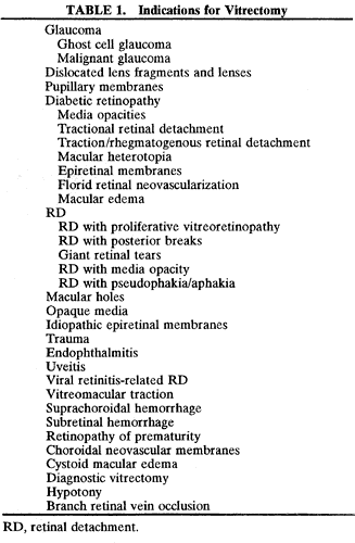
1. Kasner D: A new approach to the management of vitreous. Highlights Ophthalmol 11:304–329, 1968

2. Machemer R et al: Vitrectomy: A pars plana approach. Trans Am Acad Ophthalmol Otolaryngol 75:813–820, 1971

3. Machemer R, Parel JM, Buettner H: A new concept for vitreous surgery. I. Instrumentation. Am J Ophthalmol 74:1022–1033, 1972

4. Machemer R: Development of pars plana vitrectomy: My personal contribution. Klin Monatsbl Augenheilkd 207:147–161, 1995

5. Parel JM, Machemer R, Aumayr W: A new concept for vitreous surgery. IV. Improvements in instrumentation and illumination. Am J Ophthalmol 77:6–12, 1974

6. Machemer R: A new concept of vitreous surgery. II. Surgical technique and complications. Am J Ophthalmol 74:1022–1033, 1972

7. O'Malley C, Heintz R: Vitrectomy with an alternative instrument system. Ann Ophthalmol 7:585–594, 1975

8. Williams GA, Abrams GW, Mieler WF: Illuminated retinal picks for vitreous surgery. Arch Ophthalmol 107:1086, 1989

9. Kuhn F, Mester V, Berta A: The Tano Diamond Dusted Membrane Scraper: indications and contraindications. Acta Ophthalmol Scand 76:754–755, 1998 (letter)

10. Lewis JM et al: Diamond-dusted silicone cannula for epiretinal membrane separation during vitreous surgery. Am J Ophthalmol 124:553–554, 1997

11. Benson W, Diamond J, Tasman W: Intraocular irrigating solutions for pars plana vitrectomy. A prospective randomized, double-blind study. Arch Ophthalmol 99:1013, 1981

12. Edelhauser HF, Gonnering R, Van Horn D: Intraocular irrigating solutions: A comparative study of BSS plus and lactated Ringer's solution. Arch Ophthalmol 96:516, 1978

13. Matsuda M, Tano Y, Edelhauser HF: Comparison of intra-ocular irrigating solutions used for pars plana vitrectomy and prevention of endothelial cell loss. Jpn J Ophthalmol 28:230–238, 1984

14. DeJuan E, Hickingbotham D: Flexible iris retractor. Am J Ophthalmol 111:776–777, 1991

15. Chang S: Low viscosity liquid fluorochemicals in vitreous surgery. Am J Ophthalmol 103:38–43, 1987

16. Cibis P et al: The use of liquid silicone in retinal detachment surgery. Arch Ophthalmol 68:590–599, 1962

17. Capone A, Aaberg TM: Silicone oil in vitreoretinal surgery. Curr Opin Ophthalmol 6:33–37, 1995

18. Spitznas M, Reiner J: A stereoscopic diagonal inverter (SDI) for wide-angle vitreous surgery. Graefes Arch Clin Exp Ophthalmol 225:9–12, 1987

19. Spitznas M: A binocular indirect ophthalmomicroscope (BIOM) for non-contact wide-angle vitreous surgery. Graefes Arch Clin Exp Ophthalmol 225:13–15, 1987

20. Summanen P, Karhunen U, Laatikainen L: Characteristics and survival of diabetic patients undergoing vitreous surgery. Acta Ophthalmol (Copenh) 65:197–202, 1987

21. Blankenship G: Preoperative prognostic factors in diabetic pars plana vitrectomy. Ophthalmology 89:1246–1249, 1982

22. Ryan E, Gilbert HD: Results of surgical treatment of recent-onset full thickness idiopathic holes. Arch Ophthalmol 112:1545–1553, 1994

23. Rice TA et al: Prognostic factors in vitrectomy for epiretinal membranes of the macula. Ophthalmology 93:602–610, 1986

24. Scherfig E et al: Prognostic parameters in pars plana vitrectomy. Acta Ophthalmol (Copenh) 61:788–805, 1983

25. Landers MB et al: Temporary keratoprosthesis for use during pars plana vitrectomy. Am J Ophthalmol 91:615–619, 1981

26. Eckardt C: A new temporary keratoprosthesis for pars plana vitrectomy. Retina 7:34–37, 1987

27. Eckardt C: A new temporary keratoprosthesis. Klin Monatsbl Augenheilkd 191:243, 1987

28. de Bustros S et al: Vitrectomy for progressive proliferative diabetic retinopathy. Arch Ophthalmol 105:196–199, 1987

29. Thompson JT et al: Prognostic indicators of success and failure in vitrectomy for diabetic retinopathy. Ophthalmology 93:290–295, 1986

30. Thompson JT et al: Results and prognostic factors in vitrectomy for diabetic vitreous hemorrhage. Arch Ophthalmol 105:191–195, 1987 (published erratum appears in Arch Ophthalmol 105:496, 1987)

31. Benson WE et al: Extracapsular cataract extraction, posterior chamber lens insertion, and pars plana vitrectomy in one operation. Ophthalmology 97:918–921, 1990

32. Koenig SB et al: Combined phacoemulsification, pars plana vitrectomy, and posterior chamber intraocular lens insertion. Arch Ophthalmol 110:1101–1104, 1992

33. Kokame GT, Flynn Jr HW, Blankenship GW: Posterior chamber intraocular lens implantation during diabetic pars plana vitrectomy. Ophthalmology 96:603–610, 1989

34. Mamalis N et al: Phacoemulsification combined with pars plana vitrectomy. Ophthalmic Surg 22:194–198, 1991

35. McElvanney AM, Talbot EM: Posterior chamber lens implantation combined with pars plana vitrectomy. J Cataract Refract Surg 23:106–110, 1997

36. Menchini U et al: Combined vitrectomy, cataract extraction, and posterior chamber intraocular lens implantation in diabetic patients. Ophthalmic Surg 22:69–73, 1991

37. Smiddy WE: Intraocular lens insertion during vitrectomy. Ophthalmic Surg 23:808–810, 1992

38. Senn P, Schipper I, Perren B: Combined pars plana vitrectomy, phacoemulsification, and intraocular lens implantation in the capsular bag: A comparison to vitrectomy and subsequent cataract surgery as a two-step procedure. Ophthalmic Surg Lasers 26:420–428, 1995

39. Francese J et al: Moisture droplet formation on the posterior surface of intraocular lenses during fluid/air exchange. J Cataract Refract Surg 21:685–689, 1995

40. Hainsworth DP et al: Condensation on polymethylmethacrylate, acrylic polymer, and silicone intraocular lenses after fluid-air exchange in rabbits. Ophthalmology 103:1410–1418, 1996

41. Coleman D, Abramson D: Ocular ultrasonography. In: Duane T (ed): Clinical Ophthalmology. Hagerstown, MD: Harper & Row, 1990

42. Rubsamen PE et al: Diagnostic ultrasound and pars plana vitrectomy in penetrating ocular trauma. Ophthalmology 101:809–814, 1994

43. Hotta K et al: Ultrasound biomicroscopy for examination of the sclerotomy site in eyes with proliferative diabetic retinopathy after vitrectomy. Retina 20:52–58, 2000

44. Boker T, Spitznas M: Ultrasound biomicroscopy for examination of the sclerotomy site after pars plana vitrectomy. Am J Ophthalmol 118:813–815, 1994

45. Mandelcorn M, Blankenship G, Machemer R: Pars plana vitrectomy in severe diabetic retinopathy. Am J Ophthalmol 57:530–542, 1976

46. Sinclair S, Loebl M, Riva C: Blue field entopic test in patients with ocular trauma. Arch Ophthalmol 99:464–467, 1981

47. Algvere P, Persson HE, Wanger P: Preoperative electroretinograms and visual evoked cortical potentials for predicting outcome of vitrectomy in diabetics. Retina 5:179–183, 1985

48. Summanen P: Vitrectomy for diabetic eye disease. The prognostic value of pre-operative electroretinography and visual evoked cortical potentials. Ophthalmologica 199:60–71, 1989

49. Terasaki H et al: Focal macular electroretinograms before and after successful macular hole surgery. Am J Ophthalmol 125:204–213, 1998

50. Terasaki H et al: Focal macular electroretinogram before and after drainage of macular subretinal hemorrhage. Am J Ophthalmol 123:207–211, 1997

51. Gribomont AC, Guerit JM: Prognostic value of preoperative flash and pattern visual evoked potentials for diabetic retinopathy vitrectomy. Eur J Ophthalmol 4:218–222, 1994

52. Scherfig E et al: Flash visual evoked potential as a prognostic factor for vitreous operations in diabetic eyes. Ophthalmology 91:1475–1479, 1984

53. Bryden F, Pyott A, McGhee C: Real time ultrasound in the assessment of intraocular foreign bodies. Eye 4:727–731, 1990

54. Kuhn F, Morris R: Posterior segment intraocular foreign bodies: Management in the vitrectomy era. Ophthalmology 107:821–822, 2000 (editorial)

55. Chacko J et al: Detection and localization of steel intraocular foreign bodies using computed tomography. Ophthalmology 104:319–323, 1997

56. Puliafito CA et al: Optical Coherence Tomography of Ocular Diseases. Thorofare, NJ: Slack, 1996

57. Gobel W et al: Findings of optical coherence tomography (OPT) before and after macular hole surgery. Ophthalmologe 97:251–256, 2000

58. Munuera JM et al: Optical coherence tomography in successful surgery of vitreomacular traction syndrome. Arch Ophthalmol 116:1388–1389, 1998 (letter)

59. Hee M, Puliafito CA, Wong C: Optical coherence tomography of macular holes. Ophthalmology 102:748–756, 1995

60. Hee M et al: Quantitative assessment of macular edema with optical coherence tomography. Arch Ophthalmol 113:1019–1029, 1995

61. Mikajiri K et al: Analysis of vitrectomy for idiopathic macular hole by optical coherence tomography. Am J Ophthalmol 128:655–657, 1999

62. Otani T, Kishi S: Tomographic assessment of vitreous surgery for diabetic macular edema. Am J Ophthalmol 129:487–494, 2000

63. Tripathi R et al: Fundamentals and principles of ophthalmology. In: Basic and Clinical Science Course. San Francisco: American Academy of Ophthalmology, 1999:64–65

64. Sebag J: The Vitreous: Structure, Function, and Pathobiology. New York: Springer-Verlag, 1989

65. Gass JD: Stereoscopic Atlas of Macular Diseases: Diagnosis and Treatment. St Louis: Mosby, 1997:904–909

66. Calenda E et al: Peribulbar anesthesia and sub-Tenon injection for vitreoretinal surgery: 300 cases. Acta Ophthalmol Scand 78:196–199, 2000

67. Brucker AJ, Saran BR, Maguire AM: Perilimbal anesthesia for pars plana vitrectomy. Am J Ophthalmol 117:599–602, 1994 (published erratum appears in Am J Ophthalmol 118:273, 1994)

68. Nicholson AD et al: Peribulbar anesthesia for primary vitreoretinal surgery. Ophthalmic Surg 23:657–661, 1992

69. Li HK et al: Sub-tenon's injection for local anesthesia in posterior segment surgery. Ophthalmology 107:41–46, 2000; discussion, 46–47

70. Friedberg MA et al: An alternative technique of local anesthesia for vitreoretinal surgery. Arch Ophthalmol 109:1615–1616, 1991

71. Sharma T et al: Parabulbar anesthesia for primary vitreoretinal surgery. Ophthalmology 104:425–428, 1997

72. Yepez J, Cedeno de Yepez J, Arevalo JF: Topical anesthesia in posterior vitrectomy. Retina 20:41–45, 2000

73. Yepez J, Cedeno de Yepez J, Arevalo JF: Topical anesthesia for phacoemulsification, intraocular lens implantation, and posterior vitrectomy. J Cataract Refract Surg 25:1161–1164, 1999

74. Haimann MH, Abrams GW: Prevention of lens opacification during diabetic vitrectomy. Ophthalmology 91:116–121, 1984

75. Landers D, Stefansson E, Wolbarsht M: The optics of vitreous surgery. Am J Ophthalmol 91:611–614, 1981

76. Lesnoni G et al: The use of panoramic viewing system in relaxing retinotomy and retinectomy. Retina 17:186–190, 1997

77. Virata SR, Kylstra JA, Singh HT: Corneal epithelial defects following vitrectomy surgery using hand-held, sew-on, and noncontact viewing lenses. Retina 19:287–290, 1999

78. Charles S: Vitreous surgery. In Ryan SJ (ed): Retina. St Louis: CV Mosby, 1989:193

79. DeJuan E, Landers MI: New techniques for visualization of infusion cannula during vitreous surgery. Am J Ophthalmol 97:657, 1984

80. Welch JC, Spaeth GL, Benson W: Massive suprachoroidal hemorrhage. Ophthalmology 95:1202, 1988

81. Rice TA et al: The effect of lensectomy on the incidence of iris neovascularization and neovascular glaucoma after vitrectomy for diabetic retinopathy. Am J Ophthalmol 95:1–11, 1983

82. Ryan EH Jr, Gilbert HD: Lensectomy, vitrectomy indications, and techniques in cataract surgery. Curr Opin Ophthalmol 7:69–74, 1996

83. Smiddy W: Dislocated posterior chamber intraocular lens, a new technique of management. Arch Ophthalmol 107:1678–1680, 1989

84. Campo RV, Chung KD, Oyakawa RT: Pars plana vitrectomy in the management of dislocated posterior chamber lenses. Am J Ophthalmol 108:529–534, 1989

85. Wallace RT et al: The use of perfluorophenanthrene in the removal of intravitreal lens fragments. Am J Ophthalmol 116:196–200, 1993

86. Shapiro MJ et al: Management of the dislocated crystalline lens with a perfluorocarbon liquid. Am J Ophthalmol 112:401–405, 1991

87. Flynn HW, Brod RD: Use of a soft-tipped extrusion needle for epimacular membrane peeling. Arch Ophthalmol 108:20–21, 1990

88. Mester V, Kuhn F: Internal limiting membrane removal in the management of full-thickness macular holes. Am J Ophthalmol 129:769–777, 2000

89. Moisseiev J, Glaser BM: New and previously unidentified retinal breaks in eyes with recurrent retinal detachment with proliferative vitreoretinopathy. Arch Ophthalmol 107:1152, 1989

90. McLeod D, James C: Viscodelamination at the vitreoretinal junction in severe diabetic eye disease. Br J Ophthalmol 72:413–419, 1988

91. Ryan E: An illuminated cannula for viscodelamination. Arch Ophthalmol 107:1085, 1989

92. Abrams GW, Williams GA: “En bloc” excision of diabetic membranes. Am J Ophthalmol 103(3 Pt 1):302–308, 1987

93. Han DP, Murphy ML, Mieler WF: A modified en bloc excision technique during vitrectomy for diabetic traction retinal detachment. Results and complications. Ophthalmology 101:803–808, 1994

94. Meier P, Wiedemann P: Vitrectomy for traction macular detachment in diabetic retinopathy. Graefes Arch Clin Exp Ophthalmol 235:569–574, 1997

95. Williams DF et al: Results of vitrectomy for diabetic traction retinal detachments using the en bloc excision technique. Ophthalmology 96:752–758, 1989

96. Charles S: Vitrectomy for retinal detachment. Trans Ophthalmol Soc UK 100:542–549, 1980

97. Machemer R: Surgical approaches to subretinal bands. Am J Ophthalmol 90:81–85, 1980

98. Steinmetz RL, Masket S, Sidikaro Y: The successful removal of a subretinal cysticercus by pars plana vitrectomy. Retina 9:276–280, 1989

99. Natarajan S et al: Management of intraocular cysticercosis. Graefes Arch Clin Exp Ophthalmol 237:812–814, 1999

100. Wade EC et al: Subretinal hemorrhage management by pars plana vitrectomy and internal drainage. Arch Ophthalmol 108:973–978, 1990

101. Zhao P et al: Vitrectomy for macular hemorrhage associated with retinal arterial macroaneurysm. Ophthalmology 107:613–617, 2000

102. Lim JI et al: Submacular hemorrhage removal. Ophthalmology 102:1393–1399, 1995

103. Lewis H: Intraoperative fibrinolysis of submacular hemorrhage with tissue plasminogen activator and surgical drainage. Am J Ophthalmol 118:559–568, 1994

104. Thomas MA et al: Surgical management of subfoveal choroidal neovascularization. Ophthalmology 99:952–966, 1992

105. Berger A, Kaplan HJ: Clinical experience with the surgical removal of subfoveal neovascular membranes. Ophthalmology 99:969–976, 1992

106. Lambert HM et al: Surgical excision of subfoveal neovascular membranes in age-related macular degeneration. Am J Ophthalmol 114:241–242, 1992

107. Thomas MA, Kaplan HJ: Surgical removal of subfoveal neovascularization in the presumed ocular histoplasmosis syndrome. Am J Ophthalmol 111:1–7, 1991

108. Machemer R: Retinotomy. Am J Ophthalmol 92:768, 1981

109. Machemer R, McCuen B, DeJuan E: Relaxing retinotomies and retinectomies. Am J Ophthalmol 102:7–12, 1986

110. Aaberg TM: Management of anterior and posterior proliferative vitreoretinopathy. XLV Edward Jackson Memorial Lecture. Am J Ophthalmol 106:519, 1988

111. Lewis H, Aaberg TM, Abrams GW: Causes of failure after initial vitreoretinal surgery for severe proliferative vitreoretinopathy. Am J Ophthalmol 111:8, 1991

112. Lewis H, Aaberg TM: Causes of failure after repeat vitreoretinal surgery for recurrent proliferative vitreoretinopathy. Am J Ophthalmol 111:15, 1991

113. Blumenkranz M et al: Relaxing retinotomy with silicone oil or long-acting gas in eyes with severe proliferative vitreoretinopathy. Silicone Study Report 5. Am J Ophthalmol 116:557–564, 1993

114. Han DP et al: Vitrectomy for traumatic retinal incarceration. Arch Ophthalmol 106:640–645, 1988

115. Mathis A: Fluid-air exchange. J Fr Ophthalmol 15:367–371, 1992

116. Charles S: Ancillary techniques to trans pars plana vitrectomy. In McPherson A (ed): New and Controversial Aspects of Vitreoretinal Surgery. St Louis: CV Mosby, 1977:196–197

117. Charles S: A motorized gas injector for vitreous surgery. Arch Ophthalmol 99:1981, 1981

118. McCuen B et al: Automated fluid-gas exchange. Am J Ophthalmol 95:717, 1983

119. Hueneke RL, Aaberg TM: Instrumentation for continuous fluid-air exchange during vitreous surgery. Am J Ophthalmol 96:547–548, 1983

120. Doft BH: Intentional retinotomy for internal drainage of subretinal fluid. Arch Ophthalmol 104:807–808, 1986

121. Landers D: Sodium hyaluronate (Healon) as an aid to internal fluid-gas exchange. Am J Ophthalmol 94:557–558, 1982

122. Friberg TR: Clinical experience with a binocular indirect ophthalmoscope laser delivery system. Retina 7:28–31, 1987

123. Campochiaro PA et al: Cryotherapy enhances intravitreal dispersion of viable retinal pigment epithelial cells. Arch Ophthalmol 103:434–436, 1985

124. Bishara S, Buzney SM: Dispersion of retinal pigment epithelial cells from experimental retinal holes. Arch Ophthalmol 229:195–199, 1991

125. Singh A, Michels R, Glaser BM: Scleral indentation following cryotherapy and repeat cryotherapy enhance release of viable retinal pigment epithelial cells. Retina 6:176–178, 1986

126. Peyman GA, Grisolano J, Palacio M: Intraocular photocoagulation with argon-krypton laser. Arch Ophthalmol 98:2062–2064, 1980

127. Chang S: Perfluorocarbon liquids in vitreoretinal surgery. Int Ophthalmol Clin 32:153–163, 1992

128. Peyman GA, Schulman J, Sullivan B: Perfluorocarbon liquids in ophthalmology. Surv Ophthalmol 39:375–395, 1995

129. Loewenstein A et al: Perfluoroperhydrophenanthrene versus perfluoro-n-octane in vitreoretinal surgery. Ophthalmology 107:1078–1082, 2000

130. Scott IU et al: Outcomes and complications associated with perfluoro-n-octane and perfluorperhydrophenanthrene in complex retinal detachment repair. Ophthalmology 107:860–865, 2000

131. Rosengren B: Results of treatment of detachment of the retina with diathermy and injection of air into the vitreous. Arch Ophthalmol 16:573–579, 1938

132. Cekic O, Ohji M: Intraocular gas tamponades. Semin Ophthalmol 15:3–14, 2000

133. Chang S: Vitreous substitutes. In Guyer D et al (eds): Retina, Vitreous, Macula. Philadelphia: WB Saunders, 1999:1320–1327

134. DeJuan E, McCuen B, Tiedeman J: Intraocular tamponade and surface tension. Surv Ophthalmol 30:47–51, 1985

135. Thompson JT: Kinetics of intraocular gases. Arch Ophthalmol 107:687–691, 1989

136. Williams D et al: A two-stage technique for intraoperative fluid-gas exchange following pars plana vitrectomy. Arch Ophthalmol 108:1484, 1990

137. Cibis P et al: The use of silicone oil in retinal detachment surgery. Arch Ophthalmol 68:590–599, 1962

138. Yamamoto S, Takeuchi S: Silicone oil and fluorosilicone. Semin Ophthalmol 15:15–24, 2000

139. Regillo C et al: Repair of retinitis-related retinal detachments with silicone oil in patients with acquired immunodeficiency syndrome. Am J Ophthalmol 113:21–27, 1992

140. Lim JI et al: Improved visual results after surgical repair of cytomegalovirus-related retinal detachments. Ophthalmology 101:264–269, 1994

141. Azen SP et al: Silicone oil in the repair of complex retinal detachments: A prospective observational multicenter study. Ophthalmology 105:1587–1597, 1998

142. Barr CC et al: Postoperative intraocular pressure abnormalities in the Silicone Study. Silicone Study Report 4. Ophthalmology 100:1629–1635, 1993

143. Lewis H et al: Perisilicone proliferation after vitrectomy for proliferative vitreoretinopathy. Ophthalmology 95:583–591, 1988

144. Laroche L et al: Ocular findings following intravitreal silicone oil injection. Arch Ophthalmol 101:1422–1425, 1983

145. Ni C et al: Intravenous silicone injection. Arch Ophthalmol 101:1399–1401, 1983

146. Parmley V et al: Foreign-body giant cell reaction to liquid silicone. Am J Ophthalmol 101:680–683, 1986

147. Gonvers M, Hornung J, de Courten C: The effect of liquid silicone on the rabbit retina. Arch Ophthalmol 104:1057–1062, 1986

148. Chan CC, Okun E: The question of ocular tolerance to intravitreal liquid silicone: A long-term analysis. Ophthalmology 93:651–659, 1986

149. Federman J, Schubert HD: Complications associated with the use of silicone oil in 150 eyes after retina-vitreous surgery. Ophthalmology 95:870–876, 1988

150. Brourman N, Blumenkranz M, Cox MS: Silicone oil for the treatment of severe proliferative diabetic retinopathy. Ophthalmology 96:759–764, 1989

151. McCuen BW, Rinkoff JS: Silicone oil for progressive anterior ocular neovascularization after failed diabetic vitrectomy. Arch Ophthalmol 107:677–682, 1989 (published erratum appears in Arch Ophthalmol 107:1030, 1989)

152. Gonvers M: Temporary silicone oil tamponade in the management of retinal detachment with proliferative vitreoretinopathy. Am J Ophthalmol 100:239–245, 1985

153. Cox MS, Trese MT, Murphy P: Silicone oil for advanced proliferative vitreoretinopathy. Ophthalmology 93:646–650, 1986

154. McCuen B et al: Silicone oil in vitreoretinal surgery. Part 2: Results and complications. Retina 5:198–205, 1985

155. McCuen BW, Landers MB, Machemer R: The use of silicone oil following failed vitrectomy for retinal detachment with advanced proliferative vitreoretinopathy. Ophthalmology 92:1029–1034, 1985

156. Kirchhof B: The contribution of vitreoretinal surgery to the management of refractory glaucomas. Curr Opin Ophthalmol 10:117–120, 1999

157. Abu el-Asrar AM, al-Obeidan SA: Pars plana vitrectomy in the management of ghost cell glaucoma. Int Ophthalmol 19:121–124, 1995

158. Brucker AJ, Michels R, Green WR: Pars plana vitrectomy in the management of blood-induced glaucoma with vitreous hemorrhage. Ann Ophthalmol 10:1427–1437, 1978

159. Epstein DL: The malignant glaucoma syndromes. In Epstein DL, Allingham R, Schuman JS (eds): Chandler and Grant's Glaucoma. Baltimore: Williams & Wilkins, 1997:285–303

160. Harbour JW, Rubsamen PE, Palmberg P: Pars plana vitrectomy in the management of phakic and pseudophakic malignant glaucoma. Arch Ophthalmol 114:1073–1078, 1996

161. Lynch MG et al: Surgical vitrectomy for pseudophakic malignant glaucoma. Am J Ophthalmol 102:149–153, 1986

162. Gandham SB et al: Aqueous tube-shunt implantation and pars plana vitrectomy in eyes with refractory glaucoma. Am J Ophthalmol 116:189–195, 1993

163. Luttrull JK, Avery RL: Pars plana implant and vitrectomy for treatment of neovascular glaucoma. Retina 15:379–387, 1995

164. Scott IU et al: Combined pars plana vitrectomy and glaucoma drainage implant placement for refractory glaucoma. Am J Ophthalmol 129:334–341, 2000

165. Varma R et al: Pars plana Baerveldt tube insertion with vitrectomy in glaucomas associated with pseudophakia and aphakia. Am J Ophthalmol 119:401–407, 1995

166. Luttrull JK et al: Initial experience with pneumatically stented Baerveldt implant modified for pars plana insertion for complicated glaucoma. Ophthalmology 107:143–150, 2000

167. Wallace RT et al: The use of perfluorophenanthrene in the removal of intravitreal lens fragments. Am J Ophthalmol 116:196–200, 1993

168. Mello M et al: Surgical management and outcomes of dislocated intraocular lenses. Ophthalmology 107:62–67, 2000

169. Lambrou FH, Stewart M: Management of dislocated lens fragments during phacoemulsification. Ophthalmology 99:1260–1262, 1992

170. Kapusta MA, Chen CJ, Lam WC: Outcomes of dropped nucleus during phacoemulsification. Ophthalmology 103:1184–1187, 1996

171. Fastenberg DM et al: Management of dislocated nuclear fragments after phacoemulsification. Am J Ophthalmol 112:535–539, 1991

172. Blodi BA et al: Retained nuclei after cataract surgery. Ophthalmology 99:41–44, 1992

173. Margherio RR et al: Vitrectomy for retained lens fragments after phacoemulsification. Ophthalmology 104:1426–1432, 1997

174. Kim JE et al: Retained lens fragments after phacoemulsification. Ophthalmology 101:1827–1832, 1994

175. Gilliland GD, Hutton WL, Fuller DG: Retained intravitreal lens fragments after cataract surgery. Ophthalmology 99:1263–1267, 1992; discussion, 1268–1269

176. Borne MJ et al: Outcomes of vitrectomy for retained lens fragments. Ophthalmology 103:971–976, 1996

177. Vilar NF et al: Removal of retained lens fragments after phacoemulsification reverses secondary glaucoma and restores visual acuity. Ophthalmology 104:787–791, 1997; discussion, 791–792

178. Diabetic Retinopathy Vitrectomy Study Research Group: Two-year course of visual acuity in severe proliferative diabetic retinopathy with conventional management. Diabetic Retinopathy Vitrectomy Study (DRVS) Report 1. Ophthalmology 92:492–502, 1985

179. Diabetic Retinopathy Vitrectomy Study Research Group: Early vitrectomy for severe vitreous hemorrhage in diabetic retinopathy. Two-year results of a randomized trial. Diabetic Retinopathy Vitrectomy Study Report 2. Arch Ophthalmol 103:1644–1652, 1985

180. Diabetic Retinopathy Vitrectomy Study Research Group: Early vitrectomy for severe vitreous hemorrhage in diabetic retinopathy: Four-year results of a randomized trial. Diabetic Retinopathy Vitrectomy Study Report 5. Arch Ophthalmol 108:958–964, 1990 (published erratum appears in Arch Ophthalmol 108:1452, Oct 1990)

181. Benson WE et al: Complications of vitrectomy for non-clearing vitreous hemorrhage in diabetic patients. Ophthalmic Surg 19:862–864, 1988

182. Blankenship GW, Machemer R: Long-term diabetic vitrectomy results: Report of 10 year follow-up. Ophthalmology 92:503–506, 1985

183. Ramsay RC, Knobloch WH, Cantrill HL: Timing of vitrectomy for active proliferative diabetic retinopathy. Ophthalmology 93:283–289, 1986

184. O'Hanley GP, Canny CL: Diabetic dense premacular hemorrhage: A possible indication for prompt vitrectomy. Ophthalmology 92:507–511, 1985

185. Packer AJ: Vitrectomy for progressive macular traction associated with proliferative diabetic retinopathy. Arch Ophthalmol 105:1679–1682, 1987

186. Charles S, Flinn C: The natural history of diabetic extramacular traction retinal detachment. Arch Ophthalmol 99:66–68, 1981

187. Thompson JT et al: Results and prognostic factors in vitrectomy for diabetic traction-rhegmatogenous retinal detachment. Arch Ophthalmol 105:503–507, 1987

188. Rice TA, Michels R, Rice E: Vitrectomy for diabetic rhegmatogenous retinal detachment. Am J Ophthalmol 95:34–44, 1983

189. Yeo JH, Glaser BM, Michels R: Silicone oil in the treatment of complicated retinal detachments. Ophthalmology 94:94, 1987

190. Sato Y et al: Vitrectomy for diabetic macular heterotopia. Ophthalmology 101:63–67, 1994

191. Bresnick G, Haight B, de Venecia G: Retinal wrinkling and macular heterotopia in diabetic retinopathy. Arch Ophthalmol 97:1890, 1979

192. Diabetic Retinopathy Vitrectomy Study Research Group: Early vitrectomy for severe proliferative diabetic retinopathy in eyes with useful vision: Results of a randomized trial. Diabetic Retinopathy Vitrectomy Study Report 3. Ophthalmology 95:1307–1320, 1988

193. Diabetic Retinopathy Vitrectomy Study Research Group: Early vitrectomy for severe proliferative diabetic retinopathy in eyes with useful vision: Clinical application of results of a randomized trial, Diabetic Retinopathy Vitrectomy Study Report 4. Ophthalmology 95:1321–1334, 1988

194. Spencer R, McMeel J, Franks E: Visual outcome in moderate and severe proliferative diabetic retinopathy. Arch Ophthalmol 99:1551, 1981

195. Favard C et al: Full panretinal photocoagulation and early vitrectomy improve prognosis of florid diabetic retinopathy. Ophthalmology 103:561–574, 1996

196. Chaudhry NA et al: Early vitrectomy and endolaser photocoagulation in patients with type I diabetes with severe vitreous hemorrhage. Ophthalmology 102:1164–1169, 1995 (published erratum appears in Ophthalmology 102:1740, 1995)

197. Capone A, Panozzo G: Vitrectomy for refractory diabetic macular edema. Semin Ophthalmol 15:78–80, 2000

198. Lewis H et al: Vitrectomy for diabetic macular traction and edema associated with posterior hyaloidal traction. Ophthalmology 99:753–759, 1992

199. Harbour JW et al: Vitrectomy for diabetic macular edema associated with a thickened and taut posterior hyaloid membrane. Am J Ophthalmol 121:405–413, 1996

200. Ikeda T et al: Vitrectomy for cystoid macular oedema with attached posterior hyaloid membrane in patients with diabetes. Br J Ophthalmol 83:12–14, 1999

201. Tachi N, Ogino N: Vitrectomy for diffuse macular edema in cases of diabetic retinopathy. Am J Ophthalmol 122:258–260, 1996

202. Brazitikos PD: The expanding role of primary pars plana vitrectomy in the treatment of rhegmatogenous noncomplicated retinal detachment. Semin Ophthalmol 15:65–77, 2000

203. Escoffery RF et al: Vitrectomy without scleral buckling for primary rhegmatogenous retinal detachment. Am J Ophthalmol 99:275–281, 1985

204. Heimann H et al: Primary vitrectomy without scleral buckling for rhegmatogenous retinal detachment. Graefes Arch Clin Exp Ophthalmol 234:561–568, 1996

205. Wolfensberger TJ, Gonvers M: Long-term follow-up for retinal detachment due to macular hole in myopic eyes treated by temporary silicone oil tamponade and laser photocoagulation. Ophthalmology 106:1786–1791, 1999

206. Verstraeten T et al: Lens-sparing vitrectomy with perfluorocarbon liquid for the primary treatment of giant retinal tears. Ophthalmology 102:17–20, 1995

207. Campo RV et al: Pars plana vitrectomy without scleral buckle for pseudophakic retinal detachments. Ophthalmology 106:1811–1815, 1999; discussion, 1816

208. Brazitikos PD et al: Primary vitrectomy with perfluoro-n-octane use in the treatment of pseudophakic retinal detachment with undetected retinal breaks. Retina 19:103–109, 1999

209. Desai UR, Strassman IB: Combined pars plana vitrectomy and scleral buckling for pseudophakic and aphakic retinal detachments in which a break is not seen preoperatively. Ophthalmic Surg Lasers 28:718–722, 1997

210. Pastor JC: Proliferative vitreoretinopathy: An overview. Surv Ophthalmol 43:3–18, 1998

211. Committee RST: The classification of retinal detachment with proliferative vitreoretinopathy. Ophthalmology 90:121–125, 1983

212. Lean J et al: Classification of proliferative vitreoretinopathy used in the silicone study. Ophthalmology 96:765–771, 1989

213. Lean J et al: The prognostic utility of the Silicone Study Classification System. Silicone Study Report 9. Arch Ophthalmol 114:286–292, 1996

214. McCuen BW et al: Vitrectomy with silicone oil or perfluoropropane gas in eyes with severe proliferative vitreoretinopathy. Silicone Study Report 3. Retina 13:279–284, 1993

215. Group SS: Vitrectomy with silicone oil or perfluoropropane gas in eyes with severe proliferative vitreoretinopathy: Results of a randomized clinical trial. Silicone Study Report 2. Arch Ophthalmol 110:780–792, 1992

216. Group SS: Vitrectomy with silicone oil or sulfur hexafluoride gas in eyes with severe proliferative vitreoretinopathy: Results of a randomized clinical trial. Silicone Study Report 1. Arch Ophthalmol 110:770–779, 1992

217. Abrams GW et al: Vitrectomy with silicone oil or long-acting gas in eyes with severe proliferative vitreoretinopathy: Results of additional and long-term follow-up. Silicone Study Report 11. Arch Ophthalmol 115:335–344, 1997

218. Lucke KH, Foerster MH, Laqua H: Long-term results of vitrectomy and silicone oil in 500 cases of complicated retinal detachments. Am J Ophthalmol 104:624–633, 1987

219. Sell CH et al: Long-term results of successful vitrectomy with silicone oil for advanced proliferative vitreoretinopathy. Am J Ophthalmol 103:24–28, 1987

220. Campochiaro PA: A small piece of the PVR puzzle is put into place. Silicone Study. Arch Ophthalmol 115:407–408, 1997

221. Abrams GW et al: The incidence of corneal abnormalities in the Silicone Study. Silicone Study Report 7. Arch Ophthalmol 113:764–769, 1995

222. Kampik A, Gandorfer A: Silicone oil removal strategies. Semin Ophthalmol 15:88–91, 2000

223. Jonas JB, Budde WM, Knorr HL: Timing of retinal redetachment after removal of intraocular silicone oil tamponade. Am J Ophthalmol 128:628–631, 1999

224. Coll G et al: Perfluorocarbon liquid in the management of retinal detachment with proliferative vitreoretinopathy. Ophthalmology 102:630–639, 1995

225. Wiedemann P et al: Adjunctive daunorubicin in the treatment of proliferative vitreoretinopathy: Results of a multicenter clinical trial. Daunomycin Study Group. Am J Ophthalmol 126:550–559, 1998

226. Yang CS et al: An intravitreal sustained release triamincinolone and 5-fluorouracil codrug in treatment of experimental proliferative vitreoretinopathy. Arch Ophthalmol 116:69–77, 1998

227. Chang S et al: Giant retinal tears: Surgical techniques and results using perfluorocarbon liquids. Arch Ophthalmol 107:761–766, 1989

228. Kreiger AE, Lewis H: Management of giant retinal tears without scleral buckling: Use of radical dissection of the vitreous base and perfluoro-octane and intraocular tamponade. Ophthalmology 99:491–497, 1992

229. Ie D et al: The use of perfluoro-octane in the management of giant retinal tears without proliferative vitreoretinopathy. Retina 14:323–328, 1994

230. Glaser BM et al: Perfluoro-octane in the treatment of giant retinal tears with proliferative vitreoretinopathy. Ophthalmology 98:1613–1621, 1991

231. Chang S et al: Perfluorocarbon liquids in the management of traumatic retinal detachments. Ophthalmology 96:785–791, 1989

232. Gass JD: Reappraisal of biomicroscopic classification of stages of development of a macular hole. Am J Ophthalmol 119:752–759, 1995

233. de Bustros S: Vitrectomy for prevention of macular holes: Results of a randomized multicenter clinical trial. Vitrectomy for Prevention of Macular Hole Study Group. Ophthalmology 101:1055–1059, 1994; discussion, 1060

234. Kim JW et al: Prospective randomized trial of vitrectomy or observation for stage 2 macular holes. Vitrectomy for Macular Hole Study Group. Am J Ophthalmol 121:605–614, 1996

235. Kelly NE, Wendel RT: Vitreous surgery for idiopathic macular holes. Results of a pilot study. Arch Ophthalmol 109:654–659, 1991

236. Thompson JT et al: Comparison of recombinant transforming growth factor-beta-2 and placebo as an adjunctive agent for macular hole surgery. Ophthalmology 105:700–706, 1998

237. Freeman WR et al: Vitrectomy for the treatment of full-thickness stage 3 or 4 macular holes: Results of a multicentered randomized clinical trial. The Vitrectomy for Treatment of Macular Hole Study Group. Arch Ophthalmol 115:11–21, 1997 (published erratum appears in Arch Ophthalmol 115:636, May 1997)

238. Smiddy WE, Pimentel S, Williams GA: Macular hole surgery without using adjunctive additives. Ophthalmic Surg Lasers 28:713–717, 1997

239. Wendel RT et al: Vitreous surgery for macular holes. Ophthalmology 100:1671–1676, 1993

240. Park DW et al: Macular hole surgery with internal-limiting membrane peeling and intravenous air. Ophthalmology 106:1392–1398, 1999

241. Spraul C, Grossniklaus H: Vitreous hemorrhage. Surv Ophthalmol 42:3–39, 1997

242. Smiddy WE et al: Vitrectomy for nondiabetic vitreous hemorrhage: Retinal and choroidal vascular disorders. Retina 8:88–95, 1988

243. Isernhagen RD et al: Vitrectomy for nondiabetic vitreous hemorrhage: Not associated with vascular disease. Retina 8:81–87, 1988

244. Pournaras CJ et al: Macular epiretinal membranes. Semin Ophthalmol 15:100–107, 2000

245. Poliner LS et al: Surgical management of premacular fibroplasia. Arch Ophthalmol 106:761–764, 1988

246. de Bustros S et al: Vitrectomy for idiopathic epiretinal membranes causing macular pucker. Br J Ophthalmol 72:692–695, 1988

247. Cherfan GM et al: Nuclear sclerotic cataract after vitrectomy for idiopathic epiretinal membranes causing macular pucker. Am J Ophthalmol 111:434–438, 1991

248. de Bustros S et al: Nuclear sclerosis after vitrectomy for idiopathic epiretinal membranes. Am J Ophthalmol 105:160–164, 1988

249. Williams D et al: Results and prognostic factors in penetrating ocular injuries with retained intraocular foreign bodies. Ophthalmology 95:911–916, 1988

250. DeJuan E, Sternberg P Jr, Michels R: Penetrating ocular injuries: Types of injuries and visual results. Ophthalmology 90:1318–1322, 1983

251. Greven CM et al: Intraocular foreign bodies. Management, prognostic factors and visual outcomes. Ophthalmology 107:608–612, 2000

252. Brown G, Tasman W, Benson W: BB-gun injuries to the eye. Ophthalmic Surg 16:505–508, 1985

253. Cleary PE, Ryan SJ: Vitrectomy in penetrating eye injury. Results of a controlled trial of vitrectomy in an experimental posterior penetrating injury in the rhesus monkey. Arch Ophthalmol 106:287–292, 1981

254. Abrams GW, Topping TM, Machemer R: Vitrectomy for injury: The effect on intraocular proliferation following perforation of the posterior segment of the rabbit eye. Arch Ophthalmol 97:743–750, 1979

255. Endophthalmitis Vitrectomy Study Group: Results of the Endophthalmitis Vitrectomy Study: A randomized trial of immediate vitrectomy and of intravenous antibiotics for the treatment of postoperative bacterial endophthalmitis. Arch Ophthalmol 113:1479–1496, 1995

256. Clark WL et al: Treatment strategies and visual acuity outcomes in chronic postoperative Propionibacterium acnes endophthalmitis. Ophthalmology 106:1665–1670, 1999

257. Aldave A et al: Treatment strategies for postoperative Propionibacterium acnes endophthalmitis. Ophthalmology 106:2395–2401, 1999

258. Smiddy WE: Treatment outcomes of endogenous fungal endophthalmitis. Curr Opin Ophthalmol 9:66–70, 1998

259. Essman TF et al: Treatment outcomes in a 10-year study of endogenous fungal endophthalmitis. Ophthalmic Surg Lasers 28:185–194, 1997

260. Mieler W et al: Retained intraocular foreign bodies and endophthalmitis. Ophthalmology 97:1532–1538, 1990

261. Diamond J, Kaplan HJ: Uveitis: Effect of vitrectomycombined with lensectomy. Ophthalmology 86:1320–1327,1979

262. Foster CS, Fong L, Singh G: Cataract surgery and intraocular lens implantation in patients with uveitis. Ophthalmology 96:281–288, 1989

263. Michelson J, Friedlaender M, Nozik R: Lens implantsurgery in pars planitis. Ophthalmology 97:1023–1026,1990

264. Belmont J et al: Vitrectomy in ocular toxocariasis. Arch Ophthalmol 100:1912–1915, 1982

265. Jabs D et al: Retinal detachments in patients with cytomegalovirus retinitis. Arch Ophthalmol 109:794–799, 1991

266. Clarkson J et al: Retinal detachment following the acute retinal necrosis syndrome. Ophthalmology 91:1665–1668, 1984

267. Fisher J et al: The acute retinal necrosis syndrome. Ophthalmology 89:1309–1316, 1982

268. Freeman HM et al: Surgical repair of rhegmatogenousretinal detachment in immunosuppressed patients withcytomegalovirus retinitis. Ophthalmology 99:466–474,1992

269. Blumenkranz M et al: Visual results and complications after retinal after retinal reattachment in the ARN syndrome: The influence of operative technique. Retina 9:170–174, 1989

270. Fung WE: Vitrectomy for chronic aphakic cystoid macular edema: Results of a national, collaborative, prospective, randomized investigation. Ophthalmology 92:1102–1111, 1985

271. Harbour JW et al: Pars plana vitrectomy for chronic pseudophakic cystoid macular edema. Am J Ophthalmol 120:302–307, 1995

272. Pendergast SD et al: Vitrectomy for chronic pseudophakic cystoid macular edema. Am J Ophthalmol 128:317–323, 1999

273. Hikichi T, Yoshida A, Trempe C: Course of vitreomacular traction syndrome. Am J Ophthalmol 119:55–61, 1995

274. Smiddy WE et al: Vitrectomy for macular traction caused by incomplete vitreous separation. Arch Ophthalmol 106:624–628, 1988

275. Lakhanpal V et al: A new modified vitreoretinal approach in the management of massive suprachoroidal hemorrhage. Ophthalmology 96:793–800, 1989

276. Hochman MA et al: Subretinal hemorrhage. Surv Ophthalmol 42:197–213, 1997

277. Koshibu A: Ultrastructural studies on absorption of experimentally produced subretinal hemorrhage. III. Absorption of erythrocyte breakdown products and retinal hemosiderosis at the last stage. Nippon Ganka Gakkai Zasshi 83:386–400, 1979

278. Toth CA et al: Fibrins directs early retinal damage after experimental subretinal hemorrhage. Arch Ophthalmol 109:723–729, 1991

279. Glatt H, Machemer R: Experimental subretinal hemorrhage in rabbits. Am J Ophthalmol 94:762–773, 1982

280. Berrocal M, Lewis M, Flynn HW: Variations in the clinical course of submacular hemorrhage. Am J Ophthalmol 122:486–493, 1996

281. Shiraga F et al: Surgical treatment of submacular hemorrhage associated with idiopathic polypoidal choroidal vasculopathy. Ophthalmology 128:147–154, 1999

282. The International Committee for the Classification of the Late Stages of Retinopathy of Prematurity: An international classification of retinopathy of prematurity. II. The classification of retinal detachment. Arch Ophthalmol 105:906–912, 1987

283. Maguire AM, Trese MT: Lens-sparing vitreoretinal surgery in infants. Arch Ophthalmol 110:284–286, 1992

284. Trese MT: Management of early ROP detachments: Lens sparing vitrectomy. Presented at Retina 2000: Management of Posterior Segment Disease. Dallas, 2000

285. Hirose T et al: Vision in stage 5 retinopathy of prematurity after retinal reattachment by open-sky vitrectomy. Arch Ophthalmol 111:345–349, 1993

286. Fuchino Y et al: Long-term follow up of visual acuity in eyes with stage 5 retinopathy of prematurity after closed vitrectomy. Am J Ophthalmol 120:308–316, 1995

287. Trese MT, Droste P: Long-term postoperative results of a consecutive series of stages 4 and 5 retinopathy of prematurity. Ophthalmology105:992–997, 1998

288. Macular Photocoagulation Study Group: Laser photocoagulation of subfoveal neovascular lesions in age-related macular degeneration: Updated findings from two clinical trials. Arch Ophthalmol 111:1200–1209, 1993

289. Photodynamic Therapy Study Group: Photodynamictherapy of subfoveal choroidal neovascularization in age-related macular degeneration: One year results of two randomized clinical trials. TAP Report 1. Treatment of age-related macular degeneration with photodynamic therapy (TAP) study group. Arch Ophthalmol 117:1329–1345,1999

290. Merrill P et al: Surgical removal of subfoveal choroidal neovascularization in age-related macular degeneration. Ophthalmology 106:782–789, 1999

291. Lewis H, Vanderburg Medendorp S: Tissue plasminogen activator-assisted surgical excision of subfoveal choroidal neovascularization in age-related macular degeneration: A randomized, double-masked trial. Ophthalmology 104:1847–1852, 1997

292. Thomas MA et al: Visual results after surgical removal of subfoveal choroidal neovascular membranes. Ophthalmology 101:1384–1396, 1994

293. Holekamp NM et al: Surgical removal of subfoveal choroidal neovascularization in presumed ocular histoplasmosis: Stability of early visual results. Ophthalmology 104:22–26, 1997

294. Lambert HM: The management of subfoveal choroidal neovascular membranes and hemorrhage. Semin Ophthalmol 15:92–99, 2000

295. Vander JF: Macular translocation. Curr Opin Ophthalmol 11:159–165, 2000

296. DeJuan E, Fujii G: Further experience with limited macular translocation. Presented at Retina 2000: Management of posterior segment disease. Dallas, 2000

297. Lewis H et al: Macular translocation for subfoveal choroidal neovascularization in age-related macular degeneration. A prospective study. Am J Ophthalmol 128:135–146, 1999

298. Haller JA et al: Limited macular translocation for neovascular maculopathy. Semin Ophthalmol 15:81–88, 2000

299. Eckardt C: Further experience with 360-degree retinal rotation. Presented at Retina 2000: Management of posterior segment disease. Dallas, 2000

300. Schwartz E: Corneal sensitivity in diabetics. Arch Ophthalmol 91:174–178, 1974

301. Ohashi Y et al: Aldose reductase inhibitor (CT-112) eyedrops for diabetic corneal epithelialopathy. Am J Ophthalmol 105:233–238, 1988

302. Foulks G et al: Factors related to corneal epithelial complications after closed vitrectomy in diabetics. Arch Ophthalmol 97:1076–1078, 1979

303. Edelhauser HF et al: The effect of phenylephrine on the cornea. Arch Ophthalmol 97:937–947, 1979

304. Carter JB et al: Iatrogenic retinal breaks complicating pars plana vitrectomy. Ophthalmology 97:848–854, 1990

305. Sjaarda RN et al: Distribution of iatrogenic retinal breaksin macular hole surgery. Ophthalmology 102:1387–1392,1995

306. Stern W: Complications of vitrectomy. Semin Ophthalmol 15:205–212, 2000

307. Oyakawa RT et al: Complications of vitreous surgery for diabetic retinopathy. I. Intraoperative complications. Ophthalmology 90:517–521, 1983

308. Piper JG et al: Perioperative choroidal hemorrhage at pars plana vitrectomy: A case-control study. Ophthalmology 100:699–704, 1993

309. Tabandeh H et al: Suprachoroidal hemorrhage during pars plana vitrectomy: Risk factors and outcomes. Ophthalmology 106:236–242, 1999

310. McDonald HR, Harris MJ: Operating microscope-induced retinal phototoxicity during pars plana vitrectomy. Arch Ophthalmol 106:521–523, 1988

311. Michels M et al: Macular phototoxicity caused by fiberoptic endoillumination during pars plana vitrectomy. Am J Ophthalmol 114:287–296, 1992

312. Postel E et al: Long-term follow-up of iatrogenic phototoxicity. Arch Ophthalmol 116:753–757, 1998

313. Tolentino FI, Freeman HM, Tolentino FL: Closed vitrectomy in the management of diabetic traction retinal detachment. Ophthalmology 87:1078–1089, 1980

314. Chung H et al: Reevaluation of corneal complicationsafter closed vitrectomy. Arch Ophthalmol 106:916–919,1988

315. Schachat AP et al: Complications of vitreous surgery from diabetic retinopathy. II. Post-operative complications. Ophthalmology 90:522–530, 1983

316. Helbig H et al: Rubeosis iridis after vitrectomy for diabetic retinopathy. Graefes Arch Clin Exp Ophthalmol 236:730–733, 1998

317. Blankenship G: Pre-operative iris rubeosis and diabetic vitrectomy results. Ophthalmology 87:176–182, 1980

317a. Stefanssen E, Landers MB III, Wolbarsyat ML: Increased retinal oxygen supply following panretinal photocoagulation and vitrectomy and lensectomy. Trans Am Ophthalmol Soc 79:307, 1981

318. Blankenship G: The lens influence on diabetic vitrectomy results. Arch Ophthalmol 98:2196–2198, 1980

319. Lewis H, Abrams GW, Williams GA: Anterior hyaloidal fibrovascular proliferation after diabetic vitrectomy. Am J Ophthalmol 104:607–613, 1987

320. Campbell D et al: Glaucoma occurring after closed vitrectomy. Am J Ophthalmol 83:63–69, 1977

321. Leaver P, Grey R, Garner A: Silicone oil injection in the treatment of massive preretinal traction. II. Late complications in 93 eyes. Br J Ophthalmol 63:361–367, 1979

322. Ando F: Intraocular hypertension resulting from pupillary block by silicone oil. Am J Ophthalmol 99:87–88, 1985

323. Chang S, Sparrow JR: Vitreous substitutes. In Guyer D et al (eds): Retina, Vitreous, Macula. Philadelphia: WB Saunders, 1999:1328

324. Desai UR et al: Intraocular pressure elevation after simple pars plana vitrectomy. Ophthalmology 104:781–786,1997

325. Park SS et al: Posterior segment complications after vitrectomy for macular hole. Ophthalmology 102:775–781,1995

326. Thompson JT et al: Progression of nuclear sclerosis and long-term visual results of vitrectomy with transforming growth factor beta-2 for macular holes. Am J Ophthalmol 119:48–54, 1995

327. Krzystolik M, D'Amico DJ: Complications of intraocular tamponade: Silicone oil versus intraocular gas. Int Ophthalmol Clin 40:197, 2000

328. Novak MA et al: Vitreous hemorrhage after vitrectomyfor diabetic retinopathy. Ophthalmology 91:1485–1489,1984

329. Blankenship GW: Management of vitreous cavity hemorrhage following pars plana vitrectomy for diabetic retinopathy. Ophthalmology 93:39–44, 1986

330. Margherio RR, Cox MS, Trese MT: Removal of epimacular membranes. Ophthalmology 92:1075–1083, 1985

331. Michels RG: Vitrectomy for macular pucker. Ophthalmology 91:1384–1388, 1984

332. McDonald HR, Verre W, Aaberg TM: Surgical management of idiopathic epiretinal membranes. Ophthalmology 93:978–983, 1986

333. Banker AS et al: Vision-threatening complications of surgery for full-thickness macular holes. Vitrectomy for Macular Hole Study Group. Ophthalmology 104:1442–1452, 1997; discussion, 1452–1453

334. Buettner H, Machemer R: Histopathologic findings in human eyes after pars plana vitrectomy and lensectomy. Arch Ophthalmol 95:2029–2033, 1977

335. Cohen SM et al: Endophthalmitis after pars plana vitrectomy. The Postvitrectomy Endophthalmitis Study Group. Ophthalmology 102:705–712, 1995

336. Gass JD: Sympathetic ophthalmia following vitrectomy. Am J Ophthalmol 93:552–558, 1982

337. Croxalto J et al: Sympathetic ophthalmia after pars plana vitrectomy-lensectomy for endogenous bacterial endophthalmitis. Am J Ophthalmol 91:342–346, 1981

338. Gasch A et al: Postoperative sympathetic ophthalmia. Int Ophthalmol Clin 69–84, 2000

Back to Top