Chapter 63 Photodynamic Therapy BRANDON G. BUSBEE and CAROLINE R. BAUMAL Table Of Contents |
While the technique of photodynamic therapy (PDT) was recently
approved for treatment of specific types of exudative maculopathy, it
is not a new concept. In the 1970s, the investigational use of PDT to
treat systemic malignancies in animal models was reported.1 The mechanism of PDT tumor destruction occurs as a result of vascular
occlusion, stasis, and hemorrhage, as well as direct tumor cell death.2 The vaso-occlusive effect of PDT led to its investigation in animal
models as a potential therapy for various types of ocular neovascularization
and tumors.3, 4 The ability to visualize ocular structures and to apply directed light
to a targeted area using a contact lens makes the eye an ideal site for
the modality of PDT. The U.S. Food and Drug Administration (FDA) approved PDT in the year 2000 for treatment of subfoveal choroidal neovascularization (CNV) with specific features in age-related macular degeneration (AMD), and it was subsequently approved for myopia and ocular histoplasmosis syndrome. Its efficacy to treat other vascular disorders and tumors of the eye is currently being investigated. |
MECHANISM OF PHOTODYNAMIC THERAPY |
PDT involves two steps: the administration of a photosensitizing agent
that localizes in the target tissue, followed by the application of low-intensity
light to the target tissue to produce localized tissue
damage. The photosensitizer may be administered by various methods, including
topical, local, or systemically. For example, photosensitizers
are often given topically for dermatologic disorders.5, 6 In ophthalmology, these agents are typically administered intravenously
to reach the retinal or choroidal circulation, although liposomal release
and other experimental methods of administration have been considered. The
photosensitizing agent must be delivered and preferentially
concentrated in the target tissue compared to normal adjacent tissue
in order to have a specific treatment effect. It has been demonstrated
that certain photosensitizers have an affinity for neovascular tissue
and tumor cells.7 After the drug has reached the desired tissue, low-energy light, which
is supplied by a laser source and is within the absorption band
of the photosensitizer, is applied directly to the area of interest. The
interaction of the photosensitizer with light of a specific wavelength
induces a photochemical reaction that liberates by-products that are toxic to the target tissue. Neither
the drug nor the light alone (at the irradiance in the range
used to PDT) appears to have any significant biological effect.8 It is the combination of these factors, utilizing light of a specific
wavelength to activate the photosensitizer drug, which creates the photochemical
reaction. Sequestering of the photosensitizing agent in the
target tissue and direct application of low-energy, focused light
by a contact lens to activate the photosensitizer within the lesion
are the main features that limit induced damage to the surrounding tissue. The mechanism of PDT action resides in the photosensitized oxidation of biological matter. When the photosensitizing agent is activated by the delivered light energy, the agent is transformed from its ground state to a higher energy singlet activated molecule. For the more efficient photosensitizers, the higher energy state decays into a lower energy, more stable molecule referred to as a triplet sensitizer. This triplet sensitizer can undergo either a Type I or Type II reaction. The Type I reaction generates superoxide anions, whereas Type II reactions result in production of singlet oxygen.9 These by-products are believed to be responsible for the localized tissue damage produced by PDT. In vivo studies after PDT show the immediate onset of vascular stasis and hemorrhage.2 These observations, along with work done on PDT for systemic tumors, provided a framework for initial studies of PDT for ocular tumors and neovascularization.8,10–14 |
CLINICAL EFFECT OF PHOTODYNAMIC THERAPY | |||||||
PDT with commercially available verteporfin (Visudyne) of CNV
in AMD produces choroidal hypofluorescence on fluorescein angiography
within a few days following treatment.15–17 This corresponds to the vascular occlusion noted in animal studies.3,4 The fluorescein hypofluorescence is a result of transient, choroidal hypoperfusion, which
has been demonstrated by indocyanine green (ICG) angiography.16,18 This hypoperfusion of the underlying choroidal vasculature is most pronounced
within the first week after PDT. The vascular occlusive effect
of PDT is not permanent, and reperfusion of the choroidal vessels typically
recurs between 4 and 12 weeks after PDT.16 By 12 weeks after PDT, approximately two thirds of CNV lesions had recurred
in one series.15 In another series at 12 weeks following verteporfin-PDT, the recurrence
of hyperfluorescence in CNV approached the fluorescein hyperfluorescence
seen before any PDT therapy in 90% of eyes.19 This recurrent leakage of CNV after PDT occurred at both the very low
and high-energy light treatment doses in the early phase I and
II verteporfin studies. This led to the concept of repeating PDT treatments
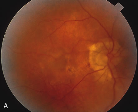
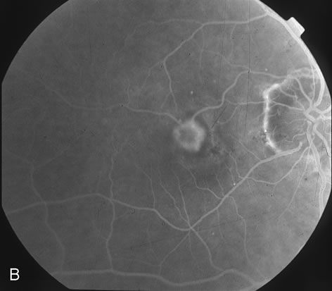
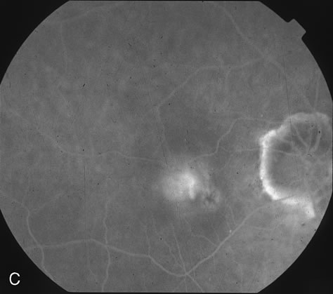
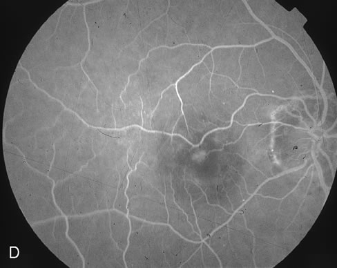
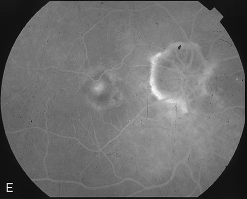
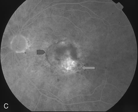
to reinduce or maintain occlusion within CNV. The ideal light energy dose, or fluence, to safely treat CNV was modeled on primate and early-phase studies that varied the parameters of laser irradiance, fluence, and treatment duration.8 Preliminary clinical studies demonstrated untoward effects of retinal vascular closure when a light energy dose of 150 J/cm2 was administered.20 This led to the recommendation in the phase III prospective clinical trials for PDT treatment at 50 J/cm2. Repetition of PDT treatment every 12 weeks was allowed if recurrent CNV leakage was present on fluorescein angiography and examination.21–23 The irradiance of 600 mW/cm2 used for PDT is markedly less than that used for thermal laser photocoagulation (which is typically in the range of 100 to 1000 W/cm2).8 Figures 1D and E demonstrate the typical CNV hypoperfusion on fluorescein angiogram noted at 4 weeks status-post PDT with verteporfin for predominantly classic CNV in AMD (Fig. 1A to C).
|
PHOTOSENSITIZING AGENTS | ||||||||||||||||||
Each photosensitizer has a specific absorption and side effect profile
that affects its clinical utility for systemic and ocular PDT. The initially
studied agents, known as the first-generation photosensitizers, were
typically activated by shorter wavelength light and have
more prolonged cutaneous photosensitivity than the newer, second-generation
photosensitizers. The most common photosensitizers and their
peak treatment wavelengths are outlined in Table 1 Most of the early experience using animal models in ophthalmology has
been with the first-generation porphyrins, hematoporphyrin derivative (HPD) and
dihematoporphyrin ether (DHE). These
photosensitizers have also been studied for a variety of systemic
diseases.24–25 DHE is commercially available as Photofrin (QLT Phototherapeutics, Vancouver, Canada). Another first-generation photosensitizer, 5-aminolevulinic acid (5-ALA), has been
used topically to treat certain cervical and cutaneous neoplasias.5,6,25
Table 1. Photosensitizing Agents Used for Photodynamic Therapy
Newer second-generation photosensitizing agents have been developed due to the side effect of prolonged cutaneous sun sensitivity with first-generation agents that required avoidance of sunlight for 4 to 6 weeks after PDT. These agents have been investigated in recent human studies for ophthalmic disease. The second-generation photosensitizers have a longer wavelength of absorption, which permits for deeper tissue penetration of the treatment effect. Additional advantages of the second-generation agents include more rapid clearance from the body, shorter duration of cutaneous light sensitivity, and greater affinity of these agents to concentrate in the proliferating cells of neovascular tissue.26,27 The agent benzoporphyrin derivative monoacid (BPD-MA or verteporfin) was approved by the FDA in April 2000 for the treatment of specific types of subfoveal CNV. This photosensitizing agent is commercially available under the trade name Visudyne (Novartis Ophthalmics, Duluth, GA). Verteporfin is administered intravenously and has a peak absorption of 692 nm.28 This agent appears to have preferential uptake in low-density lipoproteins (LDL) in human plasma. The photosensitizer-LDL complex is then delivered to neovascular tissue, which has an increased proportion of LDL-receptors compared with normal vascular endothelium. This effect may localize verteporfin in proliferating endothelial cells of ocular neovascular tissue. Experimental studies using fluorescein and ICG angiography have shown verteporfin reaches the choroidal and then retinal vasculature within 5 and 15 seconds, respectively. Clearance from the retina is completed by 30 minutes following injection of verteporfin. Experimental CNV collects verteporfin approximately 10 seconds after injection and the photosensitizer may remain in the complex for up to 1 hour.2 Verteporfin is primarily eliminated from the blood through hepatic metabolism within the first 24 hours.14 This rapid clearance of more than 50% of the injected photosensitizing agent significantly decreases the amount of time that patients are at risk of cutaneous light toxicity. This has been confirmed with favorable systemic side effect profiles using verteporfin in the following prospective studies: The Treatment of Age-related Macular Degeneration with Photodynamic Therapy (TAP) Study and The Verteporfin in Photodynamic Therapy (VIP) Study.23,30–32 Other second-generation photosensitizing agents have been investigated for treatment of exudative AMD. Tin ethyl etiopurpurin (SnET2) has been studied in animal models and human CNV as well as for ciliary body ablation.33,34 Lutetium texaphyrin (Lu-Tex) is activated by 732 nm wavelength of light. This agent has a unique property of having an emission band at 750 nm, which permits it to secondarily function as an angiographic agent to image the posterior segment circulation.35 |
PHOTODYNAMIC THERAPY IN AGE-RELATED MACULAR DEGENERATION |
AMD is the major cause of visual loss in individuals older than 65 years
in developed countries. It is subdivided into exudative (wet) and
nonexudative (dry) forms. Although the exudative form
accounts for approximately 10% of AMD cases, it produces more
severe visual loss when compared to the dry form. Most of the new investigational
therapies are directed at limiting damage from exudative
AMD. The hallmark of exudative AMD is CNV, although pigment epithelial
detachment and fibrovascular tissue are also features. CNV is classified
by the pattern of leakage on fluorescein angiography into classic, occult
or a combination of classic and occult lesions. In brief, “classic
CNV” is defined as bright, early hyperfluorescence with
late leakage in the TAP study, whereas “occult CNV” includes
irregular or stippled hyperfluorescence on fluorescein angiography.23 This classification of CNV is important when considering the potential
treatment modalities as detailed below. Before the approval of PDT for subfoveal CNV in AMD, thermal laser photocoagulation was the only treatment with efficacy demonstrated by the multicenter Macular Photocoagulation Studies (MPS). However, laser treatment of subfoveal CNV produces immediate and permanent central visual loss due to full-thickness retinal destruction induced by thermal photocoagulation. The MPS subfoveal studies demonstrated that at 2 years after randomization, the mean vision was 20/320 in subfoveal laser treated eyes compared with 20/500 in observed cases.36 Owing to the minimal potential for visual improvement for tasks involving discriminative acuity, thermal laser was recommended primarily for patients who were agreeable to the immediate visual decline demonstrated in the MPS trial, and had a purely classic subfoveal lesion no greater than 3.5 MPS disc areas.36–39 Only a limited number of eyes with subfoveal CNV lesions fulfilled these criteria. Thermal laser photocoagulation is also unsatisfactory as long-term therapy for CNV in any location owing to the high recurrence rate of CNV leakage and the full-thickness neurosensory retinal destruction that it produces. Moreover, it has only been recommended to treat classic CNV. The approval of PDT in the year 2000 offers a new therapy of CNV that is potentially less destructive. It has increased the proportion of AMD patients who are candidates for a form of treatment for subfoveal CNV, and it does not appear to produce thermal neurosensory retina damage. |
THE TAP STUDY | ||||||||||||||||||||||||||||||||||||||
The Treatment of Age-Related Macular Degeneration with Photodynamic
Therapy (TAP) Study Group was a multicenter, double-masked, placebo-controlled, randomized clinical trial. The study
enrolled over 600 AMD patients with subfoveal CNV from December 1996 to
October 1997, who were prospectively followed for 2 years. Patients
were randomized in a treatment ratio of 2:1 to either PDT with verteporfin (verteporfin-PDT group) or placebo. Inclusion
criteria included CNV secondary to AMD that extended under the geometric
center of the foveal avascular zone, lesion diameter not in excess
of 5400 μm, modified Early Treatment Diabetic Retinopathy Study (ETDRS)–equivalent visual acuity between 20/40 to 20/200, and
fluorescein angiographic evidence that the CNV contained some
classic component (Table 2). From the safety and dosing data obtained from phase I and II studies, the
cumulative treatment dose of 50 Joules/cm2 was determined optimal to reduce CNV leakage on fluorescein angiography
without producing retinal or choroidal tissue damage.21,22 To achieve this dose of 50 Joules/cm2, a diode laser delivered light at a wavelength of 689 nm for 83 seconds
through a slit-lamp and contact lens system. This was performed 15 minutes
after commencing the 10-minute intravenous infusion
of verteporfin. Patients were followed at 3-month intervals, and
could be retreated with either verteporfin or placebo (maintaining
their initial randomization) if there was recurrent leakage
present on fluorescein angiography.23 Patients were retreated with verteporfin an average of 3.4 times in the
first year, and 2.2 times during the second year. Thus, verteporfin-PDT
eyes received an average of over five treatments over the 2-year
study period.23,40 These retreatments did not appear clinically to increase the surrounding
chorioretinal atrophy in relation to the CNV area when compared to
placebo-treated eyes.40
Table 2. Characteristics of the TAP and VIP Studies
The 1-year results demonstrated an overall benefit in the primary outcome measure, which was to determine whether verteporfin-PDT could prevent moderate visual loss (defined as loss of at least 15-letters or three lines on the ETDRS chart). Sixty-one percent (61%) of the verteporfin-PDT treated eyes lost less than 15 letters on the ETDRS chart compared with 46% in the placebo group (p < 0.001). Subgroup analysis revealed the greatest benefit in eyes that had “predominantly classic” CNV at baseline, which was defined as CNV composed of equal to or greater than 50% classic component. More than two thirds of patients (67%) treated with verteporfin-PDT with “predominantly classic” CNV had less than 15-letter loss after 1 year compared with 39% in the placebo group (p < 0.001).23 The PDT treatment benefit for “predominantly classic” CNV remained at the 2-year evaluation (59% versus 31% for verteporfin-PDT vs. placebo respectively, p < 0.001).40 Severe visual loss, defined as 30 letters or six lines of visual loss, was significantly lower in the verteporfin-PDT group over the 2-year period (15% versus 36% for verteporfin PDT versus placebo respectively, p < 0.001). The findings of improved visual outcome after verteporfin-PDT were found to be independent of prior thermal laser photocoagulation, phakic status, and prior use of micronutrients.41 From these findings, PDT with verteporfin for “predominantly classic” CNV in the setting of AMD was recommended to prevent visual loss when compared with observation. For the subgroups of eyes with “minimally classic” CNV (defined as having less than 50% classic CNV) in the TAP trial, verteporfin-PDT did not appear to protect patients from losing 15 letters from baseline at both the 1 and 2-year mark. Although contrast sensitivity and fluorescein angiographic outcomes were significantly better at 1 and 2 years in the verteporfin-PDT group compared with placebo for “minimally classic” and “purely occult” CNV, over half of the patients experienced moderate visual loss (15-letter loss) in both the first and second year following treatment in both groups. Thus, the initial TAP report did not show visual acuity efficacy for verteporfin-PDT of “minimally classic” CNV in AMD.23,40 Further subgroup analysis of the “predominantly classic” CNV group demonstrated better visual outcomes in “predominantly classic” lesions without an occult component that were treated with verteporfin-PDT. It was also noted that these same lesions (classic CNV without an occult component) were smaller in size and had a lower baseline visual acuity compared with predominantly classic lesions with an occult component.41 These potential confounders, smaller lesion size and worse presenting visual acuity, were then applied to the “minimally classic” lesions that initially did not appear to benefit from verteporfin therapy compared to the “predominantly classic” CNV lesions.23,40,41 It was found in the TAP study that “minimally classic” lesions, in fact, did have larger lesions and better presenting visual acuity than the “predominantly classic' lesions. From this finding, it has been theorized that certain “minimally classic” CNV lesions with smaller size and worse presenting visual acuity may benefit from PDT. A prospective, randomized trial named Verteporfin in Minimally Classic CNV Trial (VIM) is currently in progress to evaluate verteporfin in eyes with “minimally classic” CNV with smaller size and worse presenting visual acuity. The most recent reports from the TAP trial have reported on the continued follow-up and use of verteporfin-PDT beyond 24 months of the original TAP-study design. The results of visual acuity and contrast sensitivity, along with any additional side effects with additional verteporfin-PDT administration, have been documented. The use of verteporfin-PDT appears to have minimal safety issues when used repeatedly for recurrent CNV in AMD. The patients from the original study with predominantly classic CNV received an additional 1.3 treatments between 24 and 36 months. Visual acuity appeared to stabilize with only a few patients experiencing moderate visual loss in this time period.42 Contrast sensitivity using Pelli-Robson charts was also analyzed for the subgroups within the first 24 months of the TAP study. In addition to favorable contrast sensitivity scores paralleling visual outcomes for “predominantly classic” CNV, patients with “minimally classic” CNV also lost fewer letters on the Pelli-Robson chart compared to placebo over the 24 months. The potential correlation between improved contrast sensitivity and reduced visual disability suggest that verteporfin-PDT may have a positive impact on patient's daily visual function.43 |
THE VIP STUDY |
The Verteporfin Therapy of Subfoveal Choroidal Neovascularization in Age-Related
Macular Degeneration (VIP) Study was a multi-center
study designed to evaluate further issues that arose from
the TAP Study.30 The TAP study demonstrated visual benefits using verteporfin-PDT
with “predominantly classic” CNV, but it did not address
either “purely occult” CNV or CNV with good presenting visual
acuity (20/40 or better).23 The VIP study was designed to evaluate the effect of verteporfin-PDT
for two groups of eyes: “ purely occult” CNV lesions
with no classic component that displayed recent disease progression and “presumed
early-onset predominantly classic” CNV
with relatively good visual acuity.44 An additional arm of the VIP study enrolled patients with CNV associated
with pathologic myopia (these patients are addressed in the subsection
on PDT for other ocular disorders). A total of 339 patients
with AMD were enrolled from March to September 1998, and were followed
prospectively for two years. Inclusion and treatment criteria with
regards to size, underlying diagnosis of AMD, randomization in a 2:1 ratio
to either verteporfin-PDT or placebo, treatment parameters, follow-up
examinations, and repeat treatments were identical
to the TAP study. For “purely occult” lesions with no classic
component, Snellen equivalent visual acuity had to be 20/100 or
better and show some evidence of recent hemorrhage or disease progression. For
presumed early-onset of “predominantly classic” CNV, inclusion
criteria also required visual acuity of 20/40 or
better at enrollment. Table 2 outlines the characteristics of the TAP and VIP studies. The 12-month data after randomization did not demonstrate a treatment effect for verteporfin-PDT compared with placebo for the primary outcome measure, which was moderate visual loss (defined as loss of at least 15 letters or three ETDRS lines). Approximately one half of both verteporfin-PDT and placebo groups had moderate visual loss at the 12 month examination (p=0.52). Similar rates of visual loss at the 12-month examination were noted with subgroup analysis of the eyes classified as “purely occult” CNV (with no classic CNV), which comprised over 70% of the entire cohort of patients. The visual outcomes between the verteporfin-PDT and the placebo groups did diverge, however, by the 2-year mark. Twenty-four–month results demonstrated visual benefit from verteporfin-PDT for both the entire group and the subgroup of “purely occult” CNV. Thus, a smaller proportion of the verteporfin-PDT eyes experienced moderate visual loss compared with the placebo eyes when the results were analyzed for the entire group (54% versus 67%, p = 0.023). The subgroup of “purely occult” eyes with no classic CNV show similar treatment benefit with 55% of verteporfin-PDT eyes compared with 68% of placebo patients experiencing loss of at least 15 letters (p = 0.032). In both of these groups, however, the greatest decline in visual acuity was observed in the first 12 months following the initial verteporfin-PDT treatment. This was the same time frame (12-month results) in which no difference between verteporfin-PDT and placebo groups experiencing moderate visual loss was demonstrated. One hypothesis to account for the visual benefit of verteporfin-PDT demonstrated by 24 months, but not in the initial 12-month data, is that the PDT treatment parameters used in the VIP trial may not be optimal for occult CNV. Ongoing studies address these findings and are re-evaluating the parameters used for PDT. Secondary measures of visual loss in the VIP study also demonstrated a treatment benefit. Severe vision loss of at least 30 letters or six lines was noted in 30% of the verteporfin-PDT group and 47% of the placebo group at 24 months (p = 0.001). The “purely occult” with no classic CNV also demonstrated a decreased risk of severe vision loss at 2 years with only 29% of the verteporfin group versus 47% of the placebo group (p = 0.004). Other secondary outcomes such as angiographic appearance and mean change in visual acuity appeared to favor the verteporfin-PDT treatment at both 12- and 24-month examinations. Although the VIP Study demonstrated a visual benefit for “purely occult” with no classic CNV that demonstrated signs of progression in the setting of AMD, the outcomes were not as compelling as the results of “predominantly classic” CNV in the TAP trial. However, further analysis of the lesion composition in this group did highlight a subgroup that appeared to benefit substantially from verteporfin-PDT. Patients who either had smaller initial lesions (no greater than 4 MPS disc areas36) or a presenting visual acuity of worse than 20/50-1 at baseline demonstrated a greater visual benefit from verteporfin-PDT. If the lesion was less than four disc diameters, 49% verteporfin group versus 75% of the placebo group experienced moderate visual loss (p < 0.001). If the initial visual acuity was worse than 20/50-1, only 29% of the verteporfin group versus 48% of the placebo group experienced moderate visual loss (p < 0.001). Lesions that were found to be larger than four disc diameters with visual acuity better than 20/50 did not appear to benefit from verteporfin-PDT. The VIP study also observed an adverse event after verteporfin-PDT that was not prevalent in the TAP trials. Ten patients (4%) in the verteporfin-PDT group experienced loss of at least 20 letters of vision within seven days of verteporfin-PDT. This contrasted to less than 1% of the patients losing vision in this manner after verteporfin-PDT in the TAP study.40 In the VIP study, eight of these 10 patients had pretreatment “purely occult” with no classic CNV. Nine of 10 patients experienced this visual loss after their initial treatment with verteporfin. Subretinal pigment epithelium hemorrhage or choroidal hypoperfusion were noted only in a minority of these patients, and an identifiable cause for the vision loss could not be found in more than half of these patients. Although vision marginally improved over time in half of these patients, most patients did not recover a substantial amount of their pretreatment visual acuity. The VIP Study recommended including this risk of visual loss when requesting consent from patients for verteporfin-PDT, especially if the lesion is composed of occult CNV with no classic component.30 |
PHOTODYNAMIC THERAPY FOR SELECTED EXTRAFOVEAL AND JUXTAFOVEAL CHOROIDAL NEOVASCULARIZATION |
The MPS study demonstrated the beneficial effect of thermal laser photocoagulation to treat selected classic, extrafoveal and juxtafoveal CNV in AMD.45,46 There is no current prospective trial that supports the use of PDT in these particular lesions. However, PDT may be considered as an alternative treatment in certain eyes with juxtafoveal CNV. Some of the proposed benefits of PDT over thermal photocoagulation include monocular patients who hope to avoid the permanent, paracentral scotoma induced by thermal laser, if the CNV does not meet the strict MPS criteria of purely classic CNV, if there is a hazy media precluding precise laser, or if the CNV is so close to the fovea that laser photocoagulation cannot be safely performed.47 In the majority of purely classic juxtafoveal and extrafoveal CNV lesions, the standard of care remains thermal photocoagulation as recommended by the MPS studies. |
QUALITY-OF-LIFE AND COST-EFFECTIVE MEASURES WITH PHOTODYNAMIC THERAPY |
Recent reports have demonstrated the improvement in quality of -life following PDT for predominantly classic CNV in AMD.48 Using utility values for visual acuity outcomes and quality-adjusted life-years, treatment with PDT imparted an increase in quality of life (p < 0.001). Cost-utility analysis demonstrated PDT as a moderately cost-effective procedure using the model of a patient presenting with good vision and subfoveal CNV in their better-seeing eye. This model was based on the value of money and 2001 healthcare costs in the United States. The cost-effectiveness appears to diminish in this theoretical model when PDT treatment is considered for a patient with decreased visual acuity from subfoveal CNV, and relatively good vision in the fellow eye.49 Multiple treatments with verteporfin-PDT also decreased its cost-effectiveness in this model. |
ONGOING TRIALS |
Additional studies are currently under way to build on the results from
the studies described earlier. The Verteporfin in Minimally Classic CNV (VIM) will
evaluate whether verteporfin-PDT has a
beneficial effect for “minimally classic” CNV less than 4 MPS
disc areas presenting with acuity 20/50-1 or worse. The trial follows from results in the VIP study that certain
subgroups of occult CNV had a significant treatment benefit from PDT. Phase
II of the VIM study will evaluate a reduced light dose for PDT
and will assess the CNV size at 3 months status-post PDT, which
is the endpoint of the study. If findings from the VIM appear to be safe
and effective, a larger, prospective trial assessing PDT with a reduced
light dose, and evaluating the parameters that may induce lesions
to convert from “minimally classic” to “predominantly
classic” (thus qualifying the CNV for treatment by TAP
parameters) may be considered. The Verteporfin Early Retreatment (VER) trial will evaluate whether earlier retreatment at 6 weeks, compared to 3 months after verteporfin-PDT for “predominantly classic” CNV, will be more effective to reduce the risk of moderate visual loss. This study is a product of analysis of the predominantly classic CNV group in the TAP study. In this group, 75% of the visual loss, or approximately 2 lines of acuity, occurred within the first 6 months of treatment before stabilizing.23,40 A hypothetical disadvantage of early retreatment may be potential damage to the retinal or choroidal vasculature, or to neurosensory retina cells. The effect of early retreatment on the retinal structure and visual acuity will be evaluated in this study. The Verteporfin with Altered (Delayed) Light in Occult (VALIO) CNV trial is a phase II trial to evaluate light application 30 minutes after verteporfin infusion instead of the standard 15-minute interval used in the TAP and VIP studies. The unimpressive 12-month VIP treatment results for “purely occult CNV” spurred interest to evaluate whether there were more effective treatment parameters for occult CNV. It is hypothesized that prolonging the time for light application after verteporfin infusion may be more efficacious way to treat occult CNV. The delayed application may result in less verteporfin in the choroidal circulation and more pooled verteporfin in the CNV, which could possibly produce a more selective treatment effect for occult CNV. PDT has also been suggested as an adjuvant therapy in AMD, and it has been evaluated in small studies as a combination treatment with intravitreal corticosteroid, ICG feeder vessel laser therapy, and laser photocoagulation. A recent report noted an increase of identifiable feeder vessels to neovascular complexes associated with AMD from 22% pre-PDT to 84% of persistent neovascular lesions post-PDT.50 This retrospective study of 156 eyes may lead to evaluation of PDT combined with the experimental therapy of directed laser photocoagulation of feeder vessels identified with high-speed ICG angiography. Future prospective studies may identify combination therapies with PDT that ultimately permanently close CNV complexes or prolong the time interval between PDT retreatments. |
TECHNIQUE FOR ADMINISTRATION OF PHOTODYNAMIC THERAPY |
FDA-approved clinical use of ocular PDT in the United States is
limited to the administration of verteporfin (Visudyne). The
stepwise technique of verteporfin-PDT that is based on the protocols
established by the verteporfin (TAP, VIP) studies are
outlined.23,30,31,40,41 Verteporfin-PDT is presently approved in the United States for
treatment of subfoveal CNV in AMD with predominantly classic features
on fluorescein angiography (described in the TAP study), myopic
CNV (described in the VIP study), and for CNV secondary
to ocular histoplasmosis syndrome (OHS). Fluorescein film angiography
or digital imaging is used to define the type and composition
of subfoveal CNV. The percent composition of classic or occult CNV can
be determined using the area function on digital imaging. Alternatively, standard
size reticules are available to determine the percent composition
of CNV types for film angiography. The guidelines from the
TAP and VIP studies are used to determine the indications for PDT based
on the composition of CNV23,30,40
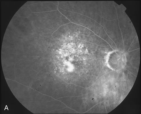
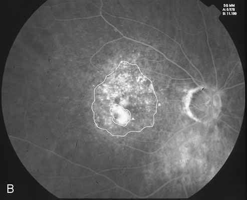
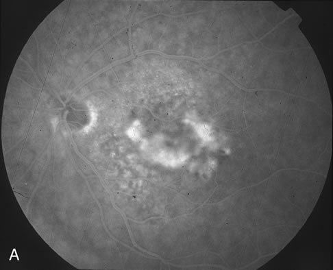
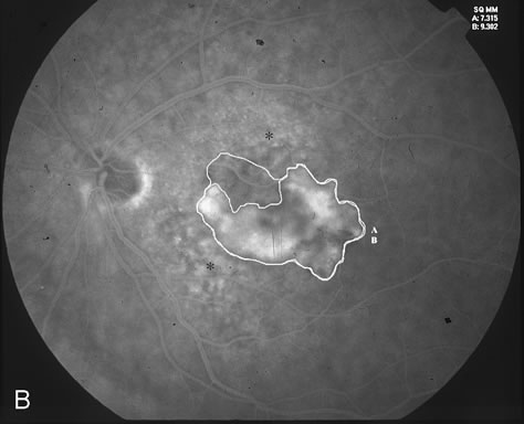
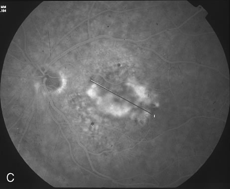
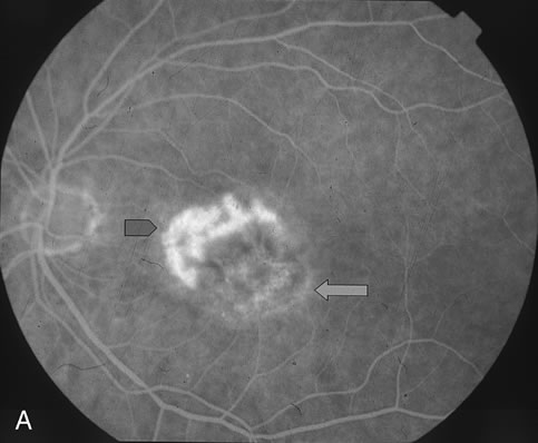
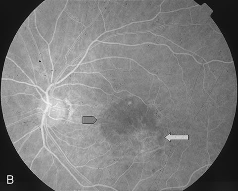
The classic and occult components of CNV are outlined in Figure 2. The percent of the CNV that is classic can then be determined as a proportion of the total CNV. In Figure 2A and B, the area of classic CNV measures 978 square microns and the total lesion measures 11,180 square microns. The classic component is thus 9% of the entire CNV and this lesion is classified as “minimally classic” CNV (defined as less than 50% classic CNV).23,40 The fluorescein angiogram demonstrates another mixed classic and occult CNV lesion in Figure 3. Using digital area measurements, the lesion in Figures 3A and B is composed of 79% classic CNV and fulfils the criteria for predominantly classic, subfoveal CNV that may benefit from verteporfin-PDT based on TAP findings.23,40 The TAP studies also demonstrated a visual benefit in eyes that had prior nonfoveal, thermal laser photocoagulation and subsequent recurrence of CNV below the fovea.23,40 In Figure 4A, subfoveal CNV recurrence in an eye with prior thermal photocoagulation is demonstrated. One month following verteporfin-PDT treatment, Figures 4B (early phase) and 4C (late phase) demonstrate characteristic hypofluorescence of the treated subfoveal CNV on fluorescein angiography. After qualifying for verteporfin-PDT based on clinical and fluorescein angiographic findings, informed consent is carefully obtained. Contraindications to PDT include porphyria, severe liver disease, pregnancy, or a known hypersensitivity to a photosensitizing agent. The greatest linear dimension (GLD) is determined with caliper measurement across the longest digital fluorescein angiographic diameter of leakage. The size of the area to be treated using the nonthermal 689 nm light should have a 500 μm border around the CNV, so a total of 1,000 μm should be added to the greatest diameter (or GLD) of CNV. The treatment area should extend no closer than 200 μm to the optic nerve, as recommended by the TAP studies.23,40 In Figure 3C, the area of CNV to be treated would be 4,184 plus 1,000 μm, or 5,184 microns. Verteporfin, which is stored as a dry powder, is reconstituted with sterile water and diluted with 5% dextrose solution immediately before use. This solution is infused intravenously over 10 minutes, preferably through a large cubital or cephalic vein. Constant monitoring during the entire infusion is imperative to avoid photosensitizer extravasation, which can have serious dermatological consequences. The calculated dose of verteporfin is 6.0 mg/m2 body surface area; thus, the accurate weight of the patient should be obtained before treatment. The 689-nm diode laser light treatment begins 15 minutes after commencing the verteporfin infusion. The laser treatment is delivered at the slit lamp through a diode-coated contact lens. Laser treatment is applied for 83 seconds, which produces a total energy dose of 50 J/cm2. In the scenario of a patient presenting with subfoveal CNV lesions in both eyes qualifying for verteporfin-PDT, guidelines have been outlined for concurrent bilateral treatment (see package insert provided by Visudyne, Novartis Ophthalmics). This technique consists of injecting an identical dose of verteporfin (6.0 mg/m2) as when treating only one eye. The CNV that appears more aggressive should be treated first at the standard 15-minute post-injection time. Immediately following the conclusion of light application in the first eye, laser settings should be adjusted to introduce the correct treatment-size parameter for the CNV in the fellow eye. Treatment for the fellow eye should commence no later than 20 minutes after commencement of verteporfin infusion. This technique is not recommended for the initial PDT treatment in patients with bilateral, eligible CNV lesions. In this case, unilateral verteporfin-PDT should be administered with follow-up in one week for treatment of the fellow eye. This can identify any safety issues that may arise from PDT. Once the fellow eye is treated, following treatments at the three-month interval can be administered concurrently if evidence of leakage is present in both eyes. Post-treatment precautions include avoidance of direct sunlight or bright indoor lights. A wide-brimmed hat, gloves, long pants, long sleeves, and protection of the eyes and face from direct sunlight are required for travel immediately after PDT. Complete avoidance of direct sunlight is strictly prescribed for 5 days following verteporfin-PDT. This is to prevent potentially serious skin burns and photosensitivity that may occur from residual, circulating photosensitizer after PDT. |
COMPLICATIONS ASSOCIATED WITH PHOTODYNAMIC THERAPY |
The TAP trial reported relatively rare adverse events associated with verteporfin-PDT. Injection site events, including extravasation, hypersensitivity, and
pain, were the most frequent side effects in the
verteporfin group, occurring in 15.9% of treated patients over
the 2-year period. Fourteen patients (3.5%) in
the verteporfin-PDT group experienced photosensitivity reactions
following treatment. This was usually mild to moderate sunburns, and
typically occurred within 24 hours following verteporfin-PDT. Other
treatment-related adverse events included visual disturbances (22% verteporfin group versus 15% placebo group), infusion-related back pain (2.5%), and
allergic reactions (3.5%). Only seven patients (1.7%) withdrew from verteporfin-PDT group due
to an adverse event.23,40 The VIP study noted less injection site events and similar instances of
back pain, but notably there were more patients (approximately 4%) who
developed severe, rapid reduction in vision in the
verteporfin-PDT treated eye. Infusion-related back pain has been reported to occur on successive infusions of verteporfin. A recent, nonrandomized study has estimated an incidence of back pain in up to 10% of infusions.51 There was no difference between patients who received oral hydration 30 minutes before injection compared with patients who did not receive this before injection. The only significant risk factor for infusion-related pain was a prior episode of pain with verteporfin injection. Also, infusion-related pain is not limited to back pain, and the episode subsides with cessation of infusion. Other sites of infusion-related pain have included leg, groin, chest, buttock, arm, and shoulder.51 It has been theorized that the mechanism of pain is related to neutrophil margination from vascular walls during the infusion.52 One small series recommended that intravenous diphenhydramine hydrochloride given before injection may alleviate this adverse event.53 |
PHOTODYNAMIC THERAPY FOR OTHER OCULAR DISORDERS |
PDT has been evaluated to treat other causes of subfoveal CNV. Pathologic
myopia was included in a separate arm of the VIP study to prospectively
examine verteporfin-PDT treatment of myopic subfoveal CNV. The
treatment parameters were similar to the regimens described in earlier
sections, with the exception that myopic CNV comprised of any proportion
of classic or occult CNV was included in the study. The main
outcome measure of loss of eight letters or 1.5 lines of visual acuity
was less rigorous than in the TAP study. One-year results revealed
a treatment benefit with fewer myopic CNV eyes that were treated
with verteporfin-PDT losing 1.5 lines of acuity compared to placebo
eyes (72% versus 44% respectively, p < 0.01). Secondary
measures of vision loss at 3 months, moderate visual
loss (>15 letters), and improvement in visual acuity (>five
letters) also favored the verteporfin-PDT myopic
group. The verteporfin-PDT myopic eyes were more likely to attain 20/40 or
better vision at 12-month examination than the placebo
group (26% versus 15%).31 The 2-year myopic VIP results did not appear to be as favorable
as findings in the first 12 months because the two study groups (verteporfin-PDT
and placebo) had similar proportions of visual
acuity loss at the end of 24 months (36% vs 51% respectively, p = .11).32 However, the verteporfin-PDT group did have a trend toward improvement
in visual acuity when compared with the placebo group at 24 months. For
this reason, verteporfin-PDT is currently approved for
subfoveal CNV secondary to myopia in the United States. It may especially
be useful in attempting to preserve vision in a patient with preexisting
ocular compromise from myopia. Subfoveal CNV due to ocular histoplasmosis syndrome was evaluated in an uncontrolled, prospective case series.54 The 1-year data on 26 patients treated with verteporfin-PDT demonstrated an overall visual acuity improvement, and more than one half of the patients treated experienced some visual improvement. A few patients had significant visual decrease with visual acuity less than 20/200 in one patient at the 12-month examination. Similar to the high myopia study, there were no cases of severe vision loss attributed to PDT as had been described in both the TAP and VIP studies. Smaller case series have now been reported using PDT for miscellaneous conditions with subfoveal CNV. The results have been variable, and no generalized recommendations have been made at this time. Some of these conditions associated with CNV treated with verteporfin-PDT include idiopathic CNV, angioid streaks, choroidal osteoma, fundus flavimaculatus, multifocal choroiditis, idiopathic polypoidal choroidal vasculopathy, and type 2 parafoveal telangectasias. 55-61 PDT was typically performed owing to a lack of any other proven treatment option. Short-term visual stabilization with no adverse events directly attributable to verteporfin-PDT was noted in some of these cases. The use of PDT for AMD in eyes with co-existing, severe nonproliferative or proliferative diabetic retinopathy has not been properly assessed, because these patients were excluded from the initial verteporfin-PDT studies. One potential concern is whether PDT could exacerbate diabetic retinovascular abnormalities.62 There are currently no long-term safety data for these diabetic eyes. Small series have also reported use PDT for non-CNV lesions. Posterior pole abnormalities including peripapillary capillary hemangiomas, circumscribed choroidal hemangiomas, and central serous chorioretinopathy have been treated with verteporfin-PDT.63–65 PDT has been investigated as an alternative to mitomycin-C to control fibrosis in eyes status-post trabeculectomy.66 Unresponsive iris melanomas have been treated with PDT using hematoporpyrin derivative as the photosensitizing agent.67 Further evaluation of PDT as a potential new treatment for various other vascular and oncologic lesions in the eye may be warranted. Moreover, combination of PDT with other treatment modalities may play a role in future therapies. A major rationale for combining PDT with another modality is to prolong or even make the vaso-occlusive effect of PDT more permanent. |
SUMMARY |
PDT is a new option to treat specific types of subfoveal CNV in AMD. Specifically, it has been approved by the FDA in the United States for treatment of subfoveal CNV in AMD that is predominantly classic, recurrent CNV after laser photocoagulation in AMD, secondary to myopia and ocular histoplasmosis (as of December 2002). The approved indications in certain European countries include progressive occult CNV secondary to AMD. Further study of the natural history of CNV in AMD and the parameters used for PDT may refine the optimal time to perform this therapy. It is notable that up to 52% of occult CNV develop classic CNV components over time, which may alter the lesions' responsiveness to PDT therapy.68 Owing to the vaso-occlusive effect of PDT, it may emerge as a potential treatment for other ocular conditions alone or combination with other CNV therapies. Further investigation will refine the indications and parameters for PDT and evaluate new uses for existing and upcoming photosensitizers. |