Chapter 69
Giant Retinal Tears
H. MacKENZIE FREEMAN
Main Menu   Table Of Contents

Search

MANAGEMENT OF A GIANT RETINAL TEAR WITH AN INVERTED RETINAL FLAP
MANAGEMENT OF FELLOW EYES OF NONTRAUMATIC GIANT RETINAL TEARS
REFERENCES

MANAGEMENT OF A GIANT RETINAL TEAR WITH AN INVERTED RETINAL FLAP
Giant retinal tears have been defined as retinal tears that extend 90° or more around the circumference of the fundus.1 Nontraumatic giant retinal tears account for approximately 77% of giant retinal tears.2 In a study involving 321 nontraumatic giant retinal tears, 84% of cases involved males; the average age was 32 years. Marfan's syndrome was present in 2.8% of patients and retinopathy of prematurity in 1.5%.3 In a recent update of that series, Wagner-Stickler syndrome was present in 9.7% of patients.4

It is important to distinguish between a giant retinal tear and a retinal dialysis greater than 90° because their surgical management is different. Most giant retinal tears occur at the posterior border of the vitreous base or along the posterior margin of lattice degeneration. A dialysis occurs along the ora serrata as a separation of the neurosensory retina from the nonpigmented ciliary epithelium. Dialyses are three times more common than giant tears and constitute approximately 10% of rhegmatogenous retinal detachments.5,6 Superior nasal dialyses may result from a traumatic avulsion of the vitreous base. Spontaneous dialyses are often bilateral and usually involve the inferotemporal quadrant. Dialyses produce a retinal detachment that develops slowly and is often characterized by concentric pigmented demarcation lines. Vitreous detachment is rare in eyes with dialyses.

The vitreous is almost always attached to the posterior edge of the dialysis, therefore inversion of the retina occurs infrequently.7,8 Scleral buckling of retinal dialyses is usually successful; anatomic reattachment rates of greater than 95% are common.5

PREOPERATIVE EXAMINATION

Indirect Ophthalmoscopy

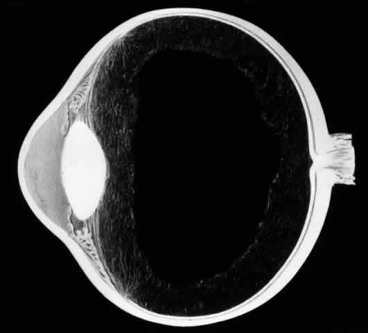
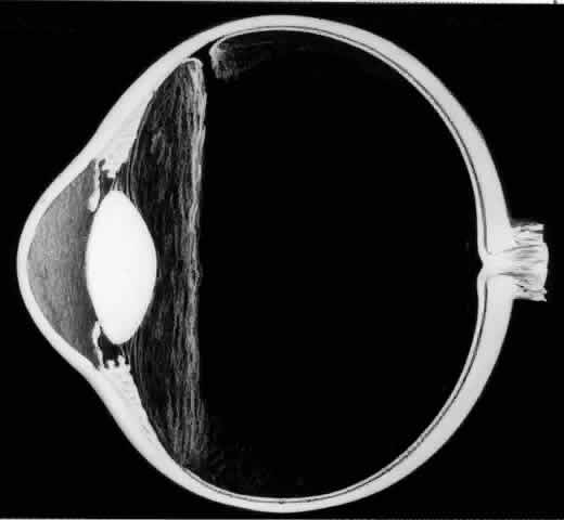
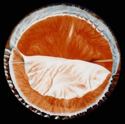
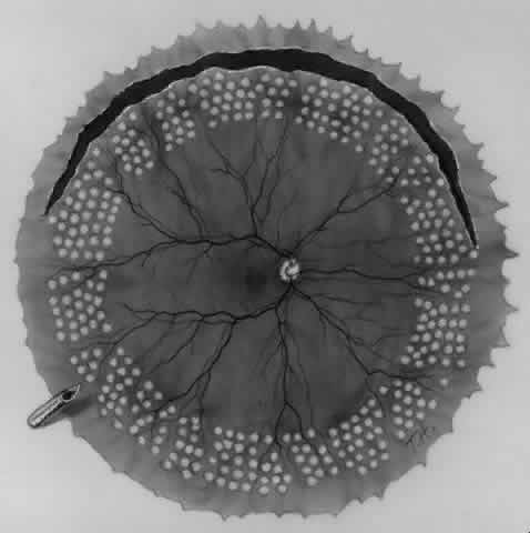
The outer surface of the inverted retinal flap (Fig. 1) should be examined for signs of epiretinal tissue proliferation, which may produce radial or star-shaped folds in the flap. Vitreous base traction produces detachment of the anterior edge of the giant tear and the pars plana epithelium anterior to it in 82% of giant retinal tears.9 It is important to identify and treat detachment of the pars plana epithelium that extends beyond the ends of the giant tear because leakage of subretinal fluid beneath it can be a potential source of postoperative posterior retinal detachment.

Fig. 1. Superior giant retinal tear extending from 9:30 to 3:30 meridian. The retina is torn at the posterior border of the vitreous base. Posterior retinal flap is invertd over the optic disc. Anterior edges of giant tear, ora serrata, and pars plana are detached from the 9:30 to 3:00 meridians as a result of vitreous base traction.

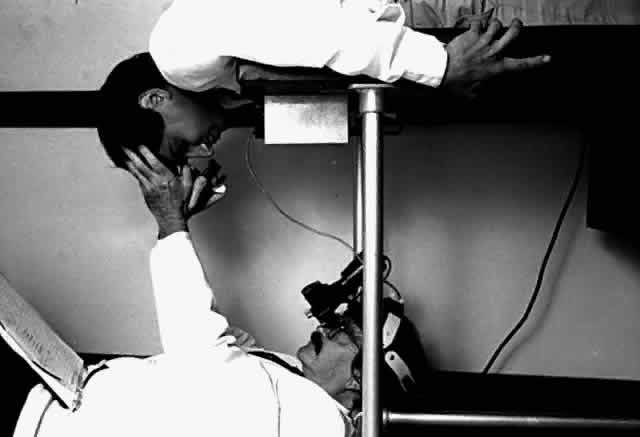
Indirect ophthalmoscopy of the patient in the prone position is helpful to determine the mobility of the inverted retinal flap and the extent to which it will unfold. During indirect ophthalmoscopy, the patient lies prone with his or her head extended over one end of the examining table, while the examiner sits cross-legged or lies on the floor (Fig. 2). If the flap unfolds poorly, either epiretinal tissue formation involving the posterior retinal flap or early proliferative vitreoretinopathy should be suspected. Preretinal organization may be present when the posterior edge of the flap is rolled in a scroll-like fashion, creating a purse-string contracture that prevents complete unfolding of the flap.

Fig. 2. Preoperative ophthalmoscopic examination of a superior giant retinal tear with an inverted retinal flap. To assess the mobility of the posterior retinal flap, the patient is examined in the prone position with his head extended over the end of the examining table.

Retinal breaks have been reported to occur in 59% of the fellow eyes of nontraumatic giant retinal tears, which is why indirect ophthalmoscopy of the fellow eye is extremely important.3

Biomicroscopy

Biomicroscopy of the vitreous is performed with the Goldmann three-mirror contact lens. Biomicroscopy reveals extensive liquefaction of the central and posterior vitreous gel and marked condensation of the anterior vitreous and vitreous base. In some eyes, a membrane attached to the anterior edge of the giant retinal tear extends across the vitreous cavity, attaching to the retina in the region of the posterior border of the vitreous base. Condensation and contraction of the vitreous base pulls the anterior edge of the giant tear toward the lens, resulting in detachment of the ora serrata and pars plana epithelium.

SURGICAL MANAGEMENT

Sclerotomies

To perform vitrectomy involving the inferior vitreous base, it may be necessary to rotate the globe so far inferiorly that the outer part of the infusion cannula is forced against the lower eyelid. This may cause the inner tip of the cannula to come in contact with, and thus damage, the peripheral retina (Fig. 3).10 To avoid this complication, the site of the infusion cannula should be located near the horizontal meridian along the inferior margin of the lateral rectus muscle. A gentian violet pen is used to make a mark on the sclera 3.5 mm posterior to the limbus. A mattress suture of 6-0 polyglactin with a spatula needle is preplaced with the arms of the suture parallel to the limbus and 1 mm anterior and posterior to the mark. As Glaser11 pointed out, a stream of infusion fluid can disrupt a perfluoro-n-octane bubble into smaller bubbles, which is why a short infusion cannula is used. To facilitate insertion of the cannula through the condensed vitreous base, thus avoiding detaching the pars plana epithelium, the slit-like opening made by the microvitreal knife is enlarged with a stiletto rotated 180°. After its insertion, the short cannula is depressed toward the center of the globe to check that the tip has entered the vitreous cavity and is not beneath a detached pars plana epithelium.

Fig. 3. When the eye is rotated far inferiorly to perform vitrectomy of the vitreous base, the outer portion of the infusion cannula can be forced against the lower eyelid (small arrow). The peripheral retina is damaged when the inner sharp tip comes in contact with it (large arrow). To avoid this complication, the infusion cannula is placed closer to the horizontal meridian (i.e., along the inferior margin of the lateral rectus muscle). (Freeman HM, Tolentino FI: Basic vitrectomy: Prevention and management of complications. In: Atlas of Vitreoretinal Surgery, pp 65–74. Stuttgart, Thieme, 1990)

Sclerotomies are made in the 2:30 and 9:30 meridians for the vitrectomy probe and the endoillumination probe, respectively. Passing the flat tip of the endoilluminator probe through the condensed vitreous base can produce a detachment of the pars plana epithelium. Therefore, the probe is slowly inserted into the sclerotomy with a rotating motion, and the tip is inspected to ensure that the pars plana epithelium or ora serrata are not being pushed in front of the probe as it enters the vitreous cavity.

Lensectomy

Recent reports indicate that scleral buckling may not be necessary in the management of a giant retinal tear, provided that traction is relieved as a result of a thorough vitrectomy involving the vitreous base.12,13 Kreiger13 believes that a lensectomy is necessary for optimal removal of the basal vitreous, and I am inclined to agree. When no nuclear sclerosis is present, lensectomy is performed with the vitrectomy probe. Phacoemulsification is used for a mildly sclerotic lens; a very hard or brunescent lens is removed intracapsularly through a corneal incision, which is closed with multiple interrupted sutures. An argument against lensectomy is that the average age of a patient presenting with a giant retinal tear is 32 years, an age at which the lens is clear. Therefore, some advocate preservation of the phakic status in these relatively young patients. Postoperative tamponade with a longacting gas may produce or accelerate cataractous changes that reduce visual acuity or hinder postoperative photocoagulation and necessitate cataract extraction. If the surgeon elects to preserve the lens, it is advisable to use perfluoroethane (C2F6) gas, which has a shorter half-life than perfluoropropane (C3F8). Additional studies will help to establish whether or not lensectomy is necessary.

Optimal Visualization During Surgery

A wide-angle fungus-viewing system such as the Advanced Visual Instrument (AVI) system is of great value during vitrectomy involving the vitreous base and during the unfolding of the inverted retinal flap. This system uses an image inverter attached to the operating microscope and a set of two contact lenses with 68° and 130° viewing angles. The 130° lens provides a panoramic view of the fundus as far anterior as the ora serrata.14

Anterior Vitrectomy

To promote maximal mydriasis, 1 mL of 1:1000 epinephrine is added to 500 mL of the balanced salt infusion solution. If a small pupil hinders visualization, four flexible nylon iris retractors are used for temporary pupillary dilation.15 Each plastic retractor measures 6 mm in length and has a 1-mm hook at one end and a small polymeric silicone (Silastic) disc to hold it in place. The iris retractors are inserted through the limbus in the 1:30, 4:30, 7:30, and 10:30 meridians (Fig. 4). Anterior vitrectomy is performed to remove the condensed anterior vitreous gel. It is important to remove the vitreous gel attached to the anterior edge of the giant retinal tear, or else this gel may be pushed posteriorly during air infusion and interfere with the unfolding of the flap or cause the posterior edge of the giant tear to slip posteriorly (Fig. 5).13 It is important to identify and remove the membrane that is attached to the anterior flap of the giant tear and extends across the vitreous cavity to the vitreous base opposite the giant tear. This membrane may be visualized best with illumination slightly eccentric to the tip of the vitrectomy probe. Removal of this membrane will decrease the incidence of retinal tears that have been observed to develop opposite the giant retinal tear.

Fig. 4. A. Iris retractor with hook at one end that retracts the pupillary margin. The hooked end is inserted into the anterior chamber through a limbal incision; then the hook is rotated 90° to engage the pupillary margin. The pupil becomes dilated as the retractor is drawn toward the limbus and held in place by the Silastic disc. B. Iris retractors are placed in the 1:30, 4:30, 7:30, and 10:30 meridians to produce a square-shaped pupil. (A and B, Freeman HM: Current management of giant retinal breaks and fellow eyes. In Glaser BM, Ryan SJ (eds): Surgical Retina, 2nd ed, pp 2313–2338. St. Louis, CV Mosby, 1994)

Fig. 5. To prevent slippage of the posterior retinal flap, the basal vitreous gel attached to the anterior edge of the giant retinal tear is removed with the vitrectomy probe. Scleral depression is used to improve visualization. (Krieger AE, Lewis H: Management of giant tears without scleral buckling. Ophthalmology 99:491, 1992)

Vitrectomy Involving the Vitreous Base

A thorough vitrectomy of the vitreous base around its entire circumference is important in the management of a giant tear without scleral buckling. Because of the strong adhesion between the peripheral retina and vitreous base, vitrectomy involving the vitreous base carries a considerable risk of causing iatrogenic retinal breaks, especially if the vitrectomy probe does not cut efficiently. Scleral depression by the assistant during the procedure allows enhanced visualization of the vitreous base. After the vitrectomy is completed, the entire fundus periphery is examined with the use of indirect ophthalmoscopy and scleral depression to determine whether the giant tear has become enlarged or to detect iatrogenic peripheral retinal breaks.

UNFOLDING THE POSTERIOR RETINAL FLAP

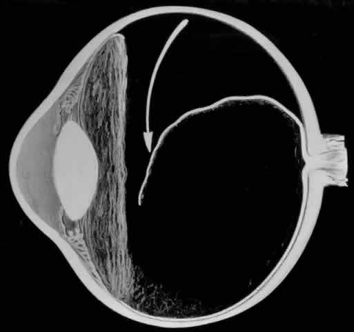
One of the most challenging problems in the management of giant retinal tears has been unfolding the inverted posterior retinal flap (Fig. 6). Schepens16 demonstrated that gravity can be used to unfold the flap by placing the patient in the prone position. This led to the development of a multipositional giant tear operating table used to rotate the patient to the prone position during surgery.17,18 Norton and associates19 and Michels20 reported the use of air injection into the vitreous cavity to unfold and tamponade the posterior retinal flap. One of the most important contributions in the management of giant retinal tears with an inverted retinal flap was made by Chang and colleagues,21–24 who demonstrated the value of liquid perfluorocarbons in unfolding the inverted retinal flap. Because this procedure is performed with the patient in the supine position, unfolding of an inverted retinal flap is accomplished more easily and with fewer complications.

Fig. 6. Giant retinal tear with a posterior retinal flap that is inverted over the optic disc. The posterior edge of the giant tear is slightly rolled as a result of epiretinal membrane proliferation. (Freeman HM: Current management of giant retinal breaks and fellow eyes. In Glaser BM, Ryan SJ (eds): Surgical Retina, 2nd ed, pp 2313–2338. St. Louis, CV Mosby, 1994)

Perfluoro-n-Octane

Perfluoro-n-octane is an ideal heavier-than-water liquid for unfolding the inverted retinal flap of a giant retinal tear. Its specific gravity of 1.76 compared with 1.03 of aqueous causes perfluoro-noctane to gravitate posteriorly, settling on the surface of the posterior retina and optic disc. The surface tension of 16.98 accounts for the tendency of injected perfluoro-n-octane to remain in one globule as the globule increases in size to unfold and tamponade the posterior retinal flap of the giant retinal tear.

Perfluoro-n-octane (C8F18) is an inert perfluorocarbon: there are no known biologic enzymes that can metabolize its carbon-fluoride bonds. It is optically clear, permitting excellent visualization of the retina; however, the edge of the bubble of perfluoro-n-octane is visible within the eye because perfluoro-n-octane has a different refractive index (1.27) than aqueous (1.33) or balanced salt infusion. Perfluoro-n-octane has a vapor pressure of 52 mmHg at 37°C, which tends to volatilize it during fluid-gas exchange. Its low viscosity (0.69 centistokes) and its surface tension account for the ease with which the gas can be removed from the eye.

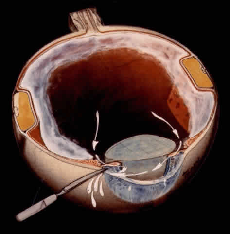
TECHNIQUE. With the tip of the vitrectomy probe or a retinal manipulator (i.e., an angulated probe with a ball-like tip), the inverted retinal flap is partially unfolded to expose the optic disc and posterior pole (Fig. 7). Perfluoro-n-octane is injected slowly through a blunt-tipped 25-gauge needle positioned 1 to 2 mm over the optic disc (Fig. 8), creating a globule. As the globule expands toward the fundus periphery, it smoothly unfolds the posterior retinal flap. To prevent the formation of multiple bubbles of perfluoro-n-octane, the tip of the injection needle must be kept within the globule during injection.

Fig. 7. The posterior retinal flap is partially unfolded with a ball-tipped retinal manipulator. (Freeman HM: Current management of giant retinal breaks and fellow eyes. In Glaser BM, Ryan SJ(eds): Surgical Retina, 2nd ed, pp 2313–2338. St. Louis, CV Mosby, 1994)

Fig. 8. Perfluoro-octane (PFO) is injected through a blunt-tipped 25-gauge needle positioned 1 to 2 mm in front of the optic disc. (Freeman HM: Current management of giant retinal breaks and fellow eyes. In Glaser BM, Ryan SJ (eds): Surgical Retina, 2nd ed, pp 2313–2338. St. Louis, CV Mosby, 1994)

If epiretinal membranes are causing the posterior edge of the flap to roll or curl, they can be removed as follows. An injection of 0.8 to 1 mL perfluoro-n-octane is used as a means of partially unfolding the flap and steadying it to enable bimanual dissection of the epiretinal membranes. After the membranes are removed, additional perfluoro-n-octane is injected to bring the level of the perfluoro-n-octane to the posterior edge of the giant tear (Fig. 9).

Fig. 9. Perfluoro-octane (PFO) is injected to the level of the posterior edge of the giant break while balanced salt solution (BSS) is infused. (Freeman HM: Current management of giant retinal breaks and fellow eyes. In Glaser BM, Ryan SJ (eds): Surgical Retina, 2nd ed, pp 2313–2338. St. Louis, CV Mosby, 1994)

Anterior Fluid-Air Exchange. Using the cannulated extrusion needle with a suction pressure of approximately 70 mmHg, the fluid anterior to the perfluoro-n-octane is removed by fluid-air exchange (Fig. 10).25 Fluid along or under the posterior edge of the giant tear promotes posterior slippage of the posterior edge of the giant tear. Therefore, it is extremely important to remove as much of this fluid as possible (Fig. 11). Slight slippage of the retinal flap can be corrected by massaging the edge of the retina anteriorly with the retinal manipulator. If this fails, the posterior edge of the flap is dragged anteriorly with minimal suction through the soft silicone tip of the cannulated extrusion needle, which is applied to the displaced posterior edge of the giant retinal tear. An effort is made to flatten the posterior edge of the giant tear completely in order to prevent perfluoro-n-octane from passing beneath it. If any perfluoro-n-octane migrates subretinally beneath the flap, it is removed with the cannulated extrusion needle or via retinotomy.

Fig. 10. Fluid-air exchange is performed with the cannulated extrusion needle in order to remove the infusion fluid anterior to the perfluoro-octane. (Freeman HM: Current management of giant retinal breaks and fellow eyes. In Glaser BM, Ryan SJ (eds): Surgical Retina, 2nd ed, pp 2313–2338. St. Louis, CV Mosby, 1994)

Fig. 11. To prevent slippage of the posterior retinal flap, the cannulated extrusion needle is used to remove as much fluid as possible along or under the posterior edge of the giant break. (Freeman HM: Current management of giant retinal breaks and fellow eyes. In Glaser BM, Ryan SJ (eds): Surgical Retina, 2nd ed, pp 2313–2338. St. Louis, CV Mosby, 1994)

Endolaser. Eight to 10 rows of endophotocoagulation are applied to the anterior third of the posterior retinal flap (Fig. 12). Five rows of endophotocoagulation are placed from the ora serrata to the equator in the fundus periphery not involved in the giant tear so that treatment extends around the entire periphery of the fundus.

Fig. 12. Endolaser photocoagulation is applied around the entire fundus periphery in 8 to 10 rows to the anterior third of the posterior retinal flap and in 5 rows between the ora serrata and the equator in the fundus periphery not involved in the giant tears. (Freeman HM, Tolentino FI: Basic vitrectomy: Prevention and management of complications. In Atlas of Vitreoretinal Surgery, pp 65–74. Stuttgart, Thieme, 1990)

Removal of Perfluoro-n-Octane. Perfluoro-noctane is removed with the cannulated extrusion needle or double-barreled vented needle. As the air meniscus approaches the retina, the layer of perfluoro-n-octane becomes flattened, making the small residual amount difficult to visualize. Approximately 0.5 to 0.8 mL of balanced salt solution is then injected into the eye to cause the perfluoro-n-octane to form small droplets in the saline phase, which can be seen and removed more easily.

Air-Gas Exchange. To allow air to be vented from the eye during an air-gas exchange, the infusion tubing is clamped and disconnected from the three-way stop cock. To produce a slow venting of air through a smaller diameter than that of the infusion tubing, and thus prevent a sudden decrease in intraocular pressure, a 25-gauge needle is attached at the end of the tubing using a female adapter. Air is exchanged for a 15% mixture of C3F8 and air that is injected from a 50-cc syringe with a 30-gauge needle inserted through the limbus in aphakic eyes and through the pars plana in phakic and pseudophakic eyes. After the needle has entered the eye, the clamp on the infusion cannula is removed and 40-cc of the gas mixture is lavaged through the eye. Air and gas is vented through the infusion tubing and needle that has been placed in balanced salt solution in a beaker. The continuous flow of bubbles in the beaker indicates a continuous lavage (Fig. 13).

Fig. 13. Gas-air exchange. A 15% mixture (50 mL) of perfluropropane (C3F8) gas and air is lavaged through a 30-gauge needle. Air is vented from the eye through the infusion cannula, to which a 25-gauge needle is attached to maintain uniform intraocular pressure. The flow of air from the eye is confirmed by bubbles in saline in a glass beaker. (Freeman HM: Current management of giant retinal breaks and fellow eyes. In Glaser BM, Michels RG (eds): Surgical Retina, 1st ed, pp 431–448. St. Louis, CV Mosby, 1989)

Air-Silicone-Fluorosilicone Oil Exchange. For infants and children in whom postoperative prone positioning is not possible and in cases of severe proliferative vitreoretinopathy, an air-oil exchange is performed with a silicone-fluorosilicone oil (SiFO) copolymer developed by Refojo, Tolentino, and associates.26 This copolymer has a viscosity of 180 centistokes, a specific gravity of 1.15 g/cm3, and a refractive index of 1.38. SiFO seems preferable to silicone oil for postoperative retinal tamponade because its higher specific gravity causes it to gravitate posteriorly against the retina, whereas silicone oil tends to float anteriorly, thus producing a higher incidence of corneal complications and therefore less tamponading of the inferior retina. SiFO is injected to fill the vitreous cavity to a level slightly posterior to the plane of the pupil. The oil is removed and replaced with balanced salt solution 3 to 6 months postoperatively or earlier if marked emulsification of the oil occurs.

Back to Top
MANAGEMENT OF FELLOW EYES OF NONTRAUMATIC GIANT RETINAL TEARS
The management of fellow eyes of nontraumatic giant retinal tears is important because of the high incidence of retinal tears and detachment in fellow eyes. In a study of fellow eyes of 321 nontraumatic giant retinal tears that were followed from 2 to 29 years, I found that retinal tears or retinal detachment developed in 59% of fellow eyes (Table 1).3


TABLE 1. Incidence of Retinal Breaks and Retinal Detachment in Fellow Eyes of Patients with Nontraumatic Giant Tears (321 Eyes)


 Initial ExamEnd of Follow-up
Type of Retinal BreakNo. of Eyes(%)No. of Eyes(%)
Retinal detachment with retinal tears, holes, breaks, or dialysis37(11.6)57(17.8)
Giant retinal tears29(9)53(16.5)
Retinal tears without detachment26(8)40(12.5)
Retinal holes without detachment22(6.8)38(11.8)
Retinal dialysis without detachment2(0.6)4(1.2)
Total116(36)192(59.8)
(Freeman HM: Fellow eye of non-traumatic giant retinal breaks. In Lewis H, Ryan SJ (eds): Medical and Surgical Retina: Advances, Controversies and Management, pp 222–225. St. Louis, CV Mosby, 1994)


RETINAL TEARS, HOLES, DIALYSIS, AND DETACHMENT

Of the 321 eyes, horseshoe retinal tears developed in 40 eyes (12.5%), retinal holes in 38 (11.8%), and retinal dialysis in 4 (1.2%). Retinal detachment caused by retinal holes, tears, or dialysis developed in 57 eyes (17.8%).

GIANT RETINAL TEARS

During the period of observation the incidence of giant retinal tears in fellow eyes increased from 9% to 16.5%. All fellow eyes that developed a giant retinal tear during the follow-up period had myopia averaging 12 diopters (range, 5 to 22 diopters). The average latent period for the development of a giant retinal tear in a fellow eye was 22 months in phakic eyes and 39 months in aphakic eyes. Ophthalmoscopic and biomicroscopic changes observed in eyes before the development of a giant retinal tear provided information regarding the pathogenesis of nontraumatic giant retinal tears and a rationale for scleral buckling to prevent giant retinal tears from developing in high-risk eyes.

Biomicroscopic Findings Preceding Development of a Giant Tear

The earliest biomicroscopic changes preceding the development of a giant retinal tear were syneresis and liquefaction of the vitreous gel, converting the posterior and central portions of the vitreous body into a large lacuna (Fig. 14). Later, the anterior gel became condensed, and its posterior aspect formed an equatorially oriented membrane extending across the vitreous cavity (Fig. 15). This membrane did not oscillate with ocular movements, suggesting that it was under tension. Marked contraction of this membrane seems responsible for tearing the retina along the posterior border of the vitreous base (Fig. 16). Liquefied vitreous gel facilitated inversion of the posterior retinal flap (Fig. 17).

Fig. 14. The earliest biomicroscopic change in the vitreous prior to the development of a nontraumatic giant retinal break is liquefaction of the vitreous gel, which converts the central portion of the vitreous gel into a large lacuna.

Fig. 15. In later biomicroscopic changes preceding a nontraumatic giant retinal break, the anterior vitreous gel becomes condensed and a membrane forms its posterior aspect.

Fig. 16. Contraction of this equatorially oriented membrane extending across the vitreous cavity produces a giant tear along the posterior border of the vitreous base.

Fig. 17. After the formation of a giant retinal tear, the posterior retinal flap inverts into the liquefied vitreous (arrow).

Ophthalmoscopic Findings Preceding Development of a Giant Tear

The earliest ophthalmoscopic finding was “white-with-pressure” extending from the ora serrata to varying distances posterior to the equator. Adjacent areas of white-with-pressure increased in size circumferentially to become confluent and more opaque, and they developed a sharp posterior edge in contrast to the hazy, ill-defined posterior margin seen earlier. In some cases a sharp ridge developed in the retina along the posterior border of the white-with-pressure.

PROPHYLACTIC SCLERAL BUCKLING IN THE MANAGEMENT OF HIGH-RISK FELLOW EYES

On the basis of this study,3 it was concluded that fellow eyes that are at high risk for developing a giant retinal tear are highly myopic, have severe lattice degeneration, or show increasing white-with-pressure and increasing condensation of the anterior vitreous and vitreous base. During the follow-up of high-risk eyes, giant retinal tears developed in 90.1% of eyes. This prompted the use of prophylactic scleral buckling in the treatment of high-risk eyes during the past 20 years. Results thus far have been encouraging: of 31 eyes treated with a prophylactic scleral buckle and followed from 6 months to 19 years, giant retinal tears developed in only 2 eyes (6.4%).

Studies of fellow eyes indicate that vitreous traction plays a major role in the pathogenesis of giant retinal tears.9 Therefore, the rationale for preventing giant retinal tears should involve measures that relieve vitreous traction. Although scleral buckling does not eliminate vitreous traction, it can be substantially reduced by a high buckling effect.

Prophylactic cryocoagulation or photocoagulation without scleral buckling does not alleviate vitreous traction. Quite the contrary, a significant number of cases have been observed in which very difficult-to-treat, posteriorly located giant retinal tears developed along the posterior edge of prophylactic photocoagulation or cryocoagulation applications.27 Taking into account the encouraging results with scleral buckling, the risks involved in a more invasive intraocular procedure such as a prophylactic vitrectomy do not justify its use in the management of high-risk eyes.

Technique

A Foley catheter is inserted into the bladder preoperatively because of the marked diuresis associated with the intraoperative use of mannitol and acetazolamide. An attempt is made to buckle 180° to 210° of the globe: an arc is selected that encompasses most of the vitreoretinal pathology (e.g., lattice degeneration, white-with-pressure, vitreous base condensation). A mark is made to localize the posterior edge of white-with-pressure or the posterior edge of lattice degeneration. In eyes with thin sclera, two rows of light cryocoagulation applications and an episcleral implant are placed in a circumferential arc straddling the localizing mark. In eyes with sclera of normal thickness, a solid silicone rubber implant 6 mm in width may be placed in an 8-mm lamellar scleral dissection. Burying the implant intrasclerally reduces the chance of implant extrusion or extraocular muscle imbalance and diplopia, which can occur with anterior episcleral buckles. To achieve a permanent buckling effect, an encircling band is placed and anchored around the greater circle of the globe. Approximately 20 minutes before the scleral buckling sutures are tightened, acetazolamide and mannitol are administered intravenously to reduce the intraocular pressure.

A large portion of the vitreous gel is liquefied in these highly myopic eyes, so a substantial amount of vitreous fluid will percolate into the anterior chamber during multiple paracenteses, thus reducing the intraocular pressure and allowing a high scleral buckling effect to be obtained (Fig. 18). The encircling band and sutures are adjusted such that the closing intraocular pressure does not exceed 20 mmHg and indirect ophthalmoscopy reveals that the ophthalmic artery is patent. Finally, an anchoring suture is placed over the encircling element near the middle of the quadrants of the globe opposite the scleral buckle.

Fig. 18. Paracentesis of aqueous and liquefied vitreous that percolates into the anterior chamber is performed to reduce the intraocular pressure so that a high scleral buckling effect can be obtained.

Back to Top
REFERENCES

1. Freeman HM, Johnson MR: Current management of giant retinal breaks. In Spaeth G (ed): Current Therapy in Ophthalmic Surgery, pp 256–264. Philadelphia, BC Dekker, 1989

2. Kanski JJ: Giant retinal tears. Am J Ophthalmol 79:846, 1975

3. Freeman HM: Fellow eye of non-traumatic giant retinal breaks. In Lewis H, Ryan SJ (eds): Medical and Surgical Retina: Advances, Controversies and Management, pp 222–225. St. Louis, CV Mosby, 1994

4. Freeman HM: Unpublished data, 1996

5. Kinyoun JL, Knoblock WH: Idiopathic retinal dialysis. Retina 4:9, 1984

6. Zion VM, Burton TC: Retinal dialysis. Arch Ophthalmol 98:1971, 1980

7. Hagler WS: In discussion. Freeman HM: Current management of giant retinal breaks: results with vitrectomy and total air-fluid exchange in 95 cases. Trans Am Ophthalmol Soc 79:29, 1981

8. Scott JD: Giant tear of the retina. Trans Ophthalmol Soc UK 95:142, 1975

9. Freeman HM: Fellow eyes of giant retinal breaks. Trans Am Ophthalmol Soc 76:343, 1976

10. Freeman HM, Tolentino FI: Basic Vitrectomy: Prevention and management of complications. In: Atlas of Vitreoretinal Surgery, pp 65–74. Stuttgart, Thieme, 1990

11. Glaser BM: Treatment of giant retinal tears combined with proliferative vitreoretinopathy. Ophthalmology 93:1193, 1986

12. Hoffman ME, Sorr EM: Management of giant retinal tears without scleral buckling. Retina 6:197, 1986

13. Krieger AE, Lewis H: Management of giant retinal tears without scleral buckling: use of radical dissection of the vitreous base and perfluoro-n-octane and intraocular tamponade. Ophthalmology 99:491, 1992

14. Corcostegui B: New developments in wide-range fundus viewing planitis. Vitreoretinal Surg Technol 6:3, 1994

15. de Juan E Jr, Hickingbotham D: Letter to the editor: Flexible iris retractor. Am J Ophthalmol 11:776, 1991

16. Schepens CL, Freeman HM: Current management of giant retinal breaks. Trans Am Acad Ophthalmol Otolaryngol 71:474, 1967

17. Schepens CL, Freeman HM, Thompson RF: A power-driven multipositional operating table. Arch Ophthalmol 73:671, 1965

18. Freeman HM: Current management of giant retinal breaks and fellow eyes. In Glaser BM, Ryan SJ (eds): Surgical Retina, 2nd ed. St. Louis, CV Mosby, 1994

19. Norton EWD, Aaberg T, Fung W, Curtin VT: Giant retinal tears: I. Clinical management with intravitreal air. Am J Ophthalmol 68:1011, 1969

20. Michels RG: Giant retinal tears. In Blankenship GW, Binder S, Gonvers M et al (eds): Basic and Advanced Vitreous Surgery, pp 179–218. New York, Springer-Verlag, 1986

21. Chang S: Low viscosity liquid fluorochemicals in vitreous surgery. Am J Ophthalmol 103:38, 1987

22. Chang S, Reppucci V, Zimmerman NJ et al: Perfluorocarbon liquids in the management of traumatic retinal detachments. Ophthalmology 96:785, 1989

23. Chang S, Lincoff H, Zimmerman NJ, Fuchs W: Giant retinal tears: surgical techniques and results using perfluorocarbon liquids. Arch Ophthalmol 107:761, 1989

24. Chang S: Giant retinal tears: Surgical management with perfluorocarbon liquids. In Lewis H, Ryan SJ (eds): Medical and Surgical Retina: Advances, Controversies and Management, pp 199–207. St. Louis, CV Mosby, 1993

25. Joondeph BC, Flynn HW Jr, Blankenship GW et al: The surgical management of giant retinal tears with the cannulated extrusion needle. Am J Ophthalmol 108:548, 1989

26. Refojo MF: Vitreous implants in retinal detachment surgery. Polymeric Materials Science and Engineering 69: 546, 1993

27. Freeman HM: The complications of light coagulation. In Schepens CL, Regan CDJ (eds): Controversial Aspects of the Management of Retinal Detachment, pp 256–260. Boston, Little Brown & Co, 1965

Back to Top