Chapter 70
Surgical Management of Posterior Uveal Melanoma
CAROL L. SHIELDS and JERRY A. SHIELDS
Main Menu   Table Of Contents

Search

GENERAL CONSIDERATIONS
PERIODIC OBSERVATION
PHOTOCOAGULATION
TRANSPUPILLARY THERMOTHERAPY
RADIOTHERAPY
LOCAL RESECTION
ENUCLEATION
ORBITAL EXENTERATION
MANAGEMENT OF SYSTEMIC METASTASIS
COUNSELING THE PATIENT
SUMMARY
REFERENCES

There have been several new developments in the management of patients with choroidal melanoma over the past decade. Depending on the clinical circumstances, observation, photocoagulation, transpupillary thermotherapy, plaque radiotherapy, charged particle irradiation, local resection, enucleation, orbital exenteration, chemotherapy, and immunotherapy have been employed.1–4 There has been a trend away from photocoagulation, with increasing interest in transpupillary thermotherapy (TTT) alone for small melanomas or TTT in combination with radiotherapy for small, medium, and some large melanomas. The recently recognized clinical risk factors for growth and metastasis of small melanocytic tumors allows for earlier detection and treatment of small choroidal melanoma. Consequently, there is a trend away from observation and a focus toward earlier treatment of small melanocytic lesions that possess risk factors. This chapter provides an overview of the methods of management for patients with choroidal melanoma.
Back to Top
GENERAL CONSIDERATIONS
Historically, enucleation of the affected eye was considered to be the only appropriate management for the patient with a posterior uveal melanoma. However, several years ago, some authorities challenged the effectiveness of enucleation for preventing metastatic disease and even proposed that enucleation may somehow promote or accelerate metastasis.5,6 The validity of these arguments was challenged by others who believed that early enucleation offered the patient the best chance of cure.7,8 This controversy over enucleation was responsible for initiating a trend away from enucleation and the increased use of more conservative therapeutic methods.

Depending upon several clinical factors, management options today include observation, photocoagulation, TTT, radiotherapy, local resection, enucleation, and various combinations of these methods. The two most frequently employed treatment methods today are enucleation and plaque radiotherapy. The Collaborative Ocular Melanoma Study (COMS) was organized and funded in 1985 to address several issues related to management of choroidal melanoma.9 More recently, there has been a documented trend of detection of smaller choroidal melanomas and increasing use of conservative treatment methods such as plaque radiotherapy.1,10–12

Back to Top
PERIODIC OBSERVATION
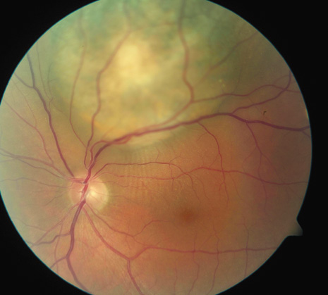
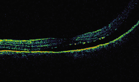
Approximately 6% of the Caucasian population manifests choroidal nevus.13 Choroidal nevi are managed by periodic observation. It is estimated that 1 in 5,000 choroidal nevi evolve into choroidal melanoma.14 Risk factors predictive of growth of small melanocytic lesions and their combined predictive values are listed in Tables 1 and 2 (Fig. 1). Documented growth of a melanocytic choroidal tumor is suggestive that the lesion is a choroidal melanoma. Because documented growth may be associated with worse systemic prognosis, some patients with small tumors that show three or more risk factors are treated promptly, without waiting for documentation of growth.12,15,16 On the basis of the few patients with medium-size choroidal melanoma who refuse treatment and are followed, natural history studies have found that there is greater mortality and higher risk of death.17
 
Fig. 1. Clinical risk factors for growth of small choroidal melanoma. (TFSOM = To Find Small Ocular Melanoma represents factors of Thickness over 2 mm, subretinal Fluid, Symptoms, Orange pigment, and Margin of tumor at optic disc) A. Thickness greater than 2 mm. Perimacular small choroidal melanoma measuring 2.9 mm in thickness and with overlying orange pigment and subretinal fluid, manifesting as retinal striae. B. Thickness greater than 2 mm. Optical coherence tomography demonstrating subfoveal fluid in the patient in A. C. Subretinal fluid. Sixteen-year-old patient with documented enlarging small choroidal melanoma and shallow subretinal fluid. D. Subretinal fluid. Optical coherence tomography demonstrating subfoveal fluid in the patient in C. E. Symptoms. Small choroidal melanoma producing symptoms of blurred vision from subfoveal fluid. Note the overlying orange pigment. F. Symptoms. Small choroidal melanoma located superiorly with trough of subretinal fluid in the foveola producing symptoms of metamorphopsia. G. Orange pigment. Macular choroidal melanoma with prominent overlying orange pigment and a rim of subretinal fluid. H. Margin at the optic disc. Minimally elevated small choroidal melanoma touching the optic disc. I. Other features. Juxtapapillary small choroidal melanoma with overlying orange pigment, shallow subretinal fluid, and irregular margins suggestive of tumor activity.

TABLE 1. Clinical features predictive of growth of small melanocytic choroidal tumors (≤3 mm thickness)12


Clinical Featurep ValueRelative RiskConfidence Intervals
Subretinal fluid
 (absent vs. present)0.051.4(1.0,1.8)
Orange pigment
 (absent vs. present)0.0041.5(1.2,2.0)
Symptoms
 (none vs. blurred vision)0.0031.6(1.2,2.2)
 (none vs. flashes/floaters)0.0021.8(1.2,2.6)
Posterior margin
 (>3.0 mm from optic disc vs. touching the disc)0.00012.6(1.9,3.6)
 (>3.0mm from optic disc vs. 0.1 to 3.0 mm from disc)0.081.4(1.0,2.0)
Thickness by ultrasound
 (0–1.0 mm vs. 1.1–2.0 mm)0.00014.3(2.9,6.4)
 (0–1.0 mm vs. 2.1–3.0 mm0.00015.2(3.5,7.8)


TABLE 2. Tumor growth in 1287 small (≤3 mm thickness) melanocytic choroidal tumors based on combination of clinical risk factors11


Clinical FeaturesKaplan-Meier 5-Year Growth (%)
No features3
One feature
 S33
 M44
 T38
 F39
 O37
Two features
 S + M60
 S + T39
 S + F45
 S + O51
 M + T68
 M + F60
 M + O56
 T + F38
 T + O38
 F + O47
Three features
 S + M + T69
 S + M + F59
 S + M + O62
 S + T + F41
 S + T + O40
 S + F + O53
 M + T + F66
 M + T + O46
 M + F + O56
 T + F + O40
Four features
 S + M + T + F64
 S + M + T + O43
 S + M + F + O60
 S + T + F + O42
 M + T + F + O49
Five features
 S + M + T + F + O56

F = subretinal fluid, O = orange pigment, M = posterior margin touch optic disc, S = symptoms, T = thickness >2 mm


Back to Top
PHOTOCOAGULATION
Photocoagulation is still an acceptable method for treating selected small choroidal melanomas.18,19 It was originally performed with xenon photocoagulation and later with argon laser photocoagulation. Studies showed that xenon achieved better tumor control but argon was associated with fewer complications.19 Low-energy long-exposure laser therapy has been advocated by some authorities.18 Recently, TTT has largely replaced argon laser for treating selected small melanomas, particularly those that are less than 3 mm in thickness and located more than 3 mm from the foveola.

Photodynamic therapy using verteporfin has been considered, but there is little evidence of its effectiveness. One report on four patients showed tumor regression for 18 months in one patient, but lack of response or continued growth in three patients.20

Back to Top
TRANSPUPILLARY THERMOTHERAPY
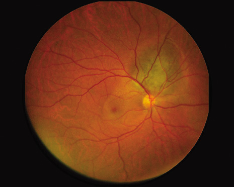
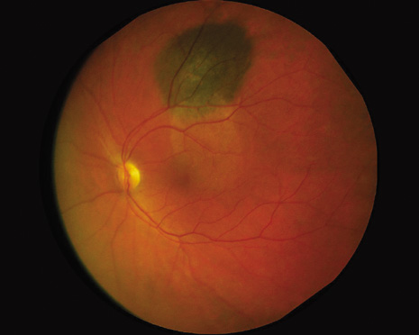
Transpupillary thermotherapy (TTT) is a recently popularized method of treating selected small- and medium-sized choroidal melanomas.21–24 With this technique, infrared radiation, using a modified diode laser system, delivers heat to the tumor, causing tumor necrosis. Transpupillary thermotherapy is typically delivered in three sessions and, at completion, leaves an atrophic chorioretinal scar at the site of the previous tumor. Tumor control is found in over 90% of properly selected cases.24 The tumors most suitable for TTT are small, heavily pigmented melanomas less than 3 mm in thickness, with minimal or no subretinal fluid, and are located in the extramacular region, not touching the optic disc (Fig. 2). Tumors at the optic disc show greater recurrence and are best managed with plaque radiotherapy combined with thermotherapy. Choroidal melanoma treated with TTT should be followed long term because delayed recurrence, even with extrascleral extension, can occur.25,26 Currently, TTT is used most frequently as a supplement to plaque radiotherapy.27 A newer technique using a transscleral thermotherapy probe has been found effective for choroidal melanoma and intrascleral tumor.28

Fig. 2. Transpupillary thermotherapy for small choroidal melanoma. A. Before treatment the heavily pigmented choroidal melanoma shows evidence of overlying orange pigment and subretinal fluid. B. Immediately after thermotherapy, the tumor appears gray white in color.

Back to Top
RADIOTHERAPY
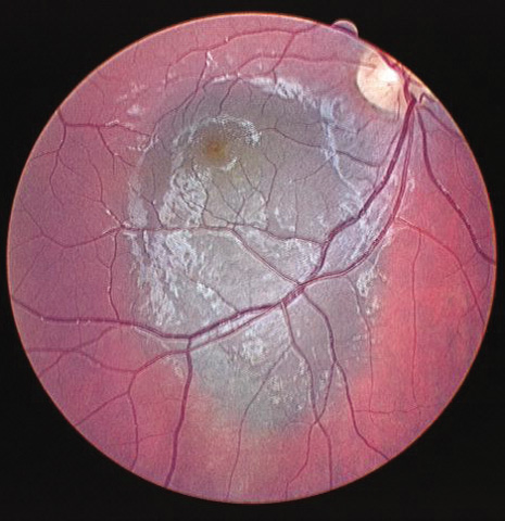
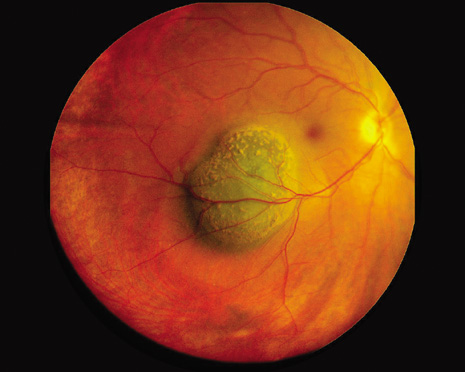
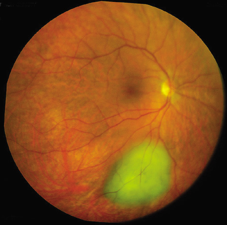
Radiotherapy is still the most widely employed treatment for posterior uveal melanoma. The most commonly employed form of radiotherapy is brachytherapy, using a radioactive plaque.2,29–35 Several years ago, Cobalt 60 plaque was popular.36 More recently, Iodine-125 and Ruthenium-106 plaques have largely replaced Cobalt-60 at most institutions.2,37 Originally, plaque radiotherapy was used for small- and medium-sized melanomas located outside the macular region and posterior to the ora serrata. Later, with innovations in radiotherapeutic plan, plaque radiotherapy can be custom designed to treat uveal melanoma at any site within the eye, including macular melanoma using a round or notched plaque, juxtapapillary melanoma using a notched plaque, ciliary body melanoma using a round or curvilinear plaque, iris melanoma using a curvilinear plaque, and even extrascleral extension of uveal melanoma.31–33,38 Plaque radiotherapy can also be custom fit to treat small, medium, and even large uveal melanoma up to approximately 12 mm in thickness (Fig. 3). 34 Shields and co-workers found that plaque radiotherapy for large melanoma was effective with satisfactory tumor control, but complications of radiation maculopathy and papillopathy often lead to poor long-term vision.34

Fig. 3. Plaque radiotherapy for choroidal melanoma. A. Before treatment, the large choroidal melanoma and extensive retinal detachment are noted. B. Following plaque radiotherapy (10 months later), the tumor has regressed and the subretinal fluid has resolved.

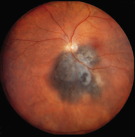
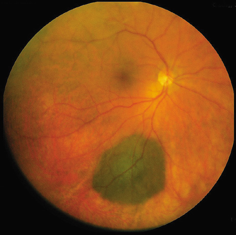
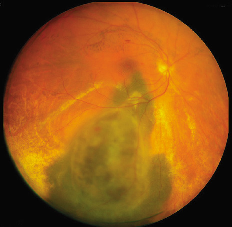
Another method of radiotherapy is charged particle irradiation.39–42 This technique provides a focused beam of radiotherapy to the region of the tumor from an external source. A recent report showed that the 5- and 10-year rates of regrowth were 3% and 4%, respectively.41 Among the 45 documented recurrences, about one-half occurred at the margin, presumably to because of treatment planning errors. Recurrence of the tumor was independently related to risk of tumor-related death. Similar to plaque radiotherapy, radiation complications in the eye and adnexa can occur.43 Tumor control with charged particle and plaque radiotherapy are similar. Plaque radiotherapy combined with thermotherapy provided excellent tumor in 270 studied patients with only 3% recurrence at 8 years (Figs. 4 and 5). 44 The high control rate of 97% was maintained for eyes with juxtapapillary choroidal melanoma despite the fact that this location is difficult for plaque placement.44 Studies have shown that 5% to 10% of patients treated with radiotherapy ultimately require enucleation of the affected eye because of tumor recurrence or radiation complications.45,46

Fig. 4. Plaque radiotherapy combined with thermotherapy for medium choroidal melanoma. A. Before treatment, the active choroidal melanoma is pigmented. B. After plaque radiotherapy and thermotherapy, the tumor is completely regressed to an atrophic scar of bare sclera.

Fig. 5. Plaque radiotherapy combined with thermotherapy for large choroidal melanoma. A. Before treatment, the active choroidal melanoma with shallow subretinal fluid is seen. B. After plaque radiotherapy and thermotherapy, the tumor is completely regressed, leaving a chorioretinal atrophic scar.

The Collaborative Ocular Melanoma Study (COMS) was designed to evaluate management of choroidal melanoma in a prospective fashion. The COMS included three substudies: (a) the large choroidal melanoma trial comparing enucleation versus enucleation preceded by external beam radiotherapy, (b) the medium choroidal melanoma trial comparing enucleation versus plaque radiotherapy, and (c) the small choroidal melanoma trial evaluating the natural history of these tumors. The results of the large tumor trial showed no difference in patient survival between enucleation and pre-enucleation radiation groups.48 Five-year Kaplan-Meier estimates for survival were 57% for the enucleation group and 62% for the pre-enucleation radiation group. The medium tumor trial showed no difference in patient survival between enucleation and plaque radiotherapy.30 Five-year Kaplan-Meier estimates for histopathologically confirmed melanoma metastasis were 11% in the enucleation group and 9% in the plaque radiotherapy group. The unadjusted estimates for 5-year survival were 81% for the enucleation group and 82% for the plaque radiotherapy group. The small tumor trial showed that small choroidal melanomas managed by observation showed tumor growth in 21% by 2 years and 31% by 5 years.49 The results of the COMS confirmed numerous previous publications regarding management of choroidal melanoma.50

Back to Top
LOCAL RESECTION
Local resection of melanomas involving the ciliary body and choroid can be performed using a partial lamellar sclerouvectomy technique.51 This surgical technique is a modification of the one popularized by Foulds, in which the tumor is removed with the aim of leaving the retina and vitreous intact.51–53

Local resection of a posterior uveal melanoma offers several theoretical advantages over enucleation and radiotherapy. In contrast to enucleation, it is designed to preserve vision and maintain a cosmetically normal eye. In contrast to radiotherapy, it has fewer long-term complications if the initial surgery is successful. However, it does have more potential immediate complications, such as vitreous hemorrhage, retinal detachment, and cataract,51 whereas radiotherapy is almost never associated with such immediate complications. However, some degree of radiation retinopathy and cataract is common in long-term complications of all forms of radiotherapy. There is no current evidence that local resection of posterior uveal melanoma is any different from enucleation or radiotherapy with regard to patient survival. There are fewer complications and better visual results for smaller, more anteriorly located tumors. More complications can be expected when larger postequatorial tumors are managed in this manner.

Back to Top
ENUCLEATION
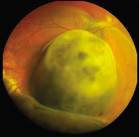
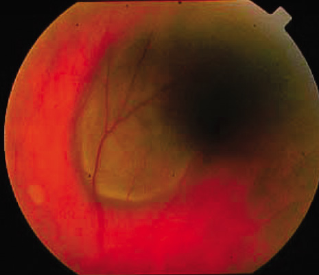
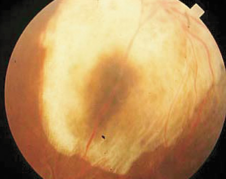
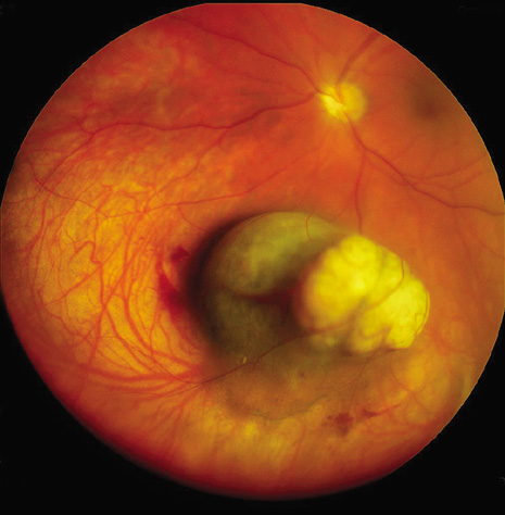
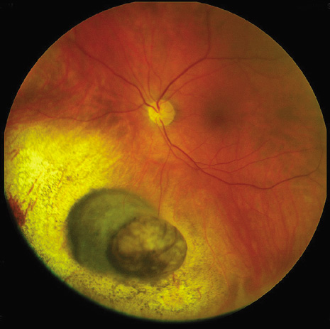
As mentioned, the traditional method of treating uveal melanomas by enucleation was challenged several years ago.5,6 Others continued to believe that enucleation was an appropriate method of management.7,8 Enucleation generally is indicated for advanced melanomas that occupy most of the intraocular structures and for those that have produced secondary glaucoma (Fig. 6). Another relative indication for enucleation is melanoma with optic nerve invasion. Enucleation with a long section of the optic nerve seems more reasonable in such cases. However, many juxtapapillary melanomas that abut the optic nerve and show no evidence of invasion often can be managed by custom-designed notched radioactive plaques rather than enucleation.31,35

Fig. 6. Enucleation for large variably nonpigmented choroidal melanoma.

The so-called “no touch enucleation” was introduced to minimize the amount of surgical trauma and theoretically to lessen the chance of tumor dissemination at the time of surgery.54 An essential aspect of this technique is to freeze the venous drainage from the tumor prior to cutting the optic nerve. The “no touch” technique has recently fallen into disuse at most centers because it is cumbersome and its benefits are only theoretical. However, a very gentle standard technique of enucleation should be employed, without clamping the optic nerve prior to cutting it.

There have been recent advances in the types of orbital implants used after enucleation. The hydroxyapatite implant, designed to improve the ocular motility in patients undergoing enucleation, is still used widely, but other motility implants have been introduced.55–57

Pre-enucleation radiotherapy (PERT) has been advocated by some authorities. In general, this involves the use of 2,000 cGy of external beam radiotherapy to the affected eye and orbit prior to enucleation. Several studies have suggested that PERT is not advantageous over standard enucleation alone.48,58

Back to Top
ORBITAL EXENTERATION
The subject of orbital exenteration for uveal melanomas with extrascleral extension also is controversial.59 Complete orbital exenteration should not be performed in cases of mild degrees of extrascleral extension. However, in the rare instance of massive orbital extension in a blind, uncomfortable eye, primary orbital exenteration probably is justified. In most instances of orbital extension of uveal melanoma, it is not necessary to sacrifice the skin of the eyelid. The eyelid-sparing exenteration skin provides a much better cosmetic appearance.59,60
Back to Top
MANAGEMENT OF SYSTEMIC METASTASIS
Ideally, the best management of uveal melanoma is to use methods of preventing metastasis in the early stages of the intraocular disease.61 Unfortunately, there is no current method of achieving this. Once a uveal melanoma has metastasized to distant organs, the patient's prognosis is poor. If the metastasis occurs as a solitary lesion, increased survival has been achieved with local resection of the metastatic focus.62 There is no current evidence that chemotherapy or immunotherapy is effective in the primary management of uveal melanomas. The vast majority of affected patients has no detectable evidence of systemic metastasis at the time of diagnosis of the uveal melanoma. Consequently, clinicians have not been inclined to employ such treatment.

Likewise, the role of chemotherapy and immunotherapy is unproved in the treatment of patients with systemic metastasis from uveal melanomas. There have been reports of tumor regression after hepatic arterial chemoembolization with cisplatin and polyvinyl sponge.63 There is a possibility that such treatment may prolong survival for a few months, but it is unlikely that it is curative. The role of monoclonal antibodies in the detection and management of metastatic uveal melanoma currently is being investigated.

Back to Top
COUNSELING THE PATIENT
Regardless of the method of management selected by the clinician, it is important that the affected patient be informed of all available options. The expected results and potential complications should be explained to the patient and family members in detail. The patient should be informed of the availability of a second opinion from other physicians familiar with uveal melanoma. The final decision regarding management should be made by the patient with guidance of the physician.64,65
Back to Top
SUMMARY
The management of choroidal melanoma depends on several factors, including patient age, tumor size and location, systemic health of the patient, and status of the opposite eye.

Small asymptomatic melanocytic tumors of the posterior uvea that exhibit dormant features probably can be observed periodically without treatment until definite evidence of growth is documented. Risk factors predictive of tumor growth are useful in identifying high-risk lesions that might benefit from early treatment. Small choroidal melanomas can be effectively treated with techniques of transpupillary thermotherapy or plaque radiotherapy combined with thermotherapy.

Radiotherapy can be employed using a variety of episcleral plaques or charged particle treatment. Although the two methods appear to be nearly equal with regard to the development of systemic metastasis, plaques seem to be associated with fewer and less severe local complications.

Selected melanomas of the ciliary body and peripheral choroid can be treated by local resection using partial lamellar sclerouvectomy. Local resection techniques have theoretical advantages, but the surgery takes longer and the immediate complications potentially are greater.

Enucleation generally is indicated for most large melanomas greater than 15 mm in diameter and greater than 10 mm in thickness. It is also indicated for tumors that have invaded the optic nerve. Pre-enucleation radiotherapy does not provide additional survival benefit to the patient.

Orbital exenteration is justified for advanced uveal melanomas with massive extraocular extension. Its value in the management of smaller degrees of extraocular extension is uncertain. It currently appears that chemotherapy and immunotherapy do not provide a therapeutic cure for uveal melanomas, but their true effectiveness awaits the results of further studies.

Back to Top
REFERENCES

1. Robertson DM: Changing concepts in the management of choroidal melanoma. Am J Ophthalmol 136:161, 2003

2. Shields CL, Shields JA, Gunduz K, et al: Radiation therapy for uveal malignant melanoma. Ophthalmic Surg Lasers 29:397, 1998

3. Shields JA, Shields CL: Surgical approach to lamellar sclerouvectomy for posterior uveal melanomas. Ophthalmic Surg 19:774, 1988

4. Shields CL, Shields JA: Recent developments in the management of choroidal melanoma. Curr Opin Ophthalmol 39:351, 2004

5. Zimmerman LE, McLean IW: An evaluation of enucleation in the management of uveal melanomas. Am J Ophthalmol 87:741, 1979

6. Zimmerman LE, McLean IW, Foster WD: Does enucleation of the eye containing a malignant melanoma prevent or accelerate the dissemination of tumour cells? Br J Ophthalmol 62:420, 1978

7. Manschot WA, Van Peperzeel HA: Choroidal melanoma: Enucleation or observation? A new approach. Arch Ophthalmol 98:71, 1980

8. Seigel D, Myers M, Ferris F, Steinhorn SC: Survival rates after enucleation of eyes with malignant melanoma. Am J Ophthalmol 87:751, 1979

9. Straatsma BR, Fine SL, Earle JD: The Collaborative Ocular Melanoma Study Research Group. Enucleation versus plaque irradiation for choroidal melanoma. Ophthalmology 95:1000, 1988

10. Collaborative Ocular Melanoma Study Group: Trends in size and treatment of recently diagnosed choroidal melanoma, 1987–1997: Findings from patients examined at collaborative ocular melanoma study (COMS) centers. Arch Ophthalmol 121:1156, 2003

11. Shields CL, Cater JC, Shields JA, et al: Combination of clinical factors predictive of growth of small choroidal melanocytic tumors. Arch Ophthalmol 118:360, 2000

12. Shields CL, Shields JA, Kiratli H, et al: Risk factors for growth and metastasis of small choroidal melanocytic lesions. Ophthalmology 102:1351, 1995

13. Sumich P, Mitchell P, Wang JJ: Choroidal nevi in a white population: The Blue Mountains Eye Study. Arch Ophthalmol 116:645, 1998

14. Ganley JP, Comstick GW: Benign nevi and malignant melanomas of the choroids. Am J Ophthalmol 76:19, 1973

15. Shields CL, Shields JA: Clinical features of small choroidal melanoma. Curr Opin Ophthalmol 13:135, 2002

16. Shields CL, Demirci H, Materin MA, et al: Clinical factors in the identification of small choroidal melanoma. Can J Ophthalmol, 39:351, 2004

17. Straatsma BR, Diener-West M, Caldwell R, Engstrom RE: Collaborative Ocular Melanoma Study Group. Mortality after deferral of treatment or no treatment for choroidal melanoma. Am J Ophthalmol 136:47, 2003

18. Foulds WS, Damato BE: Low energy long-exposure laser therapy in the management of choroidal melanoma. Graefes Arch Clin Exp Ophthalmol 224:26, 1986

19. Shields JA, Glazer LC, Mieler WF, Shields CL: Comparison of xenon arc and argon laser photocoagulation in the treatment of choroidal melanomas. Am J Ophthalmol 109:647, 1990

20. Barbazetto IA, Lee TC, Rollins IS, et al: Treatment of choroidal melanoma using photodynamic therapy. Am J Ophthalmol 135:898, 2003

21. Oosterhuis JA, Journee-de Korver HG, Kakebeeke-Kemme HM, Bleeker JC: Transpupillary thermotherapy in choroidal melanomas. Arch Ophthalmol 113:315, 1995

22. Shields CL, Shields JA, Cater J, et al: Transpupillary thermotherapy for choroidal melanoma. Tumor control and visual outcome in 100 consecutive cases. Ophthalmology 105:581, 1998

23. Shields CL, Shields JA, DePotter P, Kheterpel S: Transpupillary thermotherapy in the management of choroidal melanoma. Ophthalmology 103:1642, 1996

24. Shields CL, Shields JA, Perez N, et al: Primary transpupillary thermotherapy for small choroidal melanoma in 256 consecutive cases: Outcomes and limitations. Ophthalmology 109:225, 2002

25. Singh AD, Eagle RC Jr, Shields CL, Shields JA: Enucleation following transpupillary thermotherapy of choroidal melanoma: Clinicopathologic correlations. Arch Ophthalmol 121:397, 2003

26. Zaldivar RA, Aaberg TM, Sternberg P Jr, et al: Clinicopathologic findings in choroidal melanomas after failed transpupillary thermotherapy. Am J Ophthalmol 135:657, 2003

27. Shields CL, Cater J, Shields JA, et al: Combined plaque radiotherapy and transpupillary thermotherapy for choroidal melanoma in 270 consecutive patients. Arch Ophthalmol 120:933, 2002

28. Rem AI, Oosterhuis JA, Journee-de Korver HG, et al: Transscleral thermotherapy: Short- and long-term effects of transscleral conductive heating in rabbit eyes. Arch Ophthalmol 121:510, 2003

29. Collaborative Ocular Melanoma Study (COMS) randomized trial of I-125 brachytherapy for choroidal melanoma. III. Initial mortality findings. Ophthalmology 108:348, 2001

30. The Collaborative Ocular Melanoma Study Group: The COMS randomized trial of Iodine 125 brachytherapy for choroidal melanoma, III: Initial mortality findings. Arch Ophthalmol 119:969, 2001

31. DePotter , P, Shields CL, Shields JA, et al: The impact of enucleation versus plaque radiotherapy in the management of juxtapapillary choroidal melanoma on patient survival. Br J Ophthalmol 78:109, 1994

32. Gunduz K, Shields CL, Shields JA, et al: Plaque radiotherapy for management of ciliary body and choroidal melanoma with extrascleral extension. Am J Ophthalmol 130:97, 2000

33. Gunduz K, Shields CL, Shields JA, et al: Plaque radiotherapy of uveal melanoma with predominant ciliary body involvement. Arch Ophthalmol 117:170, 1999

34. Shields CL, Naseripour M, Cater J, et al: Plaque radiotherapy for large posterior uveal melanoma (>8 mm in thickness) in 354 consecutive patients. Ophthalmology 109:1838, 2002

35. Shields CL, Shields JA, Cater J, et al: Plaque radiotherapy for uveal melanoma. Long-term visual outcome in 1106 patients. Arch Ophthalmol 118:1219, 2000

36. Shields JA, Augsberger JJ, Brady LW, Day J: Cobalt plaque therapy of posterior uveal melanomas. Ophthalmology 89:1201, 1982

37. Shields JA, Shields CL, Donoso LA: Management of posterior uveal melanoma. Surv Ophthalmol 36:161, 1991

38. Shields CL, Naseripour M, Shields JA, et al: Custom-designed plaque radiotherapy for nonresectable iris melanoma in 38 patients: Tumor control and ocular complications. Am J Ophthalmol 135:648, 2003

39. Char DH, Kroll SM, Castro JK: Ten-year follow-up of Helium ion therapy of uveal melanoma. Am J Ophthalmol 125:81, 1998

40. Gragoudas ES, Goitein M, Verhey L, et al: Proton beam irradiation. An alternative to enucleation for intraocular melanomas. Ophthalmology 89:571, 1980

41. Gragoudas ES, Lane AM, Munzenrider J, et al: Long-term risk of local failure after proton therapy for choroidal/ciliary body melanoma. Trans Am Ophthalmol Soc 100:43; 2002

42. Seddon JM, Gragoudas ES, Albert DM, et al: Comparison of survival rates for patients with uveal melanoma after treatment with proton beam irradiation or enucleation. Am J Ophthalmol 99:282, 1985

43. Gunduz K, Shields CL, Shields JA, et al: Radiation complications and tumor control after plaque radiotherapy of choroidal melanoma with macular involvement. Am J Ophthalmol 127:579, 1999

44. Shields CL, Cater J, Shields JA, et al: Combined plaque radiotherapy and transpupillary thermotherapy for choroidal melanoma: tumor control and treatment complications in 270 consecutive patients. Arch Ophthalmol 120:933, 2002

45. Shields CL, Shields JA, Karlsson U, et al: Reasons for enucleation after plaque radiotherapy for posterior uveal melanoma. Ophthalmology 96:919, 1989

46. Shields CL, Shields JA, Karlsson U, et al: Enucleation following plaque radiotherapy: For posterior uveal melanoma. Histopathologic findings. Ophthalmology 97:1665, 1990

47. Jampol LM, Moy CS, Murray TG, et al: The COMS randomized trial of iodine 125 brachytherapy for choroidal melanoma. IV. Local treatment failure and enucleation in the first 5 years after brachytherapy. Ophthalmology 109:2197, 2002

48. The Collaborative Ocular Melanoma Study Group: The collaborative ocular melanoma study (COMS) randomized trial of pre-enucleation radiation of large choroidal melanoma II: Initial mortality findings. Am J Ophthalmol 126:779, 1998

49. The Collaborative Ocular Melanoma Study Group: Factors predictive of growth and treatment of small choroidal melanoma. Arch Ophthalmol 115:1537, 1997

50. Benson WE: The COMS: Why was it not stopped sooner? Arch Ophthalmol 120:672, 2002

51. Shields JA, Shields CL, Shah P, Sivalingam V: Partial lamellar sclerouvectomy for ciliary body and choroidal tumors. Ophthalmology 98:971, 1991

52. Foulds WS, Damato BE: Alternative to enucleation in the management of choroidal melanoma. Aust NZ J Ophthalmol 14:19, 1986

53. Shields JA, Shields CL, DePotter P: Local resection of posterior uveal tumors. In Update on Malignant Ocular Tumors. Int Ophthalmol Clin 33:137, 1993

54. Wilson RS, Fraunfelder FT: “No touch” cryosurgical enucleation: A minimal trauma technique for eyes harboring intraocular malignancy. Ophthalmology 85:1170, 1978

55. Dutton JJ: Coralline hydroxyapatite as an ocular implant. Ophthalmology 98:370, 1991

56. Shields CL, Shields JA, De Potter P: Hydroxyapatite orbital implant after enucleation. Experience with 100 consecutive cases. Arch Ophthalmol 110:333, 1992

57. Shields CL, Shields JA, DePotter P: Hydroxyapatite orbital implant after enucleation for intraocular tumors. In Update on Malignant Ocular Tumors. Int Ophthalmol Clin 33:83, 1993

58. Char DH, Phillips TL, Andejeski Y, et al: Failure of pre-enucleation radiation to decrease uveal melanoma mortality. Am J Ophthalmol 106:21, 1988

59. Shields JA, Shields CL, Suvarnamani C, et al: Orbital exenteration with eyelid sparing: indications, technique and results. Ophthalmic Surg 22:292, 1991

60. Shields JA, Shields CL, Demirci H, et al: Experience with eyelid-sparing orbital exenteration. Ophthal Plast Reconstr Surg 17:355, 2001

61. Shields JA: Future management of posterior uveal melanoma. Ophthal Pract 19:4, 2001

62. Aoyama T, Mastrangelo MJ, Berd D, et al: Protracted survival following resection of metastatic uveal melanoma. Cancer 89:1561, 2000

63. Mavligit GM, Charnsangevej C, Carrasco H, et al: Regression of ocular melanoma metastatic to the liver after hepatic arterial chemoembolization with cisplatin and polyvinyl sponge. JAMA 260:974, 1988

64. Shields JA: Counseling the patient with a posterior uveal melanoma. Am J Ophthalmol 106:88, 1988

65. Shields JA, Shields CL: The management of posterior uveal melanoma. In Intraocular Tumors. A Text and Atlas. Philadelphia: Saunders, 1992:191

Back to Top