Chapter 83
Surgical Approach to the Rectus Muscles
ALBERT W. BIGLAN
Main Menu   Table Of Contents

Search

INDICATIONS
TIMING
CONTRAINDICATIONS
SURGICAL PROCEDURE
POSTOPERATIVE CARE
RESULTS
COMPLICATIONS AND THEIR MANAGEMENT
REFERENCES

The earliest reports of the use of a procedure to correct strabismus occurred in the middle of the 18th century, when an itinerant charlatan, Chevalier, John Taylor, proceeded from city to city in Europe, with an entourage of coaches and assistants, performing procedures to treat cataracts and to correct strabismus. For the strabismus procedure, he immobilized the patient by having the subject's head held securely by a masked fool. Taylor probably performed an incidental tenotomy when taking a snip of conjunctiva. He thought that he was cutting the nerve to the muscle. The patient's eyes were patched, and instructions were given to remove the patch no sooner than 3 days after the procedure. Taylor then had sufficient time to collect his fee and leave town, placing a comfortable day or two between a dissatisfied patient and the “surgeon.”1–4 The results of Taylor's procedure are not reported.

In 1839, Johann Freidrich Dieffenbach, of Berlin, successfully performed the first myotomy of an internal rectus muscle.1–5 Surgery was performed without antisepsis or skin preparation; the patient sat in a chair during the procedure. Even though Dieffenbach's procedures were performed before general anesthesia was available, restraint of the patient was discouraged. Three assistants, and as many as seven instruments, were used to perform the procedure. An incision was made into the nasal conjunctiva, and muscle tissue was brought into the wound and severed.5 The operation was considered to be “among the most valuable surgical improvements of this age.”5 Florent Cunier performed a myotomy of a lateral rectus only 3 days after Dieffenbach's procedure.4,6

Jones,7 in an 1847 textbook on the principles and practice of ophthalmic medicine and surgery, showed the primitive level of understanding of the causes and nonsurgical treatment of strabismus. One of the treatments for convergent strabismus included a “calomel purge or two,” or mercurial chalk given orally with a laxative, followed by “tonics.” When these measures failed, “section of the internal rectus muscle” was recommended. To accomplish this treatment, a vertical incision was made through the conjunctiva, and the internal rectus muscle was grasped with toothed forceps. A bent probe or a blunt hook was placed behind the tendon of the internal rectus, and the tendon was cut from its insertion, resulting in a free tenotomy. Two assistants were recommended, one to separate the eyelids and one to sponge blood to provide better visualization. Scleral perforation with loss of vitreous was listed as an uncommon complication.7

In 1853, von Graefe recommended performing a tenotomy at the insertion rather than a myotomy. He should be credited with providing a better understanding of the anatomy of the extraocular muscles. He refined the procedure by applying information gained after cadaver dissection. He recommended preserving Tenon's capsule and the “muscle sheaths.”3 He also introduced new instrumentation to perform surgery.

In 1892, in a translated textbook of ophthalmology by Fuchs, Duane recounted recent advances in strabismus surgery.8 Topical application of cocaine was used for anesthesia. If convergent strabismus was found to be large, surgery was performed on both eyes. The surgical technique used at the end of the 19th century consisted of grasping the internal rectus muscle with forceps through a conjunctival incision made over the muscle insertion. The muscle was cut from its insertion. If an overcorrection occurred, advancement of the internal rectus muscle was recommended. This maneuver was accomplished by passing a silk suture through the conjunctiva at the limbus and attaching it to the muscle that was subsequently retrieved with a forceps. Scarring of the conjunctiva was recognized as a complication, as were exophthalmos and “sinking in of the caruncle.”8

Patients with divergent strabismus were treated with tenotomy of the lateral rectus muscle with a similar technique. Overcorrections were reported as “uncommon.” Tenotomy of the inferior rectus muscle was advocated to elevate eyes that had an inferior iris coloboma so that the visual axis could pass through the coloboma.8

In 1913, in a book titled Ophthalmic Surgery, Metter and Sweet9 recommended the use of a local anesthetic, either topical or subconjunctival, for muscle advancement procedures. To weaken the internal rectus muscle, a tenotomy was performed at the insertion of the muscle. They graded the effect by regulating the extent of the tenotomy. If an eye was undercorrected, silk support sutures were secured at the temporal limbus, passed through the upper and lower eyelids, and tied externally for 24 hours to hold the eye in an abducted position. If the operation produced an overcorrection, a “counteracting suture” was added. This suture was placed through the conjunctiva at the nasal limbus, passed back through the muscle tendon, and tied on the conjunctival surface. A tenotomy combined with an advancement procedure of the ipsilateral antagonist muscle was recommended to treat the “highest grades” of convergent strabismus.9

In 1936, in an essay on the treatment of concomitant strabismus, Travers10 stated that tenotomies of the medial rectus muscle “yielded unsatisfactory results” because “there were three times as many overcorrections as satisfactory results.” He recommended a recession of the internal rectus muscle combined with an advancement procedure of the lateral rectus muscle, to treat convergent strabismus. Surgery was performed “early”; in most cases, patients were between 3 and 6 years old. To recess a muscle, three fine chromic catgut sutures were inserted into the sclera as far behind the old insertion as one wished to displace the muscle. Sutures were passed through a portion of the muscle, which was held with a Prince forceps.10

Over the last four decades, three techniques for gaining access to a rectus muscle have been described. In 1954, Swan and Talbot11 advocated making a vertical conjunctival incision behind the insertion of the muscle. This incision was followed by a second incision into Tenon's capsule that was made parallel to the rectus muscle. “Intermuscular membranes are cut with blunt tipped scissors and fine absorbable sutures, threaded on small atraumatic cutting needles,” were used to secure the muscle to its new position.

Two approaches were described in the 1960s. von Noorden12 used illustrations to detail the steps needed to gain access to a rectus muscle through an incision made at the limbus. A conjunctival incision was made at the limbus with blunt-tipped scissors. The conjunctiva and Tenon's tissue anterior to the muscle insertion were reflected so that the muscle could be visualized directly. Following these steps, a recession or resection of the muscle could be performed.

In 1968, Parks13 described the fornix (cul-de-sac) incision for horizontal rectus muscle surgery. Previous techniques had used an approach to the rectus muscles in which incisions were made directly over or just anterior to the muscle. The fornix approach was unique because the incision was made behind the eyelid on the bulbar conjunctival surface, just above the cul-de-sac, or near the lower fornix of the lower eyelid.

The location of the rectus muscle is identified with a Stevens' hook, which is replaced with a Green's hook. The muscle is moved into the incision by reflecting the conjunctiva and Tenon's capsule over the Green's hook. At the completion of the operation, the wound is left unsutured. Both recession and resection procedures of the horizontal rectus muscles can be performed with this technique. This incision has been adopted to approach the vertical rectus muscles and the oblique muscles.

Recently, these suture techniques have been refined or improved. The introduction of adjustable sutures has provided the surgeon greater precision in achieving ocular alignment.14 The hang-back recession technique provides the ability to perform larger recessions of the rectus muscles safely, with reduced risk of scleral perforation. The augmented recession techniques have permitted large deviations to be corrected with surgery on fewer muscles.15–17 Posterior fixation or retroequatorial myopexy has enabled the surgeon to treat incomitant forms of strabismus.18,19

Over the last 40 years, it has become evident that great respect must be given to the Tenon's capsule tissue that surrounds the extraocular muscles. It is important to minimize tissue trauma if restriction of movement of the globe is to be avoided. The use of excessively deep and frequent passage of muscle hooks in search of a muscle can result in bleeding and incorporation of fat and Tenon's tissue. This damage can produce fibrosis, which later may produce restricted eye movements. Surgery now is performed with magnification and focal illumination.

There have been changes in instrumentation. Helveston has redesigned and modified instruments to perform strabismus surgery in a less traumatic and more precise fashion. (Katina Products, Inc., Denville, NJ 07834) The replacement of catgut suture with synthetic absorbable suture material has increased the reliability of securing the muscle to its new insertion. These biodegradable synthetic materials have reduced tissue reaction and decreased the tissue response to healing. The fine-wire needles with spatulated tips enable the surgeon to secure the muscle to the globe with greater safety and accuracy. The amount of muscle to be resected or recessed usually is measured to the nearest 0.5 mm. The amount of surgery performed is graded to the size of the alignment defect, the degree of restriction of movement of the globe, and the presence of paresis of the extraocular muscles.

The form of anesthesia used has come full circle; once again, ophthalmologists are returning to the use of topical and subconjunctival anesthetics to approach the extraocular muscles.20–23 For the last three decades, most strabismus surgery has been performed in outpatient or surgical short-stay facilities.

Back to Top
INDICATIONS
Surgery on the extraocular muscles is recommended when optical and other nonsurgical measures have not corrected strabismus. Surgery on a rectus muscle may be undertaken to align the eyes; eliminate diplopia; restore binocular vision;24–28 correct an abnormal head posture; reduce restricted eye movements; reduce nystagmus; enhance binocular function; prevent abnormal sensorial adaptations to strabismus; decrease asthenopia; and in rare situations, perform a biopsy on an extraocular muscle. In addition, surgery may be performed to increase the field of vision for a patient with an esotropic eye,29–31 repair trauma or disruption of an extraocular muscle, improve a patient's appearance and ability to secure employment,32 enhance a child's self-image,33,34,34a or provide patients with the ability to achieve comfortable eye-to-eye contact.
Back to Top
TIMING
Most surgeons will correct infantile forms of strabismus after a patient is 6 months old. Usually, a child takes up to 4 to 5 months to develop a strong fixation reflex. If a patient is born prematurely and has strabismus, a waiting period that is 6 months from the expected normal 40-week gestational date is recommended. For other forms of comitant strabismus, rectus muscle surgery is performed after nonsurgical measures are unsuccessful and there is stability in the size of the deviation. A waiting period of at least 6 months is suggested before incisional surgery is performed to correct paralytic strabismus. In some cases, occupational or other considerations may justify exceptions to this guideline. Further discussion of the indications for the treatment of specific problems is covered in their respective sections.
Back to Top
CONTRAINDICATIONS
Contraindications to rectus muscle surgery may be absolute or relative. An absolute contraindication is an inadequate understanding of the patient's strabismus problem or uncertainty as to which muscle or muscles must be repositioned to correct the problem. For example, surgery on a horizontal rectus muscle rarely corrects symptoms related to torsional diplopia. Another absolute contraindication is the presence of a health problem that limits life or precludes the safe administration of anesthesia. In these cases, alternative methods of treatment may be considered, such as the use of botulinum A toxin.

Relative contraindications include guidelines as to when to avoid or postpone surgery, but exceptions may occur. Surgery is relatively contraindicated when measurement of the size of a deviation is variable or inconsistent.

Patients who are young enough to be treated for amblyopia should have treatment to correct the amblyopia before undergoing surgical alignment of the eyes.

If two rectus muscles have been removed previously, detaching a third or fourth rectus muscle should be avoided because doing so may compromise the vascular circulation of the anterior segment of the eye and produce anterior segment ischemia or necrosis.35

Additional relative contraindications include operating on a patient's dominant or preferred eye and operating on a patient who has one useful eye. For example, a monocular patient may have a face turn caused by nystagmus with a null point. A recess-resect procedure or a monocular Kestenbaum operation may be considered. In these situations, the patient and the parents should be informed of the remote risk of the development of a surgically related problem that may cause reduced vision. Surgeons should be cautious about performing surgery on a child whose parents have an unrealistic expectation of the result.

Back to Top
SURGICAL PROCEDURE
Two approaches are used to access the rectus muscles: the fornix approach and the limbal approach. The advantages and disadvantages of these procedures are listed in Tables 1 and 2.


Table 1. Advantages of Surgical Approaches to the Rectus Muscles


Fornix (Cul-de-sac) IncisionLimbal Incision
Less traumaMore complete visualization of the tendon of the muscle
Less foreign body sensationEasier to understand
Dellen uncommonIncision will release conjunctival and Tenon's tissue forces
Preserves conjunctival vasculature at the limbusSuitable for en bloc recession technique
Suture closure of incision is not requiredEasier to perform adjustable suture
Incision protected by eyelid 
Better cosmesis 
Conjunctiva at limbus is preserved if a further glaucoma (filter) procedure is needed 
One incision can be used to approach two adjacent muscles 


Table 2. Disadvantages of Common Surgical Approaches to the Rectus Muscles


Fornix (Cul-de-sac) IncisionLimbal Incision
Difficult to comprehendDisrupts limbal vasculature
Does not release subconjunctival tissue bands or restrictionRequires sutures to close
Limited visualizationForeign body sensation
Limited access to muscleDellen formation
Requires more assistant participation 
More difficult to perform suture adjustment 


The type of anesthetic is chosen after consultation between the parent or patient and the surgeon. During this discussion, the advantages and disadvantages of each form of anesthesia should be explained and the most suitable form selected. The surgeon should assist the parent or patient in choosing the anesthetic. The use of a general anesthesia is most suited for the young or anxious patient. If the surgeon anticipates difficulty with the procedure, such as a re-operation, or if a “lost” muscle is going to be “found,” general anesthesia is the preferred technique. If both eyes require surgery, it is undesirable to administer a retrobulbar anesthetic that temporarily may decrease the patient's visual acuity. In these situations, topical, perimuscular, or general anesthesia is preferred.

If the result of forced duction testing is critical in determining the amount of surgery to be performed, a local or topical anesthetic may be considered. During general anesthesia, the use of a depolarizing agent, such as succinylcholine, can alter the tone of the extraocular muscles temporarily. This alteration may affect the result of forced duction testing.36 The increased tone of the extraocular muscles can be avoided with the use of a nondepolarizing agent, such as pancuronium. If postoperative suture adjustment is required, it may be several hours before the patient recovers sufficiently from general anesthesia to permit the adjustment to be performed.

Local anesthesia has been used for strabismus surgery for two centuries. Recent advances have made local anesthesia the preferred anesthetic in mature, cooperative patients.23 Because of the myotoxicity of bupivacaine, this agent should be avoided if a retrobulbar block will be used.37 Intravenous sedation with fentanyl citrate combined with midazolam will prepare cooperative adolescents for the administration of either retrobulbar anesthetic (1.0 to 2 ml of a 2% mepivacaine (Carbocaine) solution or a perimuscular block injected subconjunctivally over the muscle insertion. Topical anesthetic, either 4% lidocaine hydrochloride or a 1% to 4% solution of cocaine supplemented with intravenous sedation also may be used. The latter methods provide the patient with anesthesia of the globe without akinesia and permit the patient to participate in suture adjustment in the operating room if needed.

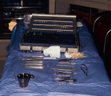
Table 3 lists instruments that are used frequently for strabismus surgery.


Table 3. Surgical Instruments Commonly Used for Strabismus Surgery


InstrumentCatalogue No.Quantity
Castroviejo caliperE2404*1
Lester fixation forcepsE1656*1
Jameson muscle forceps (right)E2331*1
Castroviejo suturing forceps (0.5-mm teeth)E1798*1
Manhattan eye and ear suturing forcepsE1831*1
Jameson muscle hookE0586*2
Stevens curved tenotomy hookE0600*2
Castroviejo needle holder (straight with lock)E3861*2
Desmarres lid retractor size 01E0981*1
Desmarres lid retractor size 0E0980*1
Westcott curved blunt-tip tenotomy scissors (right)E3320*1
Barraquer wire eyelid speculum (adult, pediatric)E4106*1 each
Large resection clampWeck 4825†1
Weck eye rakesWeck 481180†2
Heavy straight Mayo scissorsWeck†1
Small resection clampSP7/21185*1
Malleable retractorSparta 330–520‡1

* Storz Instrument, St. Louis, MO
† Linvatec, Largo, FL
‡ Sparta Surgical, Hayward, CA


PREPARATION

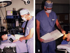
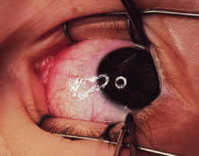
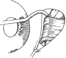
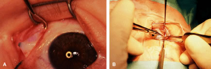
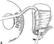
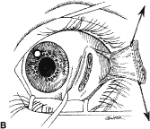
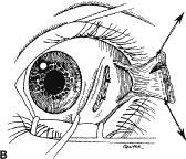
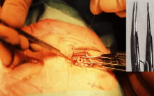
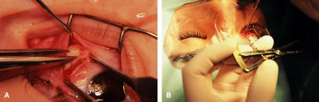
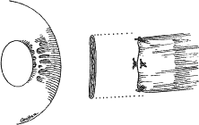
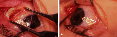
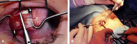
Instrument selection is reviewed by the surgeon (Fig. 1). The patient is positioned so that the head is stabilized, and a shoulder bolster or “roll” is placed under the patient's shoulders to extend the neck slightly (Fig. 2A). The patient is identified, as is the eye to receive surgery (see Fig. 2B).

Fig. 1. Instruments to be used are checked by the surgeon.

Fig. 2. A. The patient is positioned with the head slightly extended. B. The surgeon identifies the patient and the correct eye; the clinical record is checked before proceeding with the procedure.

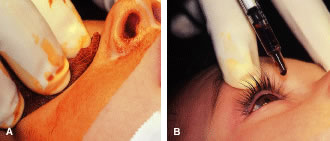
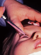
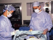
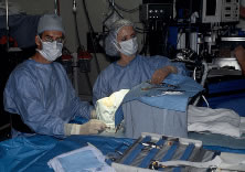
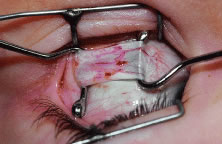
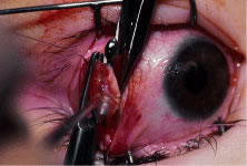
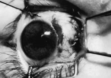
The eyelids and surrounding skin are prepared with two scrubs using a 10% povidone iodine (Betadine) solution that is diluted 50:50 with warm saline38–41 (Fig. 3A and B). The conjunctival cul-de-sacs are irrigated with a bulb syringe and warm saline. Silver protein precipitating solutions, such as Argyrol are not used. A drop of 1% phenylephrine hydrochloride may be instilled into the conjunctival cul-de-sacs to reduce conjunctival bleeding (Fig. 4). The surgical field is isolated with a sterile adhesive barrier drape (see Fig. 4 and Fig. 5). The instrument tray is positioned, and sterile cotton-tipped applicators are placed on either side of the head so that they are available for sponging the field (Fig. 6). A wet-field or bipolar cautery is available. The surgeon is seated at the head of the operating table. The assistant sits to the left of the surgeon, and the surgical technician sits to the right (Fig. 7). Consistency in the procedures that are used to prepare a patient for surgery, such as positioning of the patient and location of the surgeon and the assistants, helps to eliminate confusion among the operating room staff before each procedure. Surgical instruments can be placed on a Mayo stand if a surgical technician is not available to pass instruments to the surgeon (see Fig. 6). The clinical record is positioned where the surgeon can refer to it.

Fig. 3. A. The periorbital tissues are cleansed with two scrubs of povidone iodine (Betadine) solution diluted with warm saline. B. A drop of 5% povidone iodine is instilled into the conjunctival cul-de-sac.

Fig. 4. A drop of 1% of phenylephrine hydrochloride is instilled into the conjunctival cul-de-sac.

Fig. 5. An adhesive barrier drape is used to isolate the surgical field.

Fig. 6. The patient after the adhesive barrier drape has been applied. A Mayo stand provides easy access to instruments if a resident assistant is not available.

Fig. 7. The surgeon sits at the head of the operating room (OR) table. The assistant is to the surgeon's left. The surgical technician is positioned to his or her right.

RECESSION PROCEDURE: FORNIX INCISION

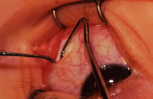
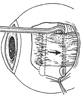
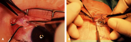
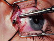
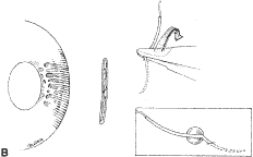
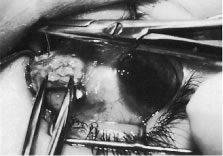
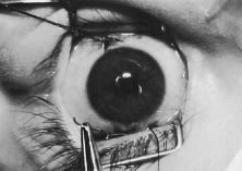
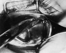
This procedure is recession of the right medial rectus muscle. The eyelids are separated with a Barraquer open-blade wire eyelid speculum (Fig. 8). The open-blade design provides more room for exposure and makes it easier to pass the suture needles, especially in small children or when large recessions are performed. The globe is grasped with a Lester forceps at the limbus. It is best to grasp the limbus with the forceps held perpendicular to the globe and then to position the forceps so that they are rotated and lie tangential to the globe. Forced ductions are performed to detect any restriction of movement of the globe (Fig. 9).

Fig. 8. The lids are separated with a Barraquer open-blade wire speculum. The eye is abducted to test for restriction.

Fig. 9. A Lester forceps grasps the limbal conjunctiva to perform a forced duction test. Care is taken not to allow the globe to move posterior during this maneuver. Restriction of globe rotation should be recorded, and, if it is significant, adjustments in the surgical procedure may be considered.

The globe then is gently elevated from the orbit or proptosed at the same time it is abducted or adducted to prepare for the incision. Elevation of the globe helps to separate the horizontal rectus muscle from the inferior rectus muscle so that the inferior rectus muscle is not cut when the incisions are made. The conjunctiva is grasped by the assistant with a Manhattan toothed forceps (Fig. 10). This forceps has teeth that are angled outward and are designed to grasp conjunctiva, as well as the deeper subconjunctival tissue, so that when the blunt-tipped Westcott scissors cuts into the tented tissue, an incision is made into the conjunctiva and Tenon's capsule (Fig. 11A and B). If the incision does not completely penetrate Tenon's capsule and the intermuscular septum to the scleral surface of the eye, additional tissue (anterior Tenon's tissue and/or intermuscular septum) is grasped with the Manhattan forceps and at least one additional cut is made.

Fig. 10. The conjunctiva is tented between two forceps.

Fig. 11. A. A blunt-tipped Westcott scissors, held perpendicular to the globe, is used to make an incision through the conjunctiva, Tenon's tissue, and intermuscular septum. B. In this view, the surgeon is spreading the scissor blades and viewing bare sclera.

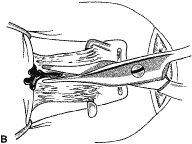
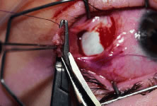
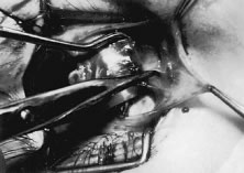
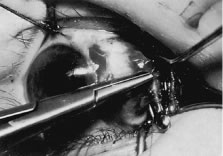
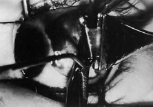
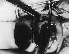
A Stevens' tenotomy hook is passed by the surgeon into the incision and is rotated so that it can be slid underneath the muscle insertion with the tip of the hook held tangential to the globe (Fig. 12). The hook should be passed 2 to 3 mm posterior to the expected location of the muscle insertion. Care is taken not to incorporate intramuscular septum or other adventitial tissue on the hook. When the lateral rectus muscle is secured with the Stevens' or Jameson muscle hook, care should be taken not to bring the inferior oblique muscle up to the insertion (Fig. 13). When the muscle is secured with the Stevens' hook, the hook and the inferior edge of the muscle insertion are elevated and a Jameson muscle hook is passed between the tented muscle tissue and the sclera to secure the muscle at its insertion (Fig. 14). Care is taken to include the entire insertion of the tendon on the hook. The tip of the Jameson hook is gently elevated by depressing the heel of the hook. This maneuver helps to ensure that the entire tendon of the muscle remains captured on the hook. A Stevens' tenotomy hook is placed in the incision, this time anterior to the insertion beneath the conjunctiva (Fig. 15). The Stevens' hook is passed posterior over the orbital surface of the muscle (Fig. 16). Gentle pressure on the Stevens' hook is directed posterior so that the check ligaments and Tenon's tissue that overlie the muscle belly are separated from the muscle capsule with blunt dissection. The hook usually is passed posterior for about 10 mm. When recessing a lateral rectus, attachments between the underside of the lateral rectus and inferior oblique are broken (Fig. 17). Two or three passes over the muscle are made, and, with simultaneous countertraction on the Stevens' hook and the Jameson hook, the conjunctiva is elevated and pulled over the tip of the Jameson hook (Fig. 18A and B).

Fig. 12. The incision is held open with a Manhattan forceps. A Stevens' tenotomy hook is introduced into the incision. The hook is being placed under the rectus muscle without dragging Tenon's tissue and intermuscular septum into the insertion.

Fig. 13. When approaching the lateral rectus, the inferior oblique muscle can be brought up to the lateral rectus insertion. In this instance, the tendon of the lateral rectus muscle should be separated from the inferior oblique muscle with blunt dissection and repositioned back into the incision.

Fig. 14. A Jameson hook is used to spread the incision open with countertraction against the Stevens' hook, which is under the rectus muscle. Then, the Stevens' hook elevates the conjunctiva and the muscle insertion. The Jameson muscle hook is inserted underneath the Stevens' hook to secure the entire insertion of the tendon of the rectus muscle.

Fig. 15. The Jameson hook has been placed under the rectus insertion. A Stevens' tenotomy hook is introduced anterior to the muscle insertion, and with an anterior-posterior movement, the conjunctiva is bluntly dissected free from the anterior extension of the rectus muscle. The Stevens' hook is moved anterior underneath the conjunctiva to approximately 3 or 4 mm anterior to the rectus muscle insertion. It is then passed over the insertion and directed posterior, following a path similar to the course of the muscle from its origin. The hook is carefully passed posterior for about 8 to 10 mm to separate adhesions between the muscle capsule and the overlying Tenon's tissue.

Fig. 16. Countertraction between the Stevens' hook and the Jameson hook will open the incision over the muscle insertion, exposing the muscle tendon on the Jameson hook. The intermuscular septum and anterior Tenon's capsule remain at the upper pole of the insertion. Exposure is provided by a Stevens' tenotomy hook.

Fig. 17. When recessing a lateral rectus, a Jameson hook is passed posterior on the orbital portion of the lateral rectus to breach attachments between the inferior oblique and the lateral rectus.

Fig. 18. A. The muscle tendon is placed on the Jameson hook, and the superior pole of the muscle insertion is exposed with the Stevens' muscle hook. B. View to show the position of the surgeon's hands (right and left) and the assistant's hands on the Jameson (superior) and Stevens' hooks (inferior).

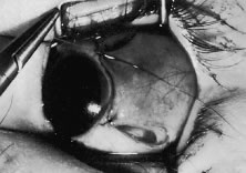
The intermuscular septum and Tenon's capsule at the ball-like tip of the Jameson hook are incised with a Westcott scissors (Fig. 19). A Manhattan forceps can be used to elevate the intramuscular and Tenon's tissue to facilitate this step. Care is taken to incorporate the entire muscle tendon on the Jameson hook before this cut is made. If it is evident that there is residual tendon that is not placed on the hook, the additional tendon, intramuscular septum, and anterior Tenon's tissue are reflected over the tip of the Jameson hook with the closed tips of the Westcott scissors or a small muscle hook.

Fig. 19. A. The intermuscular septum is grasped with a Manhattan forceps. B. A Westcott scissors is used to cut open the intermuscular septum and expose the superior pole of the rectus muscle insertion. The scissors are removed after the placement of a Stevens' hook in the opening that has been created in the intermuscular septum.

A Stevens' hook is inserted in the opening in the intermuscular septum created by the scissors and is passed anterior to the insertion. Parks has referred to this maneuver as the “pole test.” This maneuver is done to verify that the tendon of the muscle has not been split and that the complete muscle tendon is incorporated on the hook. Leaving residual slips of muscle tendon will partially or completely negate the effect of a recession procedure (Fig. 20A and B).

Fig. 20. A. After the Stevens' hook is inserted into the incision created by the Westcott scissors, the hook is moved anteriorly to pass over the upper pole of the insertion of the rectus muscle. If the hook cannot pass anterior to the rectus muscle, the insertion may have been split by the Jameson hook. Fibers of the rectus muscle tendon that are not on the hook will remain at the insertion. It is important to include these fibers to achieve a reproducible recession effect. B. Residual tendon fibers of a detached lateral rectus. These are demonstrated with the Steven's muscle hook.

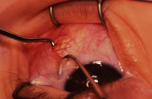
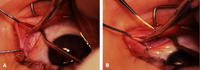
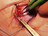
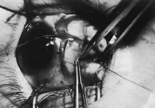
The anterior Tenon's capsule is cut free from the muscle insertion by grasping the loose tissue anterior to the insertion of the tendon with a forceps and gently tenting it (Fig. 21A and B). Care is taken not to grasp muscle capsule or muscle tendon fibers in the forceps. To best accomplish this step, the blunt tipped Westcott scissors is placed perpendicular to the globe. Cleaning the tissue anterior to the muscle permits passing the needle accurately through the rectus muscle tendon with unobstructed visualization. If the capsule of the muscle tendon is cut, the tendon may split. Two small Stevens' hooks are used to elevate the conjunctiva and expose the intermuscular septa and the tissue overlying the muscle capsule. These tissue bands are cut with the Westcott scissors (Fig. 22A and B). Care is taken not to cut into the muscle or the capsule of the muscle (Fig. 23). The intermuscular septa can be cut 3 to 4 mm back for recessions and 5 to 9 mm back for resections.42

Fig. 21. A. The tissue anterior to the insertion is advanced forward. A blunt-tipped Westcott scissors with a Lester forceps (right), held perpendicular to the globe (left), is used to cut the attachments between this subconjunctival tissue and the anterior insertion of the muscle. B. Surgeon's hand position for this maneuver.

Fig. 22. The assistant holds two Stevens' hooks. The surgeon holds the Westcott scissors and the Jameson hook. This technique will provide exposure for cutting the intermuscular septa. The intermuscular septa are cut close to the rectus muscle capsule (A, B).

Fig. 23. A. Care is taken not to cut into the rectus muscle capsule. In this frame, the scissor is aimed incorrectly at the muscle—it should be cutting parallel to the muscle. B. When cleaning the “check ligaments” care should be taken to avoid cutting into the capsule of the rectus muscle.

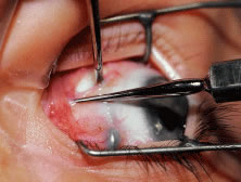
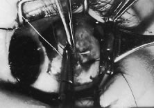
The tendon of the rectus muscle is elevated by lifting it away from the globe with the Jameson hook in a plane that is perpendicular to the surface of the globe. With gentle traction on the muscle insertion with the Jameson hook, a fine spatulated needle with a synthetic absorbable suture is woven through the tendon 1 mm from its insertion to the globe (Fig. 24). The needle exits at the superior edge of the muscle tendon and is then passed back underneath the tendon of the muscle (Fig. 25A and B). The needle is then passed back through the tendon from underneath (lock bite) and out the anterior surface of the tendon, taking a 2-mm portion of tendon that will be incorporated into the double-lock knot. Taking too large a lock bite will tend to narrow the width of the muscle tendon (Fig. 26A and B). The two sutures are brought up and held by the surgeon's thumb on the frets of the Jameson hook (Fig. 27). A blunt-tipped Westcott scissors is used to cut the muscle tendon free from its insertion. The cutting blade of the Westcott scissors is passed posterior to the muscle insertion. Care is taken not to push the tip of the scissor into the insertion but rather to pass the posterior tip of the scissor behind the muscle tendon, in the free space created by elevating the muscle with the Jameson hook. Two or three snips usually are required to remove the muscle from the globe. Care is taken to cut the scleral portion of the insertion flush with the globe. Leaving a large stump at the old insertion will leave a vertical white band of tissue that will show through the conjunctiva and leave an unsightly scar.

Fig. 24. A fine-wire spatulated needle is passed through the anterior insertion. Care is taken to pass the needle through the fibers of the tendon and not just the muscle capsule. The needle is passed back through the tendon from underneath to create a loop of suture. The loop will enable the surgeon to secure the tendon with a lock knot at the muscle insertion.

Fig. 25. A. Lock knot. A double-throw lock knot is secured at the superior and inferior pole of the tendon. B. Diagrammatic representation of the “double lock knot.”

Fig. 26. A. About 1 to 2 mm of tendon are incorporated in each lock bite (top).Too much of the tendon into the lock stitch will narrow the width of the muscle tendon (bottom). B. A narrowed insertion resulting by incorporating too much tendon in the lock bites.

Fig. 27. The surgeon holds the Jameson hook. The suture attached to the muscle insertion is pulled up and trapped between the surgeon's thumb and the frets on the hook. The assistant is exposes the upper pole of the muscle insertion with a Stevens' muscle hook so that the surgeon can cut the muscle tendon from the globe.

The muscle is suspended to verify that both the muscle tendon and the muscle capsule are incorporated with the suture and its lock bites (Fig. 28). In addition, pulling the sutures superior and inferior will assure the surgeon that the transverse suture that was woven through the tendon was not cut when the muscle was removed from the globe (Fig. 29).

Fig. 28. The surgeon suspends the rectus muscle to ensure that the muscle tissue is held securely by the sutures. This technique also will ensure that the transverse suture that was passed through the tendon has not been cut (A, B).

Fig. 29. A. After the muscle has been cut free from the globe, the surgeon should pull gently on the suture. This pull not only ensures that the suture is attached securely to the muscle but also tests whether the transverse portion of the suture placed through the tendon has been cut in the process of removing the muscle from the globe. B. When the transverse suture has been cut, an additional suture should be passed through the tendon so that the muscle will be held securely when it is recessed to its new insertion site.

The globe is stabilized with two locking Castroviejo forceps with 0.5-mm teeth (Fig. 30). Care is taken to grasp the most superior and most inferior portions (for vertical muscle, the most lateral and most nasal portions) of the insertion. For recessions, exposure of the sclera posterior to the insertion is obtained by abducting or adducting the globe while applying a gentle lifting action of the forceps at the insertion. This elevating, or proptosing the globe from the orbit, opens the edges of the conjunctival-Tenon's incision and provides improved exposure of the sclera where the muscle will be reattached.

Fig. 30. The globe is stabilized with two Castroviejo locking forceps. The forceps are held by the assistant. Inset. Magnified view of this forceps.

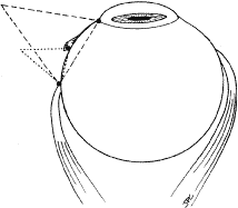
A Castroviejo caliper is used to mark a point on the sclera at a predetermined distance from the posterior portion of the muscle insertion (Fig. 31). Some surgeons prefer to mark the sclera the using the limbus as a reference point. These surgeons believe that it is more accurate to measure from the limbus than from the insertion of the rectus muscle because the insertion may have some anatomic variation. If a large recession procedure will be performed, measurement from the limbus will introduce a cord effect that will increase the amount of recession achieved (Fig. 32). To reduce this effect, William Scott has designed a curved ruler that provides a more accurate measurement of the arc. It is important for the surgeon to be careful and consistent with the measurement technique so that the effect of the surgical procedures can be critically evaluated.

Fig. 31. A. The assistant provides exposure and stabilizes the globe with the Castroviejo forceps. A Stevens' hook is used to retract the conjunctiva. The surgeon marks the sclera, taking care to make the marks directly posterior to the old insertion. B. The surgeon makes final confirmation on the number of millimeters that the caliper is set on.

Fig. 32. When recessions are large, a Castroviejo caliper is used to measure a cord and not the distance along the scleral surface. For small recessions, these effects are negligible. For large recessions, or when measurements are taken from the limbus, this technique can introduce an error in measurement.

The needle is grasped with a locking Barraquer needle holder at the middle portion of the shaft. Grasping the S wedged portion of the needle can cause the needle to roll within the jaws of the needle holder and this can increase the risk of penetration of the sclera. At the mark created by the caliper, there is usually a small dimple or depression in the sclera. The mark results from expressing water from the sclera so that the blue color of the uvea is seen. Because the muscle will attach to the globe at the point at which the needle tip enters the sclera, care is taken to start the intrascleral needle pass at the point marked by the caliper (Fig. 33). Care should be taken not to introduce any unintentional supraplacement or infraplacement effect of the tendon at the new insertion. The width of the insertion should be preserved: The width of the new insertion should equal the width of the original insertion. If the width of the insertion is not preserved, the muscle will not pull up to the new insertion evenly (Fig. 34A and B). If there is a sag in the new insertion caused by the new insertion site being too narrow, the suture can be passed back through the insertion to pull the center of the sagging muscle tendon back to the insertion (Fig. 35). The fine wire needles need to be passed following the curvature of the needle tip. Attempts to elevate the tip of the needle while it is in the sclera can cause the needle shaft to bend (Fig. 36A and B).

Fig. 33. The surgeon begins the intrascleral needle pass by engaging the tip of the needle at the point marked on the sclera by the caliper. The fine-wire, spatula-tip needle is passed through the superficial scleral fibers. Each intrascleral tract usually is 4.5 mm long and parallel to the old insertion. The second suture is passed in a similar fashion. Care is taken to bring the needle tip to the surface without cutting the previously placed suture.

Fig. 34. A. If the new insertion is wider than the old insertion, the muscle will not pull up to the new insertion. B. If the new muscle insertion is not as wide as the old insertion; the central portion of the muscle will tend to sag posteriorly.

Fig. 35. To correct muscle (tendon) sag, the muscle is secured by tying the suture at the new insertion. The needles are then passed back through the tendon, posterior to the transverse suture, and the suture is tied again over the sagging tendon.

Fig. 36. A. With fine-wire needles, the surgeon cannot lift the tip of the needle free from the sclera by trying to elevate it by rotating the needle holder. Rotating the needle holder will cause the shaft of the needle to bend. This bending is dangerous because the tip of the needle is prevented from emerging from the intrascleral bite when the needle is bent. Withdrawing the needle from the sclera will cause the tip to be directed posteriorly, and the globe may be penetrated. B. Diagram of process that can cause the needle to bend.

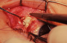
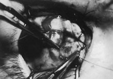
Some surgeons advocate a “crossed-swords technique” so that the second needle pass does not cut the suture from the first needle pass. With appropriate magnification, the other end of the suture is easily avoided. Once the needles have been passed through the sclera, the muscle is pulled up to its new insertion (Fig. 37). A Stevens' hook is used to keep the Tenon's and other surrounding tissue from being drawn into the insertion. The synthetic absorbable sutures tend to drag Tenon's tissue when the suture comes in contact with this tissue. Coating the suture tends to minimize this problem but does not completely eliminate it. The muscle is tied with a surgeon's (double throw) knot that is further secured with two or three square knots. The new insertion is checked and measured to verify that the muscle is in the desired position. After the measurement is checked, the locking Castroviejo forceps are removed.

Fig. 37. A. A double-throw surgeon's knot is used initially to secure the muscle tendon to the new insertion. Two or three square knots are added to complete the knot. B. When completed, the forceps are removed, and the surgical wound is swept closed with a Stevens' muscle hook.

The conjunctival edges of the wound are repositioned or swept closed with a muscle hook. The wound is not sutured. After the procedure, we do not use ointments or occlusive dressings. A drop of an antibiotic-steroid solution can be applied at the discretion of the surgeon.

RESECTION PROCEDURE: FORNIX INCISION

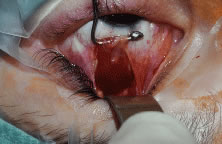
This procedure is a resection of a lateral rectus muscle. The approach to the rectus muscle to be resected is similar to that of the recession. The limbus is grasped with a Lester forceps. The globe is rotated up and away from the muscle to be resected. The conjunctiva is incised, and the rectus muscle is grasped with a Stevens' hook that is replaced with a Jameson hook. The Tenon's tissue and intermuscular septa are cut and reflected onto the Jameson hook. The intermuscular septa are cut with a Westcott scissors. The rectus muscle tendon is then suspended between the Jameson muscle hook (Fig. 38).

Fig. 38. The rectus muscle is suspended between two Jameson muscle hooks.

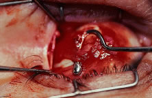
A fine-wire spatula-tip needle is woven through the muscle or tendon. Care is taken to avoid the anterior ciliary vessels on the orbital surface of the muscle (Fig. 39). The resection effect is achieved by measuring the location of the transverse suture from the posterior portion of the Jameson hook at the insertion to a predetermined point on the muscle tendon. Various locations can be used as reference points. This measurement can be taken from the anterior, middle, or posterior portion of the Jameson hook under the muscle. Periodic review of the effects of the operation will determine the amount of surgical correction that is achieved for each surgeon's individual technique. The suture is passed back through the tendon on the origin of the muscle side of the transverse suture. The suture is secured to the tendon with double-throw lock knots (Fig. 40). The muscle is clamped with a fine-tipped vascular clamp (Fig. 41A). A vascular clamp is used because it minimizes the crush damage to tissue that is held in the clamp. Care is taken not to include the transverse suture or any of the lock knots in the clamp. The clamp is stabilized on the drape with a towel clip (see Fig. 41B). A Stevens' hook is used to pull the conjunctiva and anterior Tenon's tissue away from the muscle, and a Westcott scissors is used to cut the muscle free from its insertion (Fig. 42). The muscle stump that is to be resected is grasped with forceps and elevated from the clamp. The resected tendon is then cut free from the clamp with a Westcott scissors (Fig. 43). The sutures are pulled away from the jaws of the clamp by the assistant so that they are not cut. A wet-field cautery is used to cauterize the stump of muscle that is held in the clamp (Fig. 44). The muscle insertion is grasped with a locking Castroviejo forceps in the center of the insertion. Each needle is passed from the upper or lower (for a vertical rectus, medial or lateral) pole of the insertion to exit near the center of the old insertion. The intrascleral bite is made in sclera that is just anterior to the insertion. This location is used to allow the needle to pass through the thickest portion of the sclera (Fig. 45). At the midpoint of the insertion, the tip of the needle is brought out of the sclera and the needle is regrasped and passed through the central portion of the muscle tendon on the origin side of the previously placed transverse suture (Fig. 46). A double-throw knot is tied, and the knot is pinched by the surgeon's index finger and thumb (Fig. 47A and B). The muscle clamp is removed, and the knot is pulled down to the muscle by elevating the suture and snugging it down in a stepwise fashion. Two or three additional square knots are added to secure the muscle to the new insertion (Fig. 48). The muscle is inspected to verify that Tenon's tissue is not drawn into the insertion. The knot is trimmed, and conjunctiva is reposited over the muscle with a Stevens' hook (Fig. 49).

Fig. 39. One of the arms of the Castroviejo caliper is held over the center of the Jameson hook. The other marks the position in which the transverse suture will be placed in the tendon. The surgeon can use as a reference, the anterior, middle, or posterior portion of the tendon on the Jameson muscle hook. Consistent location for the measurement will produce consistent resection results.

Fig. 40. The tendon of the rectus muscle is suspended between two Jameson muscle hooks. A synthetic absorbable suture is placed through the tendon and at each end. A double-throw lock knot placed at the upper and lower edge is used to secure the muscle tendon.

Fig. 41. A. A modified fine-tip vascular clamp is used to clamp the rectus muscle. Care is taken not to include the suture in the clamp. B. The clamp is secured to the drape with a towel clip.

Fig. 42. The surgeon elevates the rectus muscle insertion with the Jameson hook and uses a Westcott scissors to cut the muscle free from the globe. The assistant uses a Stevens' hook to retract the conjunctiva from the insertion.

Fig. 43. A forceps grasps the muscle stump, which is then resected flush to the muscle clamp.

Fig. 44. A wet-field bipolar cautery is used to cauterize the surface of the muscle stump that is held in the jaws of the resection clamp.

Fig. 45. The needle is passed into the sclera, just anterior to the muscle insertion. This area is the thickest portion of sclera, and its use will minimize the risk of penetration of the globe. The tip exits the sclera about the halfway point, near the center of the insertion.

Fig. 46. The needle is then passed underneath the clamp and back through the tendon, posterior to the transverse suture. A similar suture is passed through the superior pole of the insertion and again back through the muscle tendon.

Fig. 47. A. A double-throw knot is tied and pinched between the surgeon's index finger and thumb. B. At this point, the clamp is released. The assistant stabilizes the globe with a Castroviejo forceps. A small Stevens' hook is used to retract the conjunctiva from the insertion site.

Fig. 48. A. The muscle is secured to the new insertion site. B. The assistant is retracting the conjunctiva over the superior pole of the muscle insertion. After the positions of the muscle and surrounding tissue have been checked, the clamps are released. The conjunctiva is reposited into the cul-de-sac with a small hook.

Fig. 49. The globe is stabilized by placing two 6-0 silk sutures through the limbus. Clamps or small clips can be secured to the suture to place traction on the globe.

RECESSION PROCEDURE: LIMBAL APPROACH

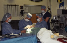
The surgeon and the assistant sit opposing each other on either side of the patient's head. The surgical technician is located between the surgeon and the assistant.

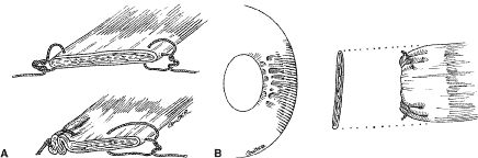
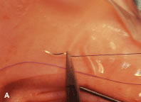
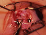
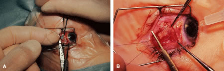
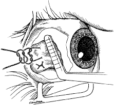
Two 6-0 silk stay sutures are passed through the conjunctiva and superficial scleral tissue at the limbus (see Fig. 49). These sutures are used to stabilize the globe during the procedure. These stabilizing sutures attached to bulldog clamps will hold the globe in adduction or abduction as required.

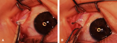
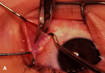
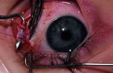
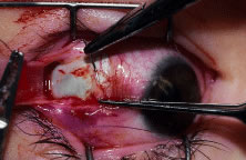
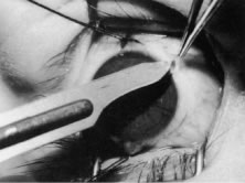
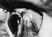
A no. 15 surgical knife or other similar instrument is used to penetrate the conjunctiva at the limbus (Fig. 50). Care is taken to approach the limbustangential to the globe so that inadvertent penetration of the sclera or cornea is avoided. A Westcott scissors also may be used to incise the conjunctiva at the limbus. To facilitate this step, conjunctiva is lifted gently with a 0.5-mm Castroviejo forceps (see Fig. 50).

Fig. 50. The surgeon is tenting the conjunctiva at the limbus. A blade is used to penetrate the tissue at the limbus.

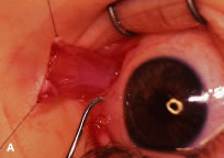
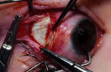
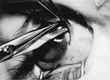
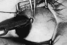
A blunt-tipped Westcott scissors is used to extend the incision for about 3 clock hours (Fig. 51). Once the fused area of conjunctiva and anterior Tenon's capsule has been penetrated, blunt dissection is performed to carry the limbal incision back toward the muscle. This maneuver is best accomplished by directing the closed tips of a blunt-tipped Westcott scissors into the tissue and letting them open to spread the tissue in a plane between the scleral surface and the underside of Tenon's tissue. The limbal incision is extended radially with a Westcott scissors (Fig. 52). Care is taken to avoid cutting into the anterior portion of the rectus muscle insertion (Fig. 53).

Fig. 51. A blunt-tipped Westcott scissors is used to expand the incision just anterior to the rectus muscle.

Fig. 52. The Westcott scissors is used to extend the incision, making radial cuts in the conjunctiva. Care is taken not to enter the muscle capsule and to place these cuts between the vertical and horizontal rectus muscles.

Fig. 53. A cut was made into the anterior muscle capsule (arrow). This error can be avoided by directing the scissor tips parallel to the plane of the muscle when cutting the check ligaments.

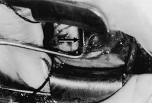
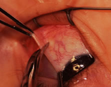
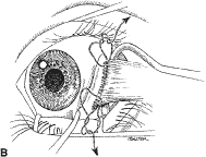
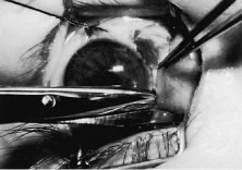
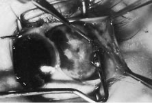
Figure 54 shows the conjunctiva being elevated by the assistant with Castroviejo forceps. The anterior extension of the muscle can be seen.

Fig. 54. The assistant tents the conjunctiva, exposing the rectus muscle insertion.

A Jameson hook is passed under the rectus muscle about 2 mm posterior to the insertion (Fig. 55). The insertion has been identified previously with a Stevens' hook that is used to lift up the tendon to facilitate passage of the Jameson hook.

Fig. 55. The surgeon has passed a Stevens' hook to secure the muscle. A Jameson muscle hook is introduced along the scleral surface to pass underneath the rectus muscle insertion, which is tented up by the Stevens' hook.

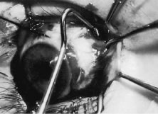
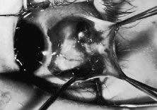
Figure 56 shows sharp dissection of the intermuscular septa and the check ligaments that extend from the orbital surface of the muscle. The assistant applies gentle traction to the conjunctiva to show the surgeon the weblike bands that are to be cut. Care is taken not to penetrate the muscle capsule because penetration can injure the muscle and will cause bleeding. This dissection is carried back about 4 to 5 mm for recessions and 5 to 9 mm for resections.

Fig. 56. In one hand, the surgeon holds the Jameson hook to stabilize the globe. In the other hand, the surgeon holds a Westcott scissors that will divide the intermuscular septa from the muscle capsule.

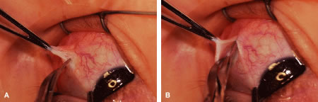
A single-armed 6-0 synthetic absorbable suture is passed through the muscle tendon near its insertion (Fig. 57). A double-lock bite is taken with the needle, and the suture is tied with two square knots to secure it to the upper and lower poles of the tendon of the rectus muscle. The muscle is lifted or tented off of the surface of the globe with a Jameson hook. A Westcott scissors is used to cut the muscle free (Fig. 58).

Fig. 57. The needle is passed through the muscle tendon about 1 mm posterior to its scleral insertion. A second bite is passed through the muscle tendon, and the suture is tied.

Fig. 58. The surgeon tents up the muscle insertion with a Jameson hook. The muscle is cut free from the globe with a Westcott scissors.

The assistant holds the Castroviejo caliper that marks the location of the new insertion (Fig. 59). Care is taken to locate it directly posterior to the stump of the old insertion and to avoid inadvertent supraplacement or infraplacement of the new insertion.

Fig. 59. The globe is stabilized with a locking Castroviejo forceps. A mark is made on the sclera with a Castroviejo caliper. This mark indicates the point of the new insertion.

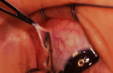
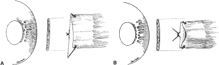
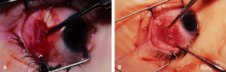
An intrascleral passage of the needle is used to secure the muscle to the globe (Fig. 60). The intrascleral bites are usually 3 to 4 mm in length. The superior and inferior pole sutures are tied to secure the muscle to its new insertion site (Fig. 61), and the wound is inspected. The new insertion is measured to ensure that the appropriate amount of recession effect has been achieved. The sutures are trimmed, and the conjunctiva is brought back to the limbus (Fig. 62). Care is taken to identify the conjunctival tissue. Confusion may occur when an attempt is made to differentiate this tissue from Tenon's tissue. A fine absorbable suture such as 8-0 collagen is used to secure the conjunctiva at the limbus (Fig. 63). If an en bloc recession technique is used, or a bare sclera closure is required, the conjunctiva is recessed 4 to 5 mm back from the limbus, leaving the sclera between the anterior conjunctiva edge and the limbus uncovered. This technique is useful when conjunctival and subconjunctival tissue is contracted or when it restricts movement of the globe.

Fig. 60. An intrascleral bite is taken with a fine-wire needle.

Fig. 61. The suture at the superior pole of the muscle is tied, bringing the muscle to its new insertion. This process is repeated for the inferior pole suture.

Fig. 62. The conjunctiva is reposited at the limbus after the muscle is checked for its new position.

Fig. 63. The conjunctiva is secured at the limbus with two 8-0 collagen sutures. The knots are cut as short as possible. The stay sutures are removed with a Westcott scissors.

RESECTION PROCEDURE: LIMBAL APPROACH

Incision is made into the conjunctiva at the limbus (Fig. 64).

Fig. 64. After the stay sutures have been placed, the conjunctiva is tented with a forceps. An incision is made at the limbus.

The assistant elevates the conjunctiva while the surgeon uses a Stevens' hook to elevate the rectus muscle (Fig. 65). A Jameson muscle hook is passed under the muscle tendon in a plane that is tangential to the scleral surface, about 3 mm behind the tented rectus muscle insertion. Figure 66 shows the rectus muscle on the Jameson hook.

Fig. 65. The rectus muscle insertion is secured with a Jameson hook by passing the hook tangential to the globe.

Fig. 66. The conjunctiva is held up with a forceps while the surgeon holds the rectus muscle tissue on the Jameson hook. The tip of the hook is exposed and is covered by intermuscular septa and anterior Tenon's capsule.

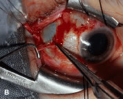
The surgeon cuts the intermuscular septa free from the muscle capsule (Fig. 67). A Castroviejo caliper is used to measure the amount of tendon that will be resected (Fig. 68). A Jameson resection clamp is placed on the muscle at the point of anticipated resection. Measurement is made from the portion of tendon that is on the posterior edge of the Jameson hook to the resection clamp. Exposure is facilitated with a Desmarres retractor.

Fig. 67. The intermuscular septa are cut with a Westcott scissors.

Fig. 68. A Jameson resection clamp is applied to the muscle tendon. A caliper is used to measure the amount of tendon that will be resected.

The muscle is cut free from its insertion (Fig. 69). The surgeon holds the Jameson clamp to elevate the muscle from the globe, and, using the other hand, the surgeon cuts the muscle free from the globe. The muscle stump is cut so that it is flush with the globe.

Fig. 69. A Desmarres retractor is used to provide improved exposure. A Westcott scissors is used to cut the muscle free from its insertion.

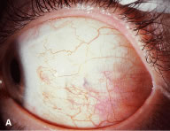
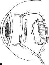
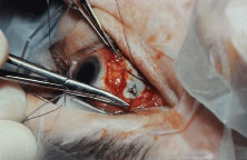
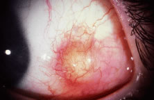
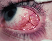
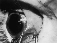
The globe is stabilized with a 0.5-mm-toothed Castroviejo forceps (Figs. 70 and 71). A mattress stitch with a double-armed 6-0 synthetic absorbable suture is placed through the most inferior portion of the insertion. The second needle is passed 1 mm below the central portion of the insertion. The initial penetration of the needle is just anterior to the insertion, and a secure bite is taken of the former insertion so that the needle exits just posterior to the old insertion. The needle is then passed underneath the Jameson resection clamp through muscle tendon, and the two sutures are held with a bulldog clamp to keep them separate from the sutures that are later passed through the upper pole of the insertion. Figure 71 shows the sutures being tied. This maneuver will effectively bring the resected muscle tendon up so that it can be reinserted at the insertion. Once the muscle is tied securely, the Jameson resection clamp is released and then advanced to the cut end of the insertion (Fig. 72). Under direct visualization, a Westcott scissors is used to trim the excess muscle stump. Care is taken to leave at least 1 mm of muscle tendon in front of the two sutures that secure the muscle to the insertion. After the muscle stump has been inspected, the conjunctiva is advanced back to the limbus (Fig. 73). Two 8-0 collagen sutures are used to secure the conjunctiva at the limbus. The small open radial portion of the incision will heal without suture closure. If the surgeon “buries the knots” under the conjunctiva near the limbus, the patient will have less foreign body sensation. Figure 74 shows the eye immediately after a recess-resect procedure. The conjunctiva should be smooth at the limbus to prevent disruption of the tear layer and formation of dellen. The eye is not patched. Antibiotic drops or antibiotic-steroid drops may be used at the discretion of the surgeon.

Fig. 70. A double-armed mattress suture is passed through the anterior portion of the insertion, exiting posterior to the severed stump of the insertion. The two needles are passed back through the muscle tendon just posterior to the Jameson clamp.

Fig. 71. Knots are tied to secure the resected muscle at its new insertion. The Jameson resection clamp has already been moved anteriorly on muscle before tying the sutures.

Fig. 72. The Jameson clamp is removed and reinserted closer to the cut portion of tendon, near the old insertion. The muscle is then resected.

Fig. 73. The conjunctiva is closed with two interrupted 8-0 collagen sutures.

Fig. 74. The immediate postoperative appearance of an eye that has had a recess-resect procedure with a limbal approach.

Back to Top
POSTOPERATIVE CARE
In the early postoperative period, patients are given antiemetic suppositories and a clear liquid diet to control nausea. If pain is experienced, acetaminophen is given. Warm compresses with a clean washcloth soaked with warm tap water are applied to the closed eye several times a day for 3 to 4 days after the operation, or until the bloody secretions have subsided. We do not routinely use antibiotic drops or occlusive dressings. The patient or family may be contacted by the surgeon's office and/or the surgical facility to ensure recovery from the procedure is proceeding as expected. The patient is re-examined 3 to 7 days after the operation and again at 4 to 6 weeks. If there is suspicion of a complication or an unexpected result, more frequent follow-up visits are indicated.
Back to Top
RESULTS
The results of operations to correct strabismus will depend on the accuracy of analysis of the causes of the ocular misalignment. The repositioning of the rectus muscles is an execution of strategy to correct the strabismus. This strategy is planned in the office, before proceeding to the operating room. This plan may be altered based on findings at the time of the operation.

Even with precise measurement and meticulous surgical technique, overcorrections and undercorrections will occur. Some will occur as a result of sensory and motor factors whose explanation is beyond the scope of this chapter. The execution of surgical repositioning of extraocular muscles should be a consistent procedure, executed with precision with the use of meticulous surgical technique. Surgeons should expect the procedure to yield an equal number of overcorrections and undercorrections. Periodically, surgeons should review their results and adjust their procedures and measurements accordingly. An acceptable result is a comitant deviation of less than 5 to 10 prism diopters as measured by the alternate prism and cover test at 6 weeks. For vertical deviations, fusion amplitudes are not as great, and deviations as small as 2 to 3 prism diopters are significant and can produce distracting diplopia.

Back to Top
COMPLICATIONS AND THEIR MANAGEMENT
All operations have complications. Some complications are avoidable, but others will occur even when the most stringent preventive measures have been taken. Complications of rectus muscle surgery may occur during the procedure or in the immediate or late postoperative period.

CORRECT EYE AND CORRECT PROCEDURE

The surgeon should identify the most probable eye or eyes that will be operated on in the office. Agreement between the surgeon and parents or patient should be confirmed when preparing the consent.

The surgeon should review the clinical record thoroughly before performing the surgery. Before the operation is scheduled, an attempt should be made to obtain old records if the surgeon believes that they will influence surgical decisions. A strategy to correct the ocular alignment with a single operation should be designed and transferred to the patient's record. The record or a copy of this record should be available in the operating room for reference in case unexpected findings occur. The surgeon should personally identify the patient and indicate the eye or eyes to be operated on before beginning the procedure. When the patient is a child, the surgeon should support and reassure the child during the induction of the anesthetic.

HEMORRHAGE

A small amount of bleeding during rectus muscle surgery is common. Bleeding is controlled with application of wet-field cautery. Small amounts of cautery will have no effect on the outcome of the procedure. Deep orbital bleeding usually occurs when a muscle hook is passed more than 10 to 12 mm posterior to the muscle insertion, where it can engage or tear a vortex vein. Excessive traction on a muscle may stretch a vortex vein to its point of rupture. Bleeding from a vortex vein is controlled by removing the instruments and the lid speculum. The globe and orbital contents are gently compressed for 3 to 4 minutes with a 4-× 4-in. gauze pad that is placed over the closed eyelids. Bleeding usually subsides with these measures. Once bleeding has stopped, the procedure can be resumed. Deep orbital bleeding can produce periorbital ecchymosis that will last 7 to 10 days.

Another common source of hemorrhage is bleeding from the anterior ciliary vessels. This bleeding may occur when a needle is passed through the tendon and penetrates one of the anterior ciliary vessels. This penetration will cause a self-limited hematoma under the muscle capsule (Fig. 75). It may also occur when the Tenon's tissue is cleaned from the muscle (Fig. 76). Hemorrhage around the insertion may make removal of the muscle from the globe difficult because it can obscure the area where the tendon of the muscle is attached to the globe. Bleeding from the anterior ciliary vessels can be controlled by cauterizing vessels with a wet-field (bipolar) cautery. Care should be taken not to cauterize tissue near a suture after it has been placed because doing so may cause structural damage to the suture.

Fig. 75. A small hematoma in the muscle capsule resulted from cutting the anterior ciliary vessel when cleaning the muscle insertion before placing the suture.

Fig. 76. The muscle capsule has been removed from the muscle tendon. Hemorrhage can occur, which can obscure the surgical field.

SLIPPED MUSCLE

If the muscle becomes completely detached from the globe and contracts posteriorly, the term “lost” muscle is used.

A muscle that moves from its desired location is called a slipped muscle.43–46 Causes of a slipped muscle include untying of the knot (Fig. 77A and B), inadvertent cutting of the suture, placing the suture too close to the insertion, or taking an inadequate bite of sclera with passage of the needle. In the latter case, the suture will “cheese wire,” or pull through the sclera. Finally, if the suture engages only the muscle capsule and not the tendon, the tendon can slide posterior within the muscle capsule. Because the maximal pull, or force, generated by an extraocular muscle is only 50 g, insufficient strength of the suture material is seldom the cause of a slipped muscle.

Fig. 77. A. This patient had a lateral rectus muscle recessed 5 days ago. The patient was referred for evaluation of overcorrection and poor abduction. The incision was explored, and the suture that was holding the rectus muscle had become untied. Careful examination of the blue suture under the conjunctiva raised suspicion of this complication. B. Schematic demonstrating the findings at surgical correction.

SCLERAL PERFORATION

Recognized and unrecognized scleral perforation will occur. Reattachment of the muscle to the globe requires passing a needle within the sclera. The scleral coat of the globe is thinnest just posterior to the insertion of the rectus muscles. If the sclera becomes dry, it will thin further, and the blue color of the underlying uveal tissue will be visible. Needles with specially designed spatula tips are used to draw the sutures through the scleral tissue. Adequate illumination and magnification will facilitate this procedure. If the operation is prolonged, periodic wetting of the cornea and sclera with saline or a balanced salt solution will reduce thinning resulting from desiccation. In addition, adequate exposure and stabilization of the globe will reduce this complication. Scleral perforation also can occur when the needle is in the sclera and the surgeon attempts to lift the tip of the needle by placing torque on the needle with the needle holder (see Fig. 36). The fine-wire needles will bend, and, when the needle is released, the tip is directed posterior or into the sclera. Unexpected movement of the patient also may cause perforation of the globe (Fig. 78).

Fig. 78. This patient unexpectedly moved when the muscle was being cut from its insertion. The sclera had a small tear that was closed with two 8-0 silk sutures.

If the surgeon suspects that a needle has penetrated the globe and passed into the choroid or through the retina, indirect ophthalmoscopy should be performed.47–49 If a hole in the retina is observed, retinal consultation should be considered. Cryotherapy can be applied over the perforation site. If there is no hemorrhage and the vitreous is solid (the patient is young), I do not use cryotherapy. However, if the patient is an older adult or there is a large myopic refractive error or the vitreous has become liquefied, it would be prudent to apply cryotherapy to the sclera over the site of the perforation. Direct visualization can be used to control the amount and extent of retina treated. Use of antibiotics and consultation with a vitreoretinal surgeon for treatment or following treatment can be considered.

CONJUNCTIVAL SCARRING AND DISTORTION OF ANATOMY

Rectus muscle surgery should not disturb the plica semilunaris. If a medial rectus muscle is lost at the time of surgery and vigorous attempts are made to find it, the tissue that attaches to the plica can be distorted as a result of tissue trauma and hemorrhage associated with the exploration. The plica may become adherent to the surface of the globe, and this adherence can result in a restrictive strabismus.

When a rectus muscle is cut free from the globe, the surgeon should attempt to cut the muscle tendon flush with the scleral surface. Failure to do so will leave a vertical white scar under the conjunctiva that overlies the old muscle insertion, especially when the lateral rectus muscle is recessed.

NAUSEA

Nausea commonly occurs after strabismus surgery despite many efforts to prevent it. Nausea occurs as a result of stimulation of the oculocardiac reflex, which produces a vagal response.50–52 When stimulated, this reflex can cause nausea or even asystole.51 Stimulation of this reflex combined with the use of general anesthesia, usually will produce 3 to 5 hours of nausea after strabismus surgery. Nausea is the most common cause of admission for overnight observation.52 To prevent nausea, the surgeon and assistant should exert only as much traction on the muscles and surrounding tissues as is needed to provide adequate visualization.

Nausea is best managed by suctioning the stomach at the end of the operation and restricting fluid intake. Light feeding is recommended in the immediate postoperative period. Care is taken to replace fluids with administration of intravenous fluids. The use of rectal suppositories such as Tigan 100 to 200 mg every 4 hours also may be helpful.

LIGHT SENSITIVITY

Light sensitivity can be caused by inadvertent trauma to the cornea during the procedure or by a combination of drying of the cornea surface and disturbance of the normal tear film by the preparation solutions. The mild keratitis caused by drying and preparation solutions can be avoided by keeping the procedure time to a minimum. Measures should be taken to protect the cornea throughout the procedure.

DELLEN

Strabismus surgery can cause a dell or dellen or a reduction in corneal thickness from deficiencies in tear film. Usually, this problem is caused by elevation of the conjunctiva at the limbus. Care should be taken to ensure that the lids will pass over a smooth conjunctival surface at the corneal-scleral junction at the conclusion of the procedure. Simply patching the eye for a few hours will cause the cornea stroma to rehydrate and return to its normal thickness.

INFECTION

Because infections after strabismus surgery are uncommon, we concur with the many surgeons who prefer not to use antibiotics or antibiotic-steroid drops after uncomplicated strabismus procedures.53–56

Conjunctivitis is common and results most commonly from irritation of the conjunctival surfaces by preparation solutions. The conjunctiva may be irritated by the procedure. If there is purulent discharge, the use of topical antibiotics should be considered.

Occasionally, a small abscess may form around a suture (Fig. 79). Orbital cellulitis and endophthalmitis are extremely rare complications of strabismus surgery. Only three cases of endophthalmitis have been reported after strabismus surgery since 1935.53 Ing55 found one case of cellulitis for every 1900 strabismus operations performed. The same author reported 1 case of endophthalmitis for every 30,000 cases of strabismus surgery.55 Cellulitis can occur if a child rubs the eyes after rubbing a runny nose. To avoid this complication, children who have a febrile illness should have elective surgery postponed.

Fig. 79. A small suture abscess present 8 weeks after strabismus surgery was treated successfully with topical applications of antibiotic-steroid eyedrops.

Endophthalmitis can result from inadvertent scleral perforation. Use of prophylactic antibiotics may be considered. Systemic intravenous therapy is used to treat cellulitis, and appropriate measures should be initiated promptly if endophthalmitis is suspected.

VASCULAR COMPLICATIONS

Anterior segment ischemia or necrosis can occur after rectus muscle surgery.35,57,58 Usually, ischemia occurs after removal of the third or, more commonly, the fourth rectus muscle. Predisposing factors are thyroid eye disease, sickle cell anemia, and prior strabismus surgery. Recovery of ocular function after ischemia usually occurs within weeks.59 To avoid this complication in patients at risk, surgeons should use a cul-de-sac approach to gain access to the rectus muscle.60 This approach to the rectus muscle preserves the conjunctival circulation at the limbus. The limbal circulation does not have an important role in nourishing the anterior segment. However, the conjunctival vessels anastomose with deeper vessels, and this approach may provide added safety in borderline cases. Patients at risk can be pretreated with aspirin 3 days before surgery. Oxygen requirements may be reduced by treating inflammation with a topical or oral corticosteroid. The surgical strategy to correct a deviation should be planned so that as few rectus muscles as possible are repositioned.

PYOGENIC GRANULOMA-CONJUNCTIVAL CYST

About 1 of every 100 strabismus operations will be complicated by the development of exuberant granulation tissue at the site of the conjunctival incision (Fig. 80). Tenon's capsule can protrude through the conjunctival incision and produce an exuberant granulation response. Granulation tissue is treated with frequent application of topical antibiotic-steroid drops. The granuloma usually will become pedunculated and drop off of the site. If the base is broad, the tissue is lifted with a forceps and removed with a scissors.

Fig. 80. Exuberant granulation tissue present 1 week after strabismus surgery.

Conjunctival cysts pose more difficult problems.61 These cysts usually form under the conjunctival surface, but some may be carried back with the recessed muscle and rest alongside the muscle or its new insertion (Fig. 81). Cysts occur more frequently after correction of strabismus in patients with strabismus following retinal detachment procedures or re-operations. Cysts do not respond to medical treatment and almost always require surgical excision. In these cases, the surgeon should attempt to remove the entire cyst structure or to marsupialize it if complete removal is impossible.

Fig. 81. A conjunctival cyst 3 months after strabismus surgery. These cysts will usually require surgical removal.

RESTRICTION

Restriction of the globe after rectus muscle surgery should be uncommon (Fig. 82). Restriction usually is caused by scar tissue that is produced by tissue trauma. Meticulous surgical technique, the use of synthetic absorbable sutures, and a clear understanding of and respect for the anatomy will minimize these complications.

Fig. 82. This globe had a Swann approach to the lateral rectus muscle. Excessive scar tissue around the muscle and underneath the conjunctiva caused restriction on attempted adduction of the eye.

EYELID POSITION

Recession or resection of a vertical rectus muscle that is larger than 3 mm may cause the lids to follow the muscle. For example, if the muscle is recessed, the lid overlying the muscle will be elevated or retracted with the muscle. The opposite effect occurs with resections. The effect of strabismus surgery on eyelid position can be reduced by dissecting the intramuscular septa and check ligaments under the inferior rectus muscle and by dissecting and severing the capsulopalpebral attachments from the inferior rectus muscle to the lower lid.62,63

OVERCORRECTION AND UNDERCORRECTION

Despite all efforts to understand a patient's problem and the meticulous execution of a surgical repositioning of the muscles, overcorrections and undercorrections occur. The surgeon should make every effort to ensure that technique failure is not the cause of the undesirable alignment. If the overcorrection and undercorrection is small, occlusion, prisms, optical, botulinum A toxin, and finally re-operation may be used to produce a satisfactory alignment. When these measures fail, and if the residual deviation is unacceptably large, re-operation should be considered as soon as 6 to 8 weeks after the initial procedure.

If a muscle is slipped or there is unexplained lack of function of a muscle during the first week after surgery, immediate exploration is indicated to correct the problem.

REFRACTIVE ERROR

An astigmatism may be produced or altered by rectus muscle surgery.64–68 Changes in children usually are transient, but in adult patients, the altered refractive error may persist.65 Once the rectus muscles resume their normal contractile force, distortion of the anterior segment and cornea is reduced and the astigmatism disappears. This effect usually occurs before 8 weeks. Change in the refraction can occur as a result of scleral necrosis67 or as a result of displacement of the lens.68

Back to Top
REFERENCES

1. Duke-Elder S: Anomalies of the ocular motility. In Duke-Elder S (ed): System of Ophthalmology. Vol 6. London, Henry Kimpton, 1973, pp 223–228

2. Evens L: History of strabismus treatment. Bull Soc Belge Ophthalmol 1981;195:19

3. Flick H: History of strabismus treatment. Historia Ophthalmol Int 1984;3:119

4. Von Noorden GK: The development of the art and science of strabismology outside North America: Part I. JAPOS 2001;5:65

5. Dix JH: Treatise on Strabismus or Squinting and the New Mode of Treatment. Boston, D. Clapp Jr, 1841, pp 3–35

6. Hirshberg J: The renaissance of ophthalmology in the eighteenth century, Part 3. The first part of the nineteenth century, Part 1. The History of Ophthalmology. Translated by FC Blodi. Vol 5, Part 1. Bonn, Germany, JP Wayenbargh, 1985, pp 167, 320, 335, 324

7. Jones TW: Strabismus convergency. In Jones TW, Hays I (eds): The Principle and Practice of Ophthalmic Medicine and Surgery. Philadelphia, Lea-Blanchard, 1847, pp 366–373

8. Fuchs E: Operations upon the adnexa bulbi. In Fuchs E, Duane A (eds): Textbook of Ophthalmology. New York, Appleton, 1892, pp 706–713

9. Metter J: The eye muscles. In Metter J, Sweet WM (eds): Ophthalmic Surgery. Philadelphia, P Blackstone & Son, 1913, pp 101–118

10. Travers TA: Operative treatment of concomitant squint. In Concomitant Strabismus. London, G Pulman, 1936, pp 79–88

11. Swan KC, Talbot T: Recession under Tenon's capsule. Arch Ophthalmol 1954;51:32

12. von Noorden GK: The limbal approach to surgery of the rectus muscles. Arch Ophthalmol 1968;80:94

13. Parks MM: Fornix incision for horizontal rectus muscle surgery. Am J Ophthalmol 1968;65:907

14. Jampolsky A: Current techniques of adjustable strabismus surgery. Am J Ophthalmol 1979;88:406

15. Repka MX, Guyton DL: Comparison of hang-back medial rectus recession with conventional recession. Ophthalmology 1988;95:782

16. Prieto-Diaz J: Large bilateral medial rectus recession in early exotropia with bilateral limitation of abduction. J Pediatr Ophthalmol Strabismus 1980;17:101

17. Nelson LB, Calhoun JC, Simon JW et al: Surgical management of large congenital exotropia. Br J Ophthalmol 1987;71:380

18. Cuppers C: The so-called “Fadenoperation.” In Fells P (ed). Second Congress of the International Strabismological Association. Marseilles, France, Diffusion Générale de Librairie, 1976, pp 395–400

19. Mühlendyck H: Diagnosis of convergent strabismus with nystagmus and its treatment with Cupper's Fadenoperation. In Moore S, Mein J, Sockbridge L (eds). Orthoptics: Past, Present and Future. New York, Stratton, 1976, p 143

20. Thorson JC, Jampolsky AJ, Scott AB: Topical anesthesia for strabismus surgery. Tr Am Acad Ophthalmol Otolaryngol 1966;70:968

21. Szmyd SM, Nelson LB, Calhoun JH et al: Retrobulbar anesthesia. Arch Ophthalmol 1984;102:1325

22. Diamond GR: Topical anesthesia for strabismus surgery. JAPOS 1989;26:86

23. Cheng KP, Larson CE, Biglan AW et al: A prospective, randomized, controlled comparison of retrobulbar and general anesthesia for strabismus surgery. Ophthalmic Surg 1992;23:585

24. Kushner BJ: Post-operation binocularity in adults with long-standing strabismus: Is surgery cosmetic only? Am Orthopt J 1990;40:64

25. Kushner BJ, Morton GV: Postoperative binocularity in adults with long-standing strabismus. Ophthalmology 1992;99:316

26. Morris RJ, Scott WE, Dickey CF: Fusion after surgical alignment of longstanding strabismus in adults. Ophthalmology 1993;100:135

27. Ohtsuki H, Hasebe S. Kobashi R et al: Critical period for restoration of normal stereoacuity in acute-onset comitant esotropia. Am J Ophthalmol 1994;118:502

28. Umazume F, Ohtsuki H, Hasbe S: Predictors of postoperative binocularity in adult strabismus. Jpn J Ophthalmol 1997;41:414

29. Greenwald ME: Expanded binocular peripheral visual fields following surgery for esotropia. JPOS 1989;26:109

30. Sullivan, TJ, Kraft SP, Burack C et al: A functional scoring method for the field of binocular single vision. Ophthalmology 1992;99:575

31. Kushner BJ: Binocular field expansion in adults after surgery for esotropia. Arch Ophthalmol 1994;112:639

32. Coats DK, Paysse EA, Towler AJ et al: Impact of large angle horizontal strabismus on ability to obtain employment. Ophthalmology 2000;107:402

33. Paysse EA, Steele EA, Brady-McCreery KM et al: Age of the emergence of negative attitudes toward strabismus. JAAPOS 2001;5:361–366

34. Costello P, Simon JW, Jia Y et al: Acquired esotropia: Subjective and objective outcomes. J AAPOS 2001;5:193

34a. Satterfield D, Keltner JL, Morrison TL: Psychosocial aspects of strabismus study. Arch Ophthalmol 1993;111:1100

35. Saunders RA, Bluestein EC, Wilson ME et al: Anterior segment ischemia after strabismus surgery. Surv Ophthalmol 1994;38:456

36. France NK, France TD, Woodburn JD et al: Succinylcholine alteration of the forced duction test. Ophthalmology 1980;87:1282

37. Rainin EA, Carlson BM: Postoperative diplopia and ptosis: A clinical hypothesis based on myotoxicity of local anesthetics. Arch Ophthalmol 1985;103:1337

38. MacRae SM, Brown B, Edelhauser HF: The corneal toxicity of presurgical skin antiseptics. Am J Ophthalmol 1984;97:221

39. Isenberg S, Apt L, Yoshimuri R: Chemical preparation of the eye in ophthalmic surgery: I. The effect of conjunctival irrigation. Arch Ophthalmol 1983;101:761

40. Isenberg S, Apt L, Yoshimuri R: Chemical preparation of the eye in ophthalmic surgery: II. Effectiveness of mild silver protein solution. Arch Ophthalmol 1983;101:764

41. Boes DA, Lindquist TD, Fritsche TR et al: Effects of povidone iodine chemical preparation and saline irrigation on the perilimbal flora. Ophthalmology 1992;99:1569

42. Friendly DS, Parelhoff ES, McKcown CA: Effect of severing the cheek ligaments and intermuscular membranes on medial rectus recessions in infantile esotropia. Ophthalmology 1993;100:945

43. Hiller TL, Brown GC, Nelson LB et al: The slipped rectus muscle. Ophthalmic Surg 1985;5:315

44. Plager DA, Parks MM: Recognition and repair of the “lost” rectus muscle. Ophthalmology 1990;97:131

45. Murray AD: Slipped and lost muscles and other tales of the unexpected. JAAPOS 1998;2:133

46. Goldberg RA: Is there a “lost” rectus muscle in strabismus surgery? Am J Ophthalmol 2001;132:101

47. Simon JW, Linninger LL, Scheraga JL: Recognize scleral perforation during eye muscle surgery: Incidence and sequelae. J Pediatr Ophthalmol Strabismus 1992;29:273

48. Mittleman D, Bakos I: The role of retinal cryopexy in the management of experimental perforation of the eye during strabismus surgery. J Pediatr Ophthalmol Strabismus 1984;21:186

49. Springer DT, Klapper SR, Bonnin JM et al: Management of experimental globe perforation during strabismus surgery. J Pediatr Ophthalmol Strabismus 1996;33:140

50. Ohashi T, Kase M, Yokoi M: Quantitative analysis of the oculocardiac reflex by traction on human extraocular muscles. Invest Ophthalmol Vis Sci 1986;27:1160

51. Gold RS, Pollard Z, Buchwald IP: Asystole due to oculocardiac reflex during strabismus surgery: A report of two cases. Ann Ophthalmol 1988;20:473

52. Isenberg SJ, Apt L, Yamada S: Overnight admission of outpatient strabismus patients. Ophthalmic Surg 1990;21:540

53. Wilson ME, Paul TO: Orbital cellulitis following strabismus surgery. Ophthalmic Surg 1987;18:92

54. Kivlin JD, Wilson ME and the Periocular Infection Study Group: Periocular infection after strabismus surgery. J Pediatr Ophthalmol Strabismus 1995;32:42

55. Ing MR: Infection following strabismus surgery. Ophthalmic Surg 1991;22:41

56. Recchia FM, Baumal CR, Sivalingam A et al: Endophthalmitis after pediatric strabismus surgery. Arch Ophthalmol 2000;118:939

57. Freeman HM, Hawkins WR, Schepens CL: Anterior segment necrosis: An experimental study. Arch Ophthalmol 1966;75:644

58. France TD, Simon JW: Anterior segment ischemia syndrome following muscle surgery: The AAPO&S experience. J Pediatr Ophthalmol Strabismus 1986;23:87

59. Oliver JM, Lee JP: Recovery of anterior segment circulation after strabismus surgery in adult patients. Ophthalmology 1992;99:305

60. Fishman PH, Repka MX, Green WR et al: A primate model of anterior segment after strabismus surgery. Ophthalmology 1990;97:456

61. Kushner BJ: Subconjunctival cysts as a complication of strabismus surgery. Arch Ophthalmol 1992;110:1243

62. Kushner BJ: A surgical procedure to minimize lower eyelid retraction with inferior rectus recession. Arch Ophthalmol 1992;110:1011

63. Pacheco EM, Guyton DL, Repka MX: Changes in eyelid position accompanying vertical rectus muscle surgery and prevention of lower lid retraction with adjustable surgery. J Pediatr Ophthalmol Strabismus 1992;29:265

64. Thompson WE, Reineche R: The changes in refractive status following routine strabismus surgery. J Pediatr Ophthalmol Strabismus 1980;17:372

65. Fix A, Baker JD: Refractive changes following strabismus surgery. Am Orthopt J 1985;35:59

66. Dottan SA, Hoffman P, Oliver MD: Astigmatism after strabismus surgery. Ophthalmic Surg 1988;19:128

67. Gross, SA, Von Noorden GK, Jones DB: Necrotizing scleritis and transient myopia following strabismus surgery. Ophthalmic Surg 1993;24:839

68. Brodsky MC: Myopia, hypotony and lenticular shift following strabismus surgery. Ophthalmic Surg Lasers 1998;29:426

Back to Top