Chapter 97
Surgery to Correct Cyclotropia
PHYLLIS E. WEINGARTEN and DAVID L. GUYTON
Main Menu   Table Of Contents

Search

ASSESSMENT OF CYCLODEVIATIONS
INDICATIONS FOR SURGICAL CORRECTION
CONTRAINDICATIONS TO TORSIONAL SURGERY
SURGICAL PROCEDURES
CONCLUSION
REFERENCES

Torsional deviations are commonly seen in patients with strabismus. Anatomic torsion is more prevalent than symptomatic torsion. Cyclodeviations may accompany cyclovertical muscle paralysis, particularly superior oblique paresis; A- and V-pattern strabismus1,2; craniofacial developmental anomalies; and restrictive deviations, including thyroid ophthalmopathy and Brown's syndrome. Few patients become symptomatic because of small amounts of torsion (Table 1); some compensate by sensory cyclofusion. Reorientation of retinal meridians at the cortical level in children, or “psychological” adaptation in adults, explains the scarcity of symptoms from torsional deviations.3,4


TABLE 1. Patient Awareness of the Presence of a Cyclodeviation Relative to the Degree of Torsion as Measured by Double Maddox Rods


Amount of Cyclodeviation (degrees)Percentage Symptomatic
15100
1086
555
(Data from Trobe J: Cyclodeviation in acquired vertical strabismus. Arch Ophthalmol 102:717, 1984.)


Back to Top
ASSESSMENT OF CYCLODEVIATIONS
Both subjective and objective cyclodeviations must be assessed for proper clinical analysis and management. Commonly used tests for measuring subjective torsion include the double Maddox rod test and the Lancaster red-green test. Double Maddox rod testing is easily performed, but environmental clues and sensory cyclofusion of the long streaks of light can affect the amount of torsion measured. Moreover, the double Maddox rod test is difficult to perform reliably away from the primary position.

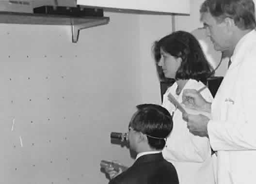
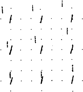
Our preferred method of testing for subjective torsion is the Lancaster red-green test (Fig. 1). This test allows the clinician to evaluate torsion in nine positions of gaze and clearly indicates where torsion is greatest and where vertical deviation is most significant. The test is performed in a darkened room so that no spatial clues in the environment can help in orienting the streaks of red and green light. By convention, a red filter is placed over the right eye and a green filter over the left. The examiner takes the light corresponding to the desired “fixing eye” and shines it on the wall successively in the nine diagnostic positions of gaze. In each position, the patient is asked whether the streak of light is straight or tilted. (We customarily do the test with vertical streaks, in that it is easier for the patient to describe tilt in a vertical streak than a horizontal streak.) If the streak is tilted, the examiner adjusts the light until the patient sees the streak exactly vertically. The patient then superimposes the second streak on the first streak. The examiner records the position and orientation of the streaks to analyze the torsional and vertical components of the deviation in the different fields of gaze (Fig. 2). The absolute horizontal deviation is difficult to judge with this test because of the poor control of accommodation provided by the streaks of colored light. The relative horizontal deviation, however, is reasonably reliable, and is used for judging A or V patterns.

Fig. 1. Performance of the Lancaster reg-green test.

Fig. 2. Lancaster red-green plot of a patient with a left superior oblique paresis. The solid line (red streak) is the projection of the subjective vertical meridian of the right, fixing eye and the dotted line (green streak) represents the left eye. Subjective extorsion appears in both eyes and a small V-pattern exotropia is present. The left hyperdeviation is greatest in upgaze and to the right.

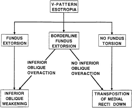
Objective torsion is best assessed by indirect ophthalmoscopy or fundus photography.2,5 For example, in a patient with V-pattern esotropia, the presence of fundus extorsion justifies inferior oblique weakening surgery in addition to horizontal surgery (Fig. 3).

Fig. 3. Contribution of fundus torsion to the surgical approach for V-pattern esotropia.

Back to Top
INDICATIONS FOR SURGICAL CORRECTION
In most cyclovertical strabismus, correction of the vertical pattern of deviation concurrently addresses the torsional deviation. For example, in a patient with superior oblique paresis with extorsion, either an inferior oblique weakening procedure or a superior oblique tuck tends to correct both vertical and torsional deviations. Contralateral inferior rectus recession likewise reduces, to a lesser extent, the relative extorsion between the two eyes. In a patient with a V-pattern esotropia and inferior oblique overaction and fundus extorsion, surgical weakening of the inferior oblique muscles decreases the fundus extorsion as well as the V pattern. A-pattern strabismus with superior oblique overaction and fundus intorsion is best treated with superior oblique weakening, which corrects the fundus intorsion as well as the A pattern and superior oblique overaction. The specific techniques for straightforward oblique muscle surgery are covered elsewhere in these volumes.

In some situations, alternative approaches to surgical correction can have very different effects on the torsional deviation, and these effects should be considered when weighing the value of the alternative approaches. For example, if a patient presents with V-pattern strabismus but no fundus extorsion, the surgeon might choose to avoid oblique surgery and instead perform vertical transposition of the horizontal rectus muscles. However, if fundus extorsion is noted, transposing the medial rectus muscles downward or the lateral rectus muscles upward theoretically makes the extorsion worse, although Metz documented only small, nonsignificant changes in subjective torsion in similar cases.6

When recessing or resecting vertical rectus muscles, the surgeon should consider the resulting torsional effect. If a patient with superior oblique paresis undergoes recession of the ipsilateral superior rectus muscle, extorsion worsens. Simultaneous temporal transposition of this muscle may help lessen the extorsional effect.

Fundus intorsion is characteristic of most cases of Brown's syndrome. Brown's syndrome responds to superior oblique weakening, which simultaneously decreases the intorsion. In some cases of Brown's syndrome, however, there is no abnormal fundus torsion in primary position, but intorsion appears only in attempted upgaze in abduction. This suggests that the tendon of the relaxed superior oblique muscle will not pull forward through the trochlea. If only a superior oblique tenotomy is performed in this case, extorsion in primary position will result. In theory, therefore, inferior oblique weakening should be added when there is no fundus intorsion preoperatively.

In thyroid ophthalmopathy with a tight inferior rectus muscle, the eye is extorted. Recession of the inferior rectus muscle corrects the extorsion as well as the hypotropia. However, performing substantial bilateral inferior rectus muscle recessions may actually lead to symptomatic intorsion. This becomes symptomatic especially in downgaze because the superior oblique muscles become the primary depressors of the eyes, producing significant intorsion when unopposed by the extorting effect of the inferior rectus muscles. Accordingly, if no extorsion is present preoperatively, prudence dictates temporal transposition at the time of recession of the inferior rectus muscles to avoid the intorsional effect.

A head tilt associated with congenital nystagmus may respond to cyclovertical surgery. One approach, described by Conrad and de Decker in 1978,7 involves advancing or recessing the insertions of the oblique muscles. It makes good sense to operate on the oblique muscles to affect the torsional position of the eye, because torsion is the primary action of these muscles. Precise oblique muscle surgery may be technically difficult, however. Horizontal transposition of the vertical rectus muscles offers an alternative technique, and von Noorden and colleagues have described such a procedure to correct a head tilt in patients with nystagmus.7 Some other head tilts, with no apparent cause, may also respond to this approach.7

Back to Top
CONTRAINDICATIONS TO TORSIONAL SURGERY
Not all cyclodeviations require surgery. If the patient has adapted sensorially4 or has dense amblyopia in one eye such that torsional diplopia is not perceived, torsion-directed surgery should not be performed. Conversely, if the torsion is implicated in causing a significant hyperdeviation on side gaze or a significant A or V pattern,8,9 appropriate surgery may be indicated. Torsional symptoms may appear after such surgery, if the patient had adapted to the abnormal torsion before. If horizontal and vertical alignment is achieved by such surgery, with fusion, the symptoms from the torsional anomalous retinal correspondence (ARC) manifest as an altered spatial sense, such as happens when new cylindrical spectacle correction is given.10 If horizontal and vertical alignment is not achieved, and diplopia is perceived, the second image often appears torted even though correct torsional alignment may have been achieved. Further surgery need not address this ARC-type, paradoxic torsional element to the diplopia, for after horizontal and vertical alignment has been achieved by further surgery, the sensory torsional component of the diplopia usually resolves spontaneously in days to weeks.

Surgeons with an incomplete understanding of torsional considerations, or inadequate equipment to evaluate cyclodeviations, should avoid surgical intervention until adequate assessment has been made.

Back to Top
SURGICAL PROCEDURES
The primary focus of this chapter is the surgical correction of significant torsional deviations in patients with minimal horizontal and vertical deviations, as may be seen in patients with bilateral superior oblique paresis.11 Surgery on the oblique muscles produces more pronounced torsional effect than surgery on the rectus muscles, with the most common torsional procedure being the Harado-Ito procedure on the superior oblique tendon. The inferior oblique muscle is rarely operated on for torsional effect alone, because of the accompanying vertical effect of such surgery. We therefore review the Harada-Ito procedure on the superior oblique tendon for correction of excyclotropia, and horizontal transposition of the vertical rectus muscles for correction or induction of torsion. Other surgical approaches have been reported but have not been widely used. Lyle in 1964 described rotating the line of insertion of the vertical rectus muscles to reduce torsion.12 De Decker described vertically displacing the horizontal rectus muscles, and Spielmann suggested slanting the insertions of the horizontal rectus muscles.7 Few surgeons have adopted these procedures. Kushner has discussed selectively weakening the anterior or posterior fibers of the oblique muscles.13 We have not obtained significant results with this approach in a few attempts.

HARADA-ITO PROCEDURE FOR EXTORSION

The Harada-Ito procedure on the superior oblique tendon is typically performed in cases with significant extorsion and little vertical deviation.14 With significant hyperdeviation and extorsion, a superior oblique tuck may be used but it is difficult to adjust. Pinchoff and coworkers described combining a tuck of one superior oblique and an adjustable Harada-Ito procedure of the fellow eye to correct asymmetric bilateral superior oblique paresis with a vertical deviation.15 Alternatively, an adjustable Harada-Ito procedure16 may be combined with adjustable vertical rectus muscle surgery.

In a typical Harada-Ito procedure, the superior oblique tendon is approached temporally to the superior rectus muscle by holding the eye down and in with a muscle hook under the superior rectus muscle. The anterior 25% to 40% of the superior oblique tendon is split from the rest of the tendon by blunt dissection with a small Stevens hook but is not disinserted. If a vertical effect is desired, a greater amount of tendon may be included. A double-armed suture is looped and tied around the anterior portion of tendon approximately 10 mm from its insertion (Fig. 4). Many surgeons use a nonabsorbable suture, but we have had good results with absorbable suture material. Apparently, the reaction incited by the resorbing suture material is sufficient to secure the superior oblique tendon to its new location.

Fig. 4. Harada-Ito procedure. Anterior portion of the superior oblique tendon is isolated.

Both needles are then passed through the sclera at a point superior to the lateral rectus muscle, 6 to 8 mm posterior to its insertion, so as to pull the anterior portion toward that point (Fig. 5). In the Fells modification of the Harada-Ito procedure, the anterior half of the superior oblique tendon is disinserted and moved temporally and anteriorly, attaching it to the sclera near the superior border of the lateral rectus muscle17,18 (Fig. 6).

Fig. 5. Harada-Ito procedure. Sutures are anchored just superior to the lateral rectus muscle and 6 to 8 mm posterior to its insertion.

Fig. 6. Fells modification of the Harada-Ito procedure. Disinsertion of the anterior portion of the superior oblique tendon.

Our usual technique is an adjustable Harada-Ito procedure with a double-armed 6-0 Vicryl suture without disinserting the anterior portion of the superior oblique tendon (Fig. 7). An adjustable noose is tied around the sutures in a double loop followed by a square knot.19 A 5-0 Mersilene suture is also passed through partial-thickness sclera for retraction of conjunctiva during the postoperative adjustment.

Fig. 7. Adjustable Harada-Ito procedure. An adjustable noose is tied around the sutures, on their exit from the sclera, in a double loop followed by a square knot. A 5-0 Mersilene suture is used for retraction of the conjunctiva during the adjustment.

Intraoperative adjustment of torsion is made by comparing anatomic torsion with the indirect ophthalmoscope before and after tightening the anterior tendon fibers. Generally, we place the fovea level with the center of the disc, finding that this amount of initial overcorrection yields good long-term results. This is our only means of adjustment in children who would be uncooperative for later adjustment at the bedside. In older children and adults, postoperative adjustment of torsion at the bedside is performed using the Lancaster red-green test in various positions of gaze. In the case of bilateral superior oblique paresis, approximately 3 to 5 degrees of immediate postoperative intorsion in primary position is desirable, more in upgaze, and less in downgaze. Each noose is adjusted, and the ends tied accordingly. This purposeful overcorrection usually disappears in the weeks following surgery, except for a small amount of consecutive intorsion in upgaze that may persist.

The Harada-Ito procedure is remarkably safe and effective, but there are several potential complications. First, although some intorsion in upgaze is usually required to achieve adequate torsional correction in downgaze, excessive intorsion in upgaze may be symptomatic. In addition, small vertical deviations may persist after bilateral Harada-Ito procedures. For this reason, we often put an adjustable suture on a vertical rectus muscle for postoperative modification. Finally, if the anterior portion of the superior oblique tendon is placed too far anteriorly, esotropia may be induced, especially in downgaze.

HORIZONTAL TRANSPOSITION OF THE VERTICAL RECTUS MUSCLES

The oblique muscles have the most torsional action in primary position, followed by the vertical rectus muscles, with essentially no torsional action from the horizontal rectus muscles. Therefore, in cases in which oblique surgery has failed and cannot be repeated, or an oblique muscle is congenitally absent, horizontal transposition of the vertical rectus muscles presents a viable alternative to oblique surgery for torsion20,21 (Fig. 8). Transposing the superior rectus muscle nasally has an extorting effect, whereas transposing it temporally has an intorting effect (Table 2). Likewise, transposing the inferior rectus muscle nasally has an intorting effect, whereas transposing it temporally has an extorting effect. Therefore, transposition of the superior rectus muscle or the opposite transposition of the antagonist inferior rectus muscle 5 to 9 mm can either correct or induce abnormal torsion. For cyclodeviations that are predominantly in downgaze,transposition of the inferior rectus muscle may be preferable. Conversely, superior rectus transposition may be preferable if torsional deviations are more evident in upgaze. Temporal transposition, in either case, is likely to be more effective than nasal transposition, because temporal transposition gives the muscle more of a torsional direction of action.

Fig. 8. Horizontal transposition of the vertical rectus muscles. The left eye is shown with temporal transposition of the superior rectus muscle and nasal transposition of the inferior rectus muscle, which will produce an intorsion effect.


TABLE 2. Obtaining Torsional Effect by Horizontal Transposition of Vertical Rectus Muscles

  To correct extorsion or create intorsion, transpose:
  Superior rectus temporally
  Inferior rectus nasally
  To correct intorsion or create extorsion, transpose:
  Superior rectus nasally
  Inferior rectus temporally


Not enough experience has been attained with this procedure to recommend specific amounts of transposition for desired degrees of torsion effect. It seems likely that its results will be less predictable than those of the Harada-Ito procedure, because the amount of transposition cannot be adjusted postoperatively.

Back to Top
CONCLUSION
Although uncommon in the absence of horizontal or vertical strabismus, cyclodiplopia can be an incapacitating symptom. Proper surgical results are obtained by appropriate preoperative evaluation and assessment of both subjective and objective torsion. Even though abnormal torsion may not be symptomatic, findings of objective torsion may be critically helpful in the evaluation and management of cyclovertical strabismus.
Back to Top
REFERENCES

1. Morton GV, Lucchese N, Kushner B: The role of fundoscopy and fundus photography in strabismus diagnosis. Ophthalmology 90:1186, 1983

2. Guyton DL: Clinical assessment of ocular torsion. Am Orthoptic J 33:7, 1983

3. Trobe J: Cyclodeviation in acquired vertical strabismus. Arch Ophthalmol 102:717, 1984

4. Guyton DL, von Noorden G: Sensory adaptations to cyclodeviations. In Reinecke RD (ed): Strabismus Proceedings of the Third Meeting of the International Strabismological Association, pp 399–403. New York: Grune & Stratton, 1978

5. Bixenman WW, von Noorden GK: Apparent foveal displacement in normal subjects and in cyclotropia. Ophthalmology 89:58, 1982

6. Metz HS: The treatment of A and V patterns by monocular surgery. Am Orthoptic J 28:64–67, 1978

7. yon Noorden G, Jenkins RH, Rosenbaum AL: Horizontal transposition of the vertical rectus muscles for treatment of ocular torticollis. J Pediatr Ophthalmol Strabismus 30:8–14,1993

8. Guyton DL, Weingarten PE: Sensory torsion as the cause of primary oblique muscle overaction/underaction and A- and V- pattern strabismus. Binoc Vis Eye Muscle Surgery Q 9:209–236, 1994

9. Guyton DL: Ocular torsion: Sensorimotor principles. Graefes Arch Clin Exp Ophthalmol 226:241, 1988

10. Guyton DL: Prescribing cylinders: The problem of distortion. Surv Ophthalmol 22: 177, 1977

11. Kushner BJ: The diagnosis and treatment of bilateral masked superior oblique palsy. Am J Ophthalmol 105: 186, 1988

12. Lyle KT: Torsional diplopia due to cyclotropia and its surgical treatment. Trans Am Acad Ophthalmol Otolaryngol 68:387, 1964

13. Kushner BJ: Surgery with respect to cyclotropia. J Ocular Ther Surg 1:44, 1981

14. Harada M, Ito Y: Surgical correction of cyclotropia. Jpn J Ophthalmol 8:88, 1964

15. Pinchoff BS, Bergstrom TJ, Sandall GS: A combined surgical approach to bilateral superior oblique palsy. Ophthalmic Surg 13:1000, 1982

16. Lerner H, Metz H: The adjustable Harada-lto procedures. Arch Ophthalmol 99:624, 1981

17. Fells P: Management of paralytic strabismus. Br J Ophthalmol 58:255, 1974

18. Mitchell PR, Parks MM: Surgery for bilateral superior oblique palsy. Ophthalmology 89:484, 1982

19. Guyton DL: Strabismus. In Gottsch JD, Stark WJ, Goldberg MF (eds): Rob and Smith's Operative Surgery: Ophthalmic Surgery, pp 64–103. London: Arnold, 1999

20. Mumma JV: Surgical procedure for congenital absence of the superior oblique. Arch Ophthalmol 92:221, 1974

21. von Noorden G, Chu MW: Surgical treatment options in cyclotropia. J Pediatr Ophthalmol Strabismus 27:291, 1990

Back to Top