Chapter 98
Reoperation of the Extraocular Muscles
JOHN S. DAVIS and ALBERT W. BIGLAN
Main Menu   Table Of Contents

Search

INDICATIONS FOR SURGERY
CONTRAINDICATIONS
PREOPERATIVE ASSESSMENT
PREOPERATIVE NONSURGICAL INTERVENTIONS
NONINCISIONAL SURGICAL INTERVENTION (CHEMOSURGERY)
TIMING OF INTERVENTION
ESOTROPIA
EXOTROPIA
VERTICAL DEVIATIONS
ANOMALOUS HEAD POSITIONS
SURGICAL PLANNING FOR REOPERATION
PROCEDURE
COMPLICATIONS
RESULTS OF SURGERY
SUMMARY
REFERENCES

No matter how much is known about a problem that causes strabismus, or how well it is evaluated, or how precisely the corrective procedure is done, with time, either early in the postoperative period or months or years after the procedure, satisfactory ocular alignment may deteriorate, and reoperation of the extraocular muscles may be required. Reoperation may be necessary to re-establish ocular alignment, to eliminate diplopia, to re-establish binocularity, or to provide improved self-image, self-esteem, and cosmesis.

The exact effect of strabismus surgery for each individual patient is difficult to predict. For any type of strabismus procedure that is performed, there will be an expected number of patients who will be overcorrected, and this should be matched by an equal number of patients who are undercorrected.1 Results of strabismus surgery should fit a gaussian distribution (Fig. 1). If enough procedures are performed, surgeons will have patients with undercorrected and overcorrected strabismus through no fault of the surgeon.

Fig. 1. A gaussian distribution curve for correction of horizontal strabismus surgery. In most situations, a result of ±8Δ of residual deviation is considered acceptable.

In 1961, Cooper,2 in an article addressing the management of secondary exotropia, listed other causes for unsatisfactory results. Some of these contributing causes are: poor surgical planning; faulty, inaccurate, or incomplete diagnosis; little-understood innervational factors; faulty technique; and unsuspected or undiscovered anatomic abnormalities.

Before surgery is performed for strabismus, the goals of surgical realignment of the eyes should be agreed upon by the surgeon and the patient. If the initial attempt to accomplish these goals results in unsatisfactory alignment, or a satisfactory alignment of the visual axes is obtained but later deteriorates, the surgeon is still committed to the initial agreement. Failure to achieve a desired result or loss of satisfactory ocular alignment is disappointing to both the patient and the ophthalmologist. Once indications are met for additional surgery, the ophthalmologist should swallow his or her pride, apply objective analysis of the problem, and recommend appropriate corrective procedures. Avoidance or delay in this decision may undermine the doctor-patient relationship and may compromise the patient's prognosis. One cause for delay in reintervention is the failure to adequately understand the cause (or complexity) of the strabismus problem. If a surgeon feels uncomfortable analyzing a problem, or a technically difficult procedure is required, referral to an ophthalmologist who has better understanding of the condition or possesses skills necessary to correct the defect in alignment is the best course of action.

The surgeon and family must have realistic expectations of the results of strabismus surgery. Some clinical presentations will have excellent prognosis for both alignment and binocularity. Other conditions, such as those with restriction, paresis/paralysis, neurologic defects, or poor vision, may not have as good a prognosis. In these latter situations, a perfect result may be too much to expect, and the surgeon should emphasize this. After analysis of the problem, realistic goals of surgery should be set, and the goals should be discussed with the patient or parents. The possibility and likelihood of postoperative nonsurgical interventions and additional procedures should be discussed.

Back to Top
INDICATIONS FOR SURGERY
The indications for reoperation will usually reflect similar indications for the initial procedure. Nonsurgical methods of managing the problem (e.g., glasses, occlusion, prisms, time) should be considered before surgery is recommended. In addition to failure of nonsurgical methods, indications will also be influenced by the age of the patient, the level of sensory development, and the cause of strabismus. Indications include: placement of the eyes in a position where binocular vision can develop, elimination of diplopia, correction of an abnormal head posture, and improvement in cosmesis and self-image.

Although it is the surgeon's responsibility to achieve a mechanical solution to a given strabismus problem, this should be undertaken only with a full understanding of the sensory status to achieve the optimal functional benefit from the procedure. The level of maturation or plasticity of the visual system, the presence of fusional control of ocular alignment, and the presence of sensory adaptations or diplopia are some factors that influence outcome. One may distinguish between situations in which the surgeon is attempting to create a favorable alignment to facilitate development of binocularity (e.g., infantile esotropia) from conditions wherein the goal of surgery is to re-establish a previously normal binocular sensory state (e.g., diplopia related to an acquired sixth nerve palsy in an adult).

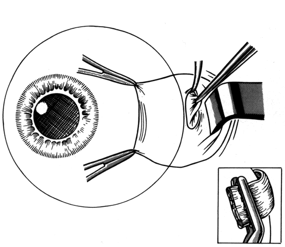
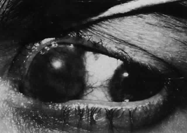
Reintervention to correct strabismus is indicated if a technical problem occurred immediately after the initial procedure. A muscle that has become disinserted or is “lost” postoperatively requires immediate exploration and correction of the problem (Figs. 2 and 3). A similar situation may occur when a previously operated muscle retracts ("slips") inside the muscle capsule (Fig. 4). The slipped muscle will appear paretic, and early surgical intervention should be considered. The clinical pictures of these technical failures are very similar. There is poor function of the affected rectus muscle, and the induced strabismus will be noncomitant. Operations may be performed on the incorrect eye muscles or eye (Fig. 5). If this occurs, reoperation should not be delayed.

Fig. 2. This 8-year-old patient had her lateral rectus recessed 4 days before referral. Twenty-five prism diopters of esotropia and absent abduction were noted. The insertion was “empty” in appearance, and an untied suture could be visualized underneath the conjunctiva (arrow).

Fig. 3. This diagram demonstrates the findings of surgical exploration of the patient in Figure 2.

Fig. 4. Failure to incorporate a portion or the entire tendon of the muscle when sutures are placed near the insertion can permit a portion or all of the muscle to slip posteriorly and produce an exaggerated recession effect. On exploration, the muscle capsule will be present but the tendon will be retracted within the muscle capsule.

Fig. 5. A. This patient was referred for correction of a left hypertropia following strabismus surgery. B. When the patient looks down, there is poor infraduction. Exploration confirmed that the left inferior rectus had been recessed instead of the left medial rectus. (Courtesy of John Simon, MD)

More commonly, reoperation is required when an unexpected unsatisfactory result is encountered. This includes the occurrence of overcorrections and undercorrections. A general guideline for recommending correction of horizontal strabismus is the presence of diplopia or the presence of a residual deviation of 12 to 15 Δ (prism diopters) or greater. Because fusional control of vertical deviations is generally less than fusional control of horizontal deviations, patients with diplopia and vertical deviations greater than 5 Δ are candidates for reoperation. A rectus muscle that has been inadvertently offset vertically when reattached to the globe may induce an unexpected hypertropia. Unexpected results may also occur from failure to recognize a problem before the initial procedure (e.g., masked bilateral superior oblique palsy). They also may occur months or years after procedures that were accompanied by an exuberant healing response due to excessive bleeding, heavy use of cautery, excessive use of blunt dissection with cotton-tipped applicators, or when the integrity of the orbital fat has been violated.3 Failure to adequately balance the forces of the extraocular muscles may also result in the creation of incomitance, unsatisfactory distance-near disparity, and overcorrections and undercorrections, and may necessitate reoperation.4,5 Other undesirable results after strabismus operations include residual head postures or tilts, and creation of ocular torsion.

Re-emergence of the original problem is another indication for reoperation. This may be due to insufficient fusional control of small degrees of residual misalignment in conditions such as infantile esotropia. It may also occur when strabismus is associated with decreased vision in one eye and fusional control is absent. The presence of a paretic muscle or muscles (e.g., third, fourth, or sixth cranial nerve palsy) may initially result in a good response to surgical alignment, but the innervation to the muscle/muscles may be so compromised that the alignment will later deteriorate and additional intervention will be necessary. The presence of scar around the extraocular muscles or in the subconjunctival tissue may cause recurrence of the original misalignment by mechanical restriction.

The presence of an underlying condition that has variable or progressive characteristics may lead to additional strabismus surgery. Conditions such as progressive external ophthalmoplegia, thyroid ophthalmopathy, myasthenia gravis, and ocular myositis will demonstrate variability in the size of the deviation. In these cases, it is important to exercise restraint. Surgery should not be recommended until stability in prism-cover measurements is obtained and any inflammatory component has resolved.

Some reoperations are necessary because a new problem, one unrelated to the underlying motility disorder, may not be present at the time of the first operation but emerges later as part of the natural history of the original problem. The occurrence of dissociated vertical deviation (DVD) and overaction of the inferior oblique muscles after correction of infantile esotropia are two examples of this. Emergence of a contralateral superior oblique palsy or unmasking a bilateral superior oblique palsy, after the first operation, is another example.6

In unusual situations, the initial operation may be the first step in a planned staged procedure. This may occur in a condition that requires correction of a complex cyclovertical muscle problem, such as third cranial nerve palsy. In such cases, reoperation is anticipated from the beginning.

In cases where repair of the strabismus is principally cosmetic, it is important that the surgeon not be satisfied with an appearance that is unsatisfactory to the patient or parent(s). Visual appearance is subjective, and care should be taken to listen to the patient's or parent's observations.

Back to Top
CONTRAINDICATIONS
When a patient has a residual deviation after a strabismus procedure, or develops a new problem, there will be situations in which the surgeon will recognize the problem but advise the patient to delay correction or even to avoid additional operations. Contraindications to strabismus surgery may be absolute or relative.

Absolute contraindications include any procedure that carries a significant risk of loss of vision. For example, if there is a significant risk of anterior segment ischemia, or if ischemia occurred in the patient previously, and alterations in surgical technique will not reduce this risk, surgery is inadvisable. If correction of a problem requires surgery on the sound eye of a patient with monocular blindness, and the patient cannot accept the remote risk of loss of vision, surgery is contraindicated (Fig. 6). Reoperation should not be attempted in situations wherein the surgeon is unable to understand the causes of the motility problem or is unable to formulate a surgical plan that has a reasonable chance for successfully achieving the desired alignment. Surgical risks and morbidity are also probably not justifiable in some cases where the patient has a very limited life expectancy. As for any surgery, unacceptable anesthetic risk is a strict contraindication to reoperation.

Fig. 6. Loss of vision after strabismus surgery is very uncommon. This 45-year-old woman had received orbital radiation prior to her strabismus surgery. The surgery was performed with retrobulbar anesthesia. Four days after the operation, vision was decreased from 20/20 to light perception. It is presumed that there was injection of the anesthetic into or around the optic nerve that caused compromise of the central retinal artery. The fundus photograph was taken 4 days after the procedure.

Relative contraindications for surgery also exist. Early overcorrection of some forms of strabismus is not a reason for surgical reintervention. Examples of this include consecutive esodeviation after surgery for intermittent exotropia, where nonsurgical measures (e.g., prisms, occlusion, abduction exercises) and additional time will frequently lead to an acceptable result.7 Similarly, a muscle that has had a large recession may initially appear paretic. This muscle will usually “take up slack” over time and regain a more normal function. Reintervention should be delayed in conditions that are unstable or variable, or when adequate measurements cannot be obtained.

The surgeon should be cautious when the patient has unreasonable expectations regarding results after surgery. Helveston1 has stated that a patient needing a reoperation will have a 30% chance of needing another operation to obtain a satisfactory result. Refusal of the patient to accept the possibility of the need for further procedures or therapeutic measures is a relative contraindication to surgery.

Back to Top
PREOPERATIVE ASSESSMENT

Whenever possible, efforts should be made to obtain information from the patient's history and from clinical records regarding the original diagnosis, preoperative measurements, and previous surgical procedures. In lieu of this information, some information can be obtained on examination. For example, slit lamp examination may disclose conjunctival scarring, which may suggest which muscles have had prior surgery (Fig. 7).

Fig. 7. Conjunctival scarring over the lateral rectus muscle and the history of correction of an exotropia suggest that the lateral rectus muscle had been recessed.

Analysis of the patient's sensorimotor development and consideration of factors such as identification of the preferred eye, evaluation of restriction, and the balance of force between the agonist/antagonist and yoke muscles will assist in developing a successful surgical strategy to correct the strabismus.1,2,4,5,8–11 The goal of evaluation of the sensory system is to determine the level of sensory fusion that can be expected when the deviation is corrected. This is important information because fusional control may effectively reduce symptoms of diplopia in the presence of a small residual deviation. In some patients, postoperative diplopia can be predicted. Diplopia may be anticipated in patients who have had an overcorrection of long-standing intermittent exotropia.7,11 In patients who are visually mature (over 8 years of age), diplopia can be expected if an overcorrected eye position causes images to fall outside of a pre-existing suppression scotoma. Prisms may be used preoperatively to evaluate this possibility. Age of the patient, chronicity of the deviation, and etiology of the original strabismus problem are all factors that will help to predict those patients who will be at risk for diplopia. Tests of binocularity, such as the Worth four dot test and Bagolini lenses, and tests to measure stereoacuity will indicate the potential for binocular cooperation in patients with intermittent deviations. In general, young children with strabismus usually have an ability to develop suppression and secondary adaptations such as anomalous retinal correspondence. In adults with acquired deviations, there is usually an inability of the sensory system to develop suppression, resulting in diplopia. In some instances, such as in closed head injury, or in long-standing unilateral visual loss that is rehabilitated (e.g., cataract removal), there may be central disruption of fusion wherein seemingly perfect realignment of the visual axes results in constant diplopia that cannot be fused by the patient.12 Disturbing and persistent diplopia may occur in patients who have orthotropia on cover testing but also have macular heterotropia related to a retinal detachment procedure or to preretinal membranes that distort the normal relationship of the fovea to the peripheral retina.

When diplopia is present, it can be documented by performing binocular visual fields using a Goldmann perimeter (Fig. 8). This technique can be used before and after surgery to outline and quantitate the area of single binocular vision and diplopia.13,14 Binocular perimetry is also useful in explaining goals of surgery to the patient. For example, in patients who have a sixth cranial nerve palsy, it can be demonstrated that the area of single binocular vision will be shifted toward primary position and that in eccentric gaze positions diplopia may persist.

Fig. 8. Binocular perimetry on a patient with a right sixth cranial nerve palsy. Diplopia is present in the right field of vision.

One of the most important components in assessing a patient's oculomotor system is accurate measurement of deviation. Meticulous prism and cover testing techniques should be done in the diagnostic positions of gaze. When gathering this information, one must ensure that refractive errors are corrected and that accommodation is controlled. If measurements are variable or tend to increase with prolonged prism and cover test, occlusion of the nondominant eye for a half hour will suspend fusion, and measurements obtained after this time will more accurately reflect the balance of forces influencing the ocular alignment.

Preoperatively, the surgeon should specifically evaluate for the presence of muscle paresis and restriction. These may be suggested by the presence of a primary and secondary deviation.15 The primary deviation refers to the size of deviation when the nonparetic (or nonrestricted) eye is forced to take up fixation. The secondary deviation, which is different in size from the primary, is measured with the paretic (or restricted) eye fixating. The secondary deviation tends to be larger because the abnormal eye is fixating “under duress” and by Hering's law causes a larger degree of innervation to the yoke of a paretic muscle or the yoke of the antagonist of a restricted muscle. Limited ocular rotations and the presence of end-point nystagmus also occur in eyes in which there is paresis or restriction.

In the case of paretic and restricted muscles, there may also be a difference in the amount of deviation measured, depending on which eye is chosen for placement of the prism. If the prism is placed in front of the eye with the paretic or restricted muscle, it will tend to measure less than when the prism is placed over the nonparetic or nonrestricted eye.

Evaluation of saccadic eye movement will also help to differentiate restrictions from paresis. Paretic muscles will show decreased saccadic velocity, whereas eyes that have restriction will have normal saccadic movements until the leash effect of the restriction is encountered. Although this can be quantitated with electro-oculogram (EOG) tracings, an experienced ophthalmologist can gain similar information through observation.

Paresis and restriction can usually be differentiated by forced duction testing. In cases where there is a paretic muscle, there will be absence of mechanical restriction on forced duction testing, and force generation testing will show reduced muscle strength. When forced duction testing is performed, it is important to make sure that the patient is relaxing the muscle being evaluated for mechanical restriction. The patient should be encouraged to look as far away as possible from the field of action of the muscle. If this is not done, the patient may “splint” or hold the eye in a position of fixation that will simulate mechanical restriction when in fact none is present.

The presence of ocular torsion may be evaluated subjectively (Maddox double rod test) and objectively (indirect ophthalmoscopy).16,17 The patient's deviation should also be evaluated for distance-near disparity, wherein there is a significant difference in the size of the deviation at distance and near. Stability of a deviation can be ensured by comparing measurements of the deviation with measurements obtained on previous examinations. For patients who have ocular deviations that have variable measurements or some degree of ptosis, a Tensilon test should be considered to evaluate for ocular myasthenia.

If the clinical findings suggest a slipped muscle or the presence of a restrictive band, magnetic resonance imaging (MRI) of the orbit with coronal and sagittal cuts may be performed. The surgeon should review studies with a radiologist experienced with orbital pathology.

If nystagmus with a null point is present, a trial of prisms can be used to normalize the head position and to quantitate the amount of anomalous head turn correction needed. This may be done by holding loose prisms in front of the eyes or by using Fresnel prisms.

Ocular fixation preference may have significance regarding which eye and muscles should have surgery. If there is a strong fixation preference and the fixating eye is found to have a paretic muscle, causing a secondary deviation, surgery should be performed based on the amount of the secondary deviation.

The final step in assessment of the patient is selection of the anesthetic that is best suited for the patient and determination of whether there is an acceptable risk for using that anesthetic. Most reoperations require moderate to extensive tissue dissection, and this is expected to be painful. In these situations, a general anesthetic or a long-acting retrobulbar anesthetic should be considered.

If the use of adjustable sutures is contemplated, it is important to establish that the patient can cooperate for postoperative adjustment. This can be assessed by performing a forced duction test or a “Q-tip test” in the office prior to surgery. This test requires placement of a “Q-tip” on the conjunctiva of the topically anesthetized eye and movement of the eye. Patients who can tolerate this test are usually good candidates for an adjustable suture procedure.

It is important that the patient have realistic expectations about the anticipated surgery and that the surgeon and patient understand, and are in agreement with, the goals, risks, and anticipated outcome of the procedure. It is important to be aware of the level of frustration and perhaps anger that exists in patients who have had unsuccessful strabismus surgery.

Back to Top
PREOPERATIVE NONSURGICAL INTERVENTIONS
Before a second or third strabismus procedure is recommended, it is necessary to consider all practical nonsurgical measures that may correct the problem. Optical means should be tried to improve the focus of the retinal image. For small degrees of exotropia, minus lenses may be used to stimulate convergence. For esotropia, plus lenses can be used to relax accommodation. Similarly, in young children with undercorrected infantile esotropia, miotics such as echothiophate 0.125% (Phospholine Iodide) instilled daily may have some use in reducing accommodative convergence. Prisms can be used temporarily to restore single binocular vision and to promote fusion. As fusional control of a residual deviation improves, the power of the prism can be reduced or eliminated.

Ocular rotation exercises can be used to increase the field of action of a muscle whose antagonist is tight or inelastic after a large resection or advancement. For example, if a recess-resect procedure was used to correct an exodeviation and the medial rectus (resected muscle) is tight, abduction exercise may improve lateral movement of the eye. This combined with occlusion of the dominant eye and correction of any residual hyperopia may be especially useful in patients who show an overcorrection (consecutive esotropia) after surgery for intermittent exotropia.

In children younger than 8 years of age, recognition and treatment of amblyopia prior to surgery is generally indicated.

Reoperation is indicated when nonsurgical measurements have been tried and have failed to rectify the problem, or when nonsurgical measures are not realistically expected to be effective.

Once the decision is made to perform surgery, prisms should be removed from the patient's optical correction prior to the procedure so that refractive errors can be corrected immediately after the procedure. Patients will benefit by prompt simultaneous stimulation of the fovea of each eye in the early postoperative period, a period when the forces of fusion will help to favorably prejudice the alignment so that the healing process will take place with the eye in optimal alignment.

Back to Top
NONINCISIONAL SURGICAL INTERVENTION (CHEMOSURGERY)
In selected patients, correction of the residual ocular misalignment may be achieved by injection of botulinum A toxin (Botox) into one or more extraocular muscles. This drug will be very useful in patients who refuse conventional incisional surgery or refuse to undergo general anesthesia. Treatment with botulinum A toxin is worthwhile in cooperative patients with small overcorrections or undercorrections (see elsewhere in these volumes). This potent neurotoxin is also used to augment transposition surgery for sixth cranial nerve palsy by injecting the toxin into the antagonist medial rectus muscle in conjunction with a transposition procedure.18,19 The effectiveness of botulinum toxin will depend on the dose of toxin, the administration technique, and the degree of function of the ipsilateral antagonistic muscle. The goal of treatment is to produce an overcorrection of the position of the eye in a direction that is away from the injected muscle. This is considered to be essential to produce the desirable changes in the antagonist muscle during the period of chemo-denervation.

Botulinum A toxin is not useful for large-angle deviations or problems in which there is significant mechanical restriction.18

Back to Top
TIMING OF INTERVENTION
The timing of the reoperation will depend on the circumstances that were responsible for the initial failure. If the need for reoperation is obvious, such as a slipped or lost muscle, immediate intervention is appropriate. It is best to reoperate early, usually within days to 1 week, before the healing response is well established. When exploration is performed within a week of the original procedure, bleeding is reduced, tissue planes are still able to be identified, and the sutures in the muscle tendons are still present. The presence of a previously placed muscle suture will be of great assistance in helping to find a muscle that has slipped.

For situations in which there is a small but unacceptable undercorrection or overcorrection, or a new undesirable secondary problem is induced, such as hypertropia, or when the healing response may correct the problem, we advise waiting at least 6 to 8 weeks after the initial procedure before reintervention. Effects of the surgical trauma, such as inflammation, edema, suture dissolution, and tightness of a resected or tucked muscle, will have subsided. The optical distortion caused by tearing and changes in refractive error, as well as the patient's discomfort, tend to improve during this period. This 6- to 8-week period also allows the surgeon an opportunity to use nonsurgical interventions to ameliorate the problem. In addition to these general recommendations, patient considerations such as diplopia, occupation, distance traveled, and vacations may enter into the decision for timing of the reoperations. In visually immature children, the goal of preserving binocularity or permitting binocularity to develop would arbitrate against excessive delay when an inadequate result from surgery is encountered.

In situations where botulinum A toxin has been used, it may be necessary to wait 3 to 4 months after an injection. This is the period of time necessary for a chemonervated muscle to recover its function. Complete recovery can be confirmed by comparing prism and cover measurements on sequential examinations.

Back to Top
ESOTROPIA
There are three acceptable methods to correct residual esotropia. The medial recti may be rerecessed,20–22 a medial rectus marginal myotomy may be combined with a resection of the lateral rectus, or both lateral recti may be resected.23–26 These procedures have similar success rates.23 If the medial recti are in a recessed position (i.e., near the arc of contact of the medial rectus with the globe), caution should be used in considering further recession. In such cases, resection of the lateral recti is the preferred procedure.25

Undercorrection of infantile esotropia occurs commonly. Increased amounts of medial rectus recessions have been advocated to improve the rate of undercorrection.27 If an esotropia greater than 15 Δ persists after 6 to 8 weeks, and it is not improved by alternate occlusion or correction of a hypermetropic refractive error, reoperation should be considered. Because the achievement of bifoveal fixation in infantile esotropia is unlikely,28 smaller deviations compatible with development of a monofixation syndrome29,30 or satisfactory cosmesis are acceptable results.

For older children with esotropia, indications for reoperation are the same as those used for the first procedure. For example, if surgery was recommended for a patient with nonaccommodative esotropia to eliminate a residual deviation, and this was not accomplished, then the same indication will apply for the reoperation. Success of the initial procedure has been improved by using prism adaptation in acquired esotropia and by using augmented amounts of surgery for this condition.31

The persistence of distance-near disparity, where the esotropia is larger at near than at distance and is not controlled by bifocals (or use of bifocals is not desired), requires further recession of the medial recti or limitation of their action by use of recession combined with posterior fixation sutures.25,32

For undercorrections of acquired, nonaccommodative esotropia when there is little distance-near disparity, the surgeon's choice of the reoperation procedure will be influenced by previous procedures performed on the medial and lateral rectus muscles and whether previous surgery involved one or both eyes. If a bilateral medial rectus recession was performed, resection of the lateral rectus(i), reweakening of the medial rectus(i) by marginal myotomy or re-recession, or a combination may be employed.

The presence of a secondary esotropia after surgery to correct intermittent exotropia is desirable and is expected to diminish with time.7 Initial overcorrection in younger children (under 3 years of age) in whom suppression and persistent esotropia may develop is a concern.11 If high-grade sensory fusion with stereopsis is lost in association with consecutive esotropia, and there is no improvement after use of adjunctive measures and an adequate period of observation, reoperation is recommended.

When an adult has a small-angle esotropia after surgery for exotropia, the condition will usually improve without surgical intervention. If the deviation persists and is greater than 10 Δ 8 weeks after surgery, and little or no improvement is shown, intervention is indicated. Injection of botulinum A toxin into the medial rectus muscle of the esotropic eye will frequently produce a satisfactory result.18 Alternatively, the medial rectus muscle may be recessed. If the lateral rectus is truly paretic from excessive recession or “slippage,” it may be advanced.

In patients with acquired sixth cranial nerve palsy, there may be a recurrence of esotropia, especially if there is minimal innervation of the lateral rectus. Creation of abducting force can be achieved by transposition of the vertical recti temporally by half of a muscle tendon width or more (Hummelscheim) or by use of a Jensen or similar plication procedure (see elsewhere in these volumes). When a full tendon transfer is performed, consideration should be given to preservation of the anterior ciliary vessels in the vertical recti. The undesirable occurrence of hypertropia when using a full tendon transfer of the vertical recti may be controlled with the use of adjustable sutures.

Back to Top
EXOTROPIA
Exotropia that occurs or persists after strabismus surgery may be either residual or consecutive. If an exotropia greater than 15 Δ exists, reoperation should be considered.

Consecutive exodeviations after repair of infantile esotropia may occur months or even years after the initial procedure. In patients who have a large esodeviation occurring during the first years of life, there are frequently overcorrections. This is especially true when there is a history of a neurologic abnormality or developmental delay. In these situations, there is frequently an unpredictable response to traditional amounts of graded surgery. Reoperation is indicated with either advancement of the previously recessed medial recti or recession of one or both lateral recti. If an adduction deficiency is noted, the former should be considered. If the medial recti previously underwent a large recession, advancement should be done cautiously because the medial recti may become inelastic over the interval of time between the first and second procedures.33

Recurrent exotropia may occur several years after correction of intermittent exotropia. To develop a treatment strategy, classification of the exotropia according to distance and near measurements in primary gaze position, as well as in lateral gaze positions, will indicate the appropriate corrective procedure. An exodeviation greater at distance fixation than at near indicates a need for (re)weakening of one or both lateral recti. If the deviation is equal at distance and near fixation, there is not a strong ocular preference, and a recess-resect procedure was previously performed, a recess-resect of the lateral and medial rectus of the fellow eye is appropriate. If a strong preference for fixation exists, a recession or marginal myotomy of the lateral rectus combined with a medial rectus resection should be considered. A correction of a convergence insufficiency pattern will require resection of one or both medial recti.

Back to Top
VERTICAL DEVIATIONS
Vertical deviations may occur as a new finding after horizontal strabismus surgery, or they may occur as a result of an overcorrection or undercorrection of a previous hypertropia. Vertical deviations may also occur de novo as part of the natural history of a strabismus condition, such as dissociated vertical deviation following horizontal correction of infantile esotropia.

In some situations, a portion of a horizontal rectus may be left at the insertion site (Figs. 9 and 10). The muscle is usually split at the time of the initial procedure, and this can cause a shift of the distribution of force at the insertion and cause vertical misalignment. The pattern of the strabismus and the details of the previous surgery should provide clues to the problem and allow a logical approach for correcting the vertical deviation. Suspicions should be confirmed by careful dissection and exploration of the region of the muscle insertion. The importance of careful evaluation and elimination of mechanical restrictions cannot be overemphasized.4,10

Fig. 9. Residual exotropia or hypertropia can occur if a portion of the lateral rectus muscle is not recessed with the first procedure. To prevent this, care must be taken to incorporate all portions of the tendon when recessing an extraocular muscle.

Fig. 10. The pole test, or sweeping a Stevens muscle hook anterior to the insertion, will ensure that the entire tendon is on the Jameson muscle hook.

Undercorrections and overcorrections of a cyclovertical muscle problem may cause or fail to correct anomalous head position or diplopia, or may produce inadequate cosmesis. Each case must be evaluated by analysis of prism-cover measurements in the appropriate gaze positions. Attention must be given to the torsional component of the deviation as well as the behavior of the problem in the primary gaze and reading position. Nonsurgical forms of treatment (e.g., prisms) are usually of little benefit if the deviation is accompanied by torsion or when incomitance contributes to the patient's complaint of diplopia.

Back to Top
ANOMALOUS HEAD POSITIONS
Common mechanisms responsible for compensatory head postures after previous strabismus surgery include: incomitant horizontal strabismus, cyclovertical muscle imbalance, and null-point nystagmus. Inadequate correction of these problems may result in a continued need to adopt an anomalous head position. If the amount of residual or new torticollis exceeds 10° to 15°, it is sufficient to warrant consideration for additional surgery. More severe degrees of torticollis, especially in younger children, should be corrected out of concern for long-term musculoskeletal problems that may ensue.34
Back to Top
SURGICAL PLANNING FOR REOPERATION
Generalized guidelines can be offered; however, the strategy and selection of the surgical procedure for each patient must be individualized. Cooper2 emphasized that the strategy for correction of the alignment should be considered as if the patient had not had prior surgery. A surgical plan should be formulated that is designed to achieve the desired result by a single operation on as few muscles as possible.
Back to Top
PROCEDURE
The goal of a reoperation is to re-establish the balance of forces of the extraocular muscles.1,4,5,8–10 This usually will require surgical repositioning of the extraocular muscles combined with removal or release of scar tissue created by prior procedures.

Sufficient exposure of the extraocular muscles and comfort of the patient and surgeon throughout a somewhat lengthy case is important. To achieve optimal exposure, a full complement of instruments to provide exposure is necessary and should include extra Stevens and Jameson muscle hooks, and instruments to retract tissue, such as <fr1/4>- and <fr1/2>-inch malleable ribbon retractors. These retractors are versatile and can be bent to enhance exposure of the muscle. Desmarres or Conway retractors are also helpful for retracting conjunctiva and Tenon's tissue, especially if the exposure is required anterior to the equator of the globe. Visualization during dissection is enhanced by using wet-field cautery to control bleeding. Sufficient illumination should be available for deep dissection to the equator of the globe. The surgeon should have a fiberoptic head lamp available. Stabilization of and traction on the globe can be facilitated by placing a locking 0.5-mm tooth Castroviejo forceps on the muscle insertion. A Jameson resection clamp is useful when working on a muscle that has been split or torn. This clamp will secure the muscle while preserving the width of the tendon so that repair can be accomplished. In difficult reoperations, the surgeon should request a pair of capable hands to assist him/her throughout the procedure.

The anesthetic level should provide the patient sufficient comfort. The new rapid-acting intravenous sedation agents provide the surgeon with sufficient sedation to perform some procedures with topical or perimuscular anesthesia. This provides an opportunity for patient cooperation for intraoperative suture adjustment. Once the anesthetic has been administered, the surgeon can proceed with forced duction testing (Fig. 11). If the patient is under general anesthesia or a retrobulbar block has been used, the globe can be moved back and forth and then released to seek its natural position of rest. The “spring back balance test,” popularized by Jampolsky, Scott and Rosenbaum, provides the surgeon with information on the extent and location of restriction (Fig. 12). Forced ductions should include torsional forced ductions to determine if the oblique muscles are restricted.35 During forced ductions, the surgeon should observe the conjunctiva for underlying restrictive tissue bands. Adhesions between these bands and the conjunctiva will cause the conjunctiva to pucker.

Fig. 11. Forced ductions are used to determine the presence and extent of restrictive forces on the globe. This photograph illustrates difficulty abducting the globe due to an inelastic medial rectus muscle resulting from previous resection of the medial rectus muscle.

Fig. 12. The spring back balance test may be performed at any time during surgery. A. The globe is moved from right to left several times and is then released. B. Restrictive bands, scar tissue, or contraction of an inelastic muscle will cause the globe to return to the position induced by the restriction.

The incision should provide sufficient exposure and should be one the surgeon feels comfortable using. Our preference is to attempt to use a cul-de-sac incision whenever possible. This incision eliminates dissection through conjunctiva and anterior Tenon's capsule. When corrections require surgery on the superior rectus, conjunctiva is preserved for the remote possibility that a glaucoma filtering procedure may be required in the future (Figs. 13 and 14). The conjunctival incision can usually be made either through the old incision or just posterior to it.

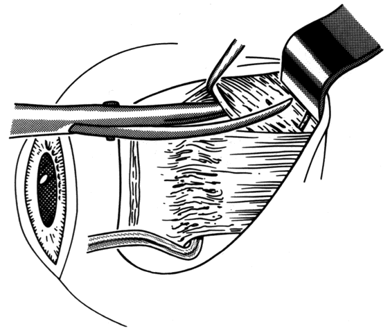
Fig. 13. Incision is made through the conjunctiva. Conjunctiva is then dissected free from any adherent Tenon's capsule that overlies the site of the old muscle insertion. A blunt-tip Wescott scissors facilitates this dissection.

Fig. 14. This dissection is carried past the insertion until a hook is able to be passed under the muscle without restriction.

Placing the conjunctival incision at the limbus provides better visualization, but the surgeon must disrupt the circulation at the limbus when using this incision. If three rectus muscles are going to be disturbed, or if there is significant risk for anterior segment ischemia, the contribution of the limbal blood supply to the anterior segment circulation may be important.36

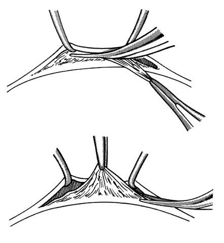
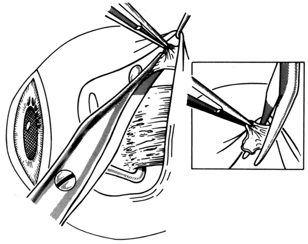
After the incision has been made and the dissection through Tenon's capsule has been completed to the surface of the sclera, muscle hooks are passed behind the rectus muscle to identify the insertion of the muscle. Once the muscle has been “hooked,” there is improved control of the globe. This will facilitate the blunt and sharp dissection that will be needed to reflect the conjunctiva over the tendon of the muscle (Fig. 15). Sharp dissection beneath the conjunctiva is used to separate fibrous tissue and restrictive bands from the surface of the muscle capsule and the sclera (Figs. 16 and 17). Frequently it will be necessary to remove small amounts of tissue to visualize the area of contact of the muscle insertion (Fig. 18).

Fig. 15. A Jameson muscle hook is under the insertion of a previously recessed horizontal rectus muscle. The insertion is cleared of any residual adhesive bands and scar tissue.

Fig. 16. Wescott scissors and toothed forceps are used to reflect conjunctiva (top) for removal of subconjunctival fibrous tissue (bottom).

Fig. 17. When there is extensive fibrous proliferation between the conjunctiva and the muscle, Wescott scissors are used to cut it free from both surfaces. An attempt is made to remove this tissue without disturbing the muscle capsule or cutting into the insertion of the rectus muscle.

Fig. 18. This fibrous tissue was removed from a patient with prior strabismus surgery. Note the connective tissue component. Thick bands of compact collagenous scar tissue infiltrate orbital fat (hematoxylin and eosin, X 100). (Courtesy of Bruce L. Johnson, MD)

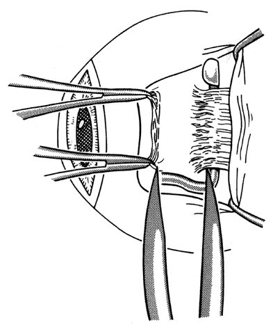
Once the muscle insertion has been “cleaned” of adherent tissue, the distance the muscle is from the original insertion and from the limbus is measured and recorded (Figs. 19 and 20).

Fig. 19. A Castroviejo caliper is used to measure the distance between the original insertion and the new insertion.

Fig. 20. Measurement of the distance between the insertion and the limbus is also helpful. This will determine how much further a rectus muscle can be recessed.

Once these measurements have been obtained, dissection is extended posterior on the muscle, removing tissue adherent to the muscle capsule. Care is taken not to enter the muscle capsule. When this happens, hemorrhage will occur (Figs. 21 and 22). Attachments between the muscle and the globe will frequently occur. These adhesions must be removed with either sharp or blunt dissection.

Fig. 21. Blunt Wescott scissors and forceps are used to dissect fibrous tissue from the surface of the rectus muscles. Two Stevens hooks are used to provide exposure of the rectus muscle to facilitate dissection.

Fig. 22. Rapid hemorrhage occurs if the muscle capsule is entered inadvertently.

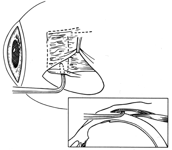
To detect and disrupt adhesions between the underside of the rectus muscle and the globe, or attachments between the lateral rectus and the inferior oblique muscle, a Jameson muscle hook is slid posteriorly.37 If an adhesion is encountered, it can usually be broken by gently disrupting the adhesion with a second Jameson hook (Figs. 23 and 24). If additional adherent tissue remains near the insertion, it should be removed prior to placement of sutures at the insertion of the tendon (Fig. 25).

Fig. 23. A Jameson muscle hook is placed under the insertion.

Fig. 24. Adhesions between the underside of the muscle and the sclera can be broken by gently passing the Jameson hook posteriorly. Failure to free up these adhesions will reduce or eliminate the effect of recession of the muscle.

Fig. 25. A strand of fibrous tissue is adherent and incorporated in the intermuscular septum. This tissue is removed with sharp dissection.

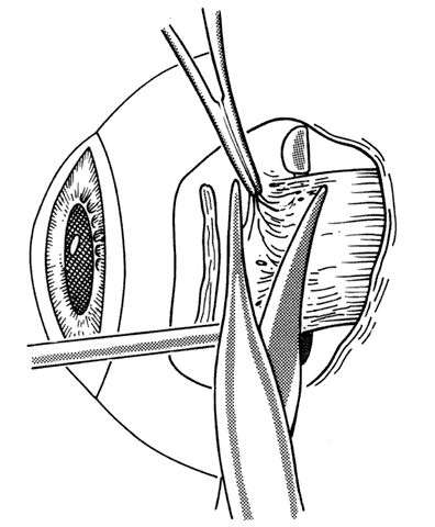
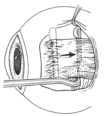
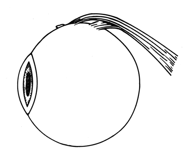
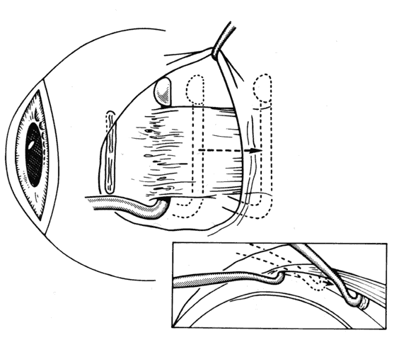
The insertion of a previously operated rectus muscle may have anterior extensions of scar tissue or even a pseudotendon. The term pseudotendon refers to fibrous tissue that extends from the surface of the true tendon (Fig. 26). If a muscle has been previously recessed, these attachments may extend anterior and become adherent to the original insertion. Care must be taken to identify “pseudotendons” and to separate them from the true tendon of the rectus muscle (Figs. 27 through 29).

Fig. 26. Care must be taken to identify the true insertion. A fibrous extension from the insertion can extend anterior to the old or original insertion. This is called a “pseudotendon” because it may simulate the tendon of the rectus muscle inserting on the globe. Inset. A Stevens hook is under a “pseudotendon.” The true tendon is posterior to this fibrous structure.

Fig. 27. After identification of the insertion of the true tendon of the rectus muscle, the “pseudotendon” is excised with sharp dissection. Dissection should be carried down to bare sclera.caption.

Fig. 28. This intraoperative photograph demonstrates tissue that may simulate the insertion of the lateral rectus muscle. The cornea is to the right, just visible under the lid. The Jameson hook is under tissue that extends from the original insertion and the inferior oblique muscle. The true insertion is further posterior. The insertion of the muscle was found to be 17.5 mm posterior to the limbus.

Fig. 29. Attachments can occur between sclera and the rectus muscle. These need to be cut or disrupted. Failure to do this will alter the desired effects of recession or resection of the rectus muscles.

During the dissection, bleeding is controlled with wet-field cautery. We try to avoid using cotton-tipped applicators, but when Q-tips are used, they are used sparingly. Cotton applicators or Q-tips may leave foreign material behind, and they tend to incite or accentuate an exuberant healing response.

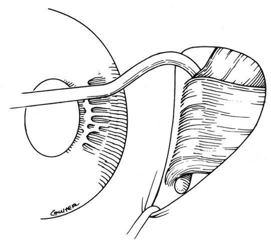
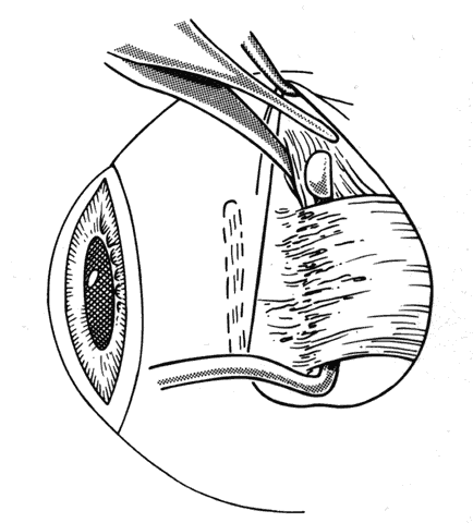
When approaching the superior and lateral rectus muscles that have had previous surgery, care should be taken to identify the superior oblique tendon and the inferior oblique muscles respectively. One should carefully free up attachments of these structures to the rectus muscle so that a normal anatomic relationship between the rectus and oblique muscles can be re-established. Frequently, the lateral rectus muscle will have adhesions to the orbital surface of the inferior oblique muscle as it passes underneath the lateral rectus. There is a propensity for the inferior oblique muscle to be drawn up into the insertion during resection procedures.37 When this occurs, the inferior oblique and lateral rectus muscle must be separated, and the inferior oblique muscle should be repositioned into its normal anatomic position (Figs. 30 through 33).

Fig. 30. To break posterior adhesions between the rectus muscle and the sclera, a second Jameson hook should be carefully pushed posteriorly along the belly of the rectus muscle. These adhesions are usually broken with little effort. If the adhesions resist blunt separation, a suture should be placed through the tendon and the muscle cut free from the insertion. With a malleable retractor, the adhesion is identified and then cut.

Fig. 31. The inferior oblique muscle has been incorporated into the insertion of the right lateral rectus.

Fig. 32. Sharp and blunt dissection is used to “free up” the inferior oblique muscle from the scleral side of the lateral rectus.

Fig. 33. If the dissection needs to be carried beyond the equator, care must be exercised to avoid avulsing or cutting a vortex vein. If this happens, a moderate hemorrhage will occur.

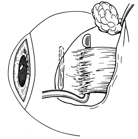
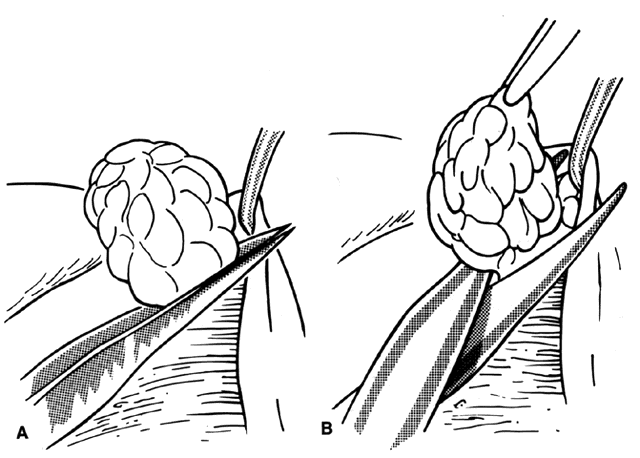
When dissections are carried posterior to the equator, fat will frequently prolapse into the operative field. When this occurs, it should be removed. The tissue should be identified as fat only. If a vortex vein is present, it can be cauterized and divided. Cautery is applied to the base of the fat and the fat is then excised with scissors (Figs. 34 and 35). Removal of excess fat is important because it will relieve restrictions on the globe and will also help to prevent additional restrictions from occurring.38

Fig. 34. Fat may prolapse into the wound.

Fig. 35. A. The tissue is identified as fat only. A wet-field cautery is applied to the stalk of fat tissue. B. After cautery, the fat is lifted with a forceps and removed with scissors.

In rare situations, patients may have fibrotic bands that extend from the muscle and attach to the orbital wall or to the sclera posterior to the equator of the globe. Restrictive bands may also attach to posterior structures in the globe. These fibrous bands may be congenital or they may be found after previous ocular surgery. One of the authors (AWB) managed a patient who had a hypertropia caused by a congenital fibrotic band that attached to the sclera 3 mm superior to the optic nerve. The eye had a persistent hypertropia even after a large recession of the ipsilateral superior rectus and a myotomy of the inferior oblique muscle. Extreme care should be taken when approaching bands this far posterior. It is important to avoid injury to the optic nerve. If restrictive bands are suspected, MRI of the orbit may assist in defining the extent and location of these bands preoperatively.

Once the muscle has been cleaned of surrounding adherent tissues, a synthetic absorbable suture is placed into the tendon and secured with lock knots at each pole. The entire muscle must be incorporated in the suture. Bands extending superior or inferior from the insertion (or medial or lateral in the case of vertical rectus muscles) should be cut so that when the muscle has been cut free from the globe, the globe is free to move without restriction. Frequently there will be residual bands of fibrous tissue located on the surface of the sclera. If the globe is pulled in a direction opposite the pull of the bands, these bands will indent the scleral surface, which will facilitate their identification. The passage of a Stevens hook across the sclera will frequently catch on the restrictive bands. Once identified, they can be cut with scissors. After this, the globe will be free to move without restriction.

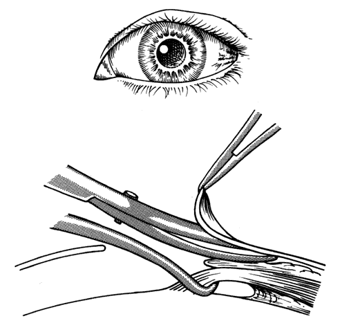
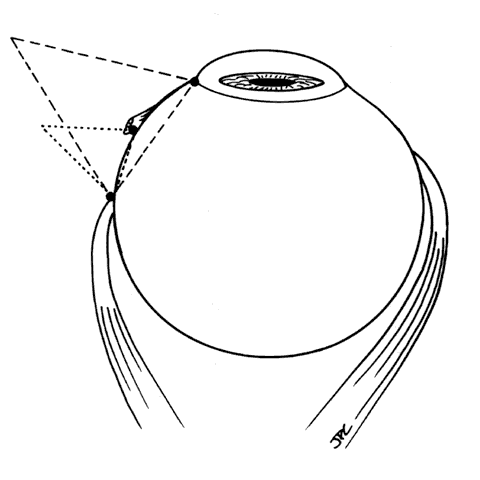
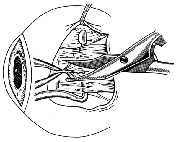
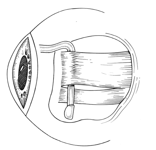
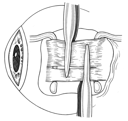
If a rectus muscle has been recessed 5 mm or more, further recession will put the new insertion at or beyond the arc of contact of the rectus muscle and the globe. This may cause decreased function of the muscle and produce noncomitant ocular rotations, especially in extreme gaze positions. To weaken a rectus muscle without disturbing its insertion site, a marginal myotomy can be performed.39,40 Usually, two separate cuts are made into the muscle near the tendon (Fig. 36) until the muscle lengthens. The weakening effect can be graded by increasing the number of cuts from two to three or by increasing the percent of the muscle that is cut. To be effective, a marginal myotomy should be accompanied by resection of the ipsilateral antagonist muscle.

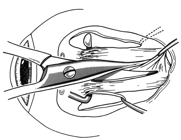
Fig. 36. To perform a marginal myotomy, the muscle tendon is gently stretched between two Jameson muscle hooks. Prophylactic hemostasis is achieved by clamping the muscle or by application of a wet-field cautery to blanch the vessels in the tendon. The muscle is cut with small “snips” using a Wescott scissors.

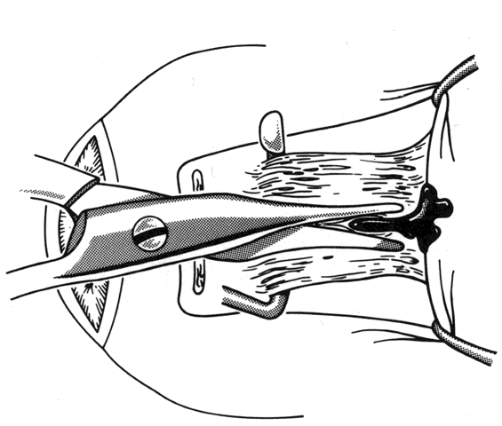
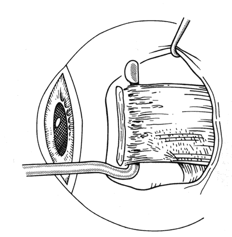
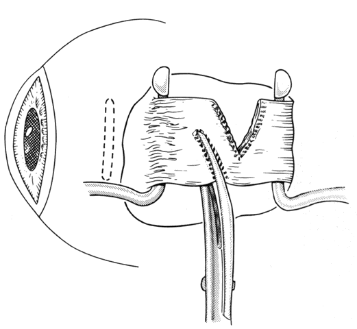
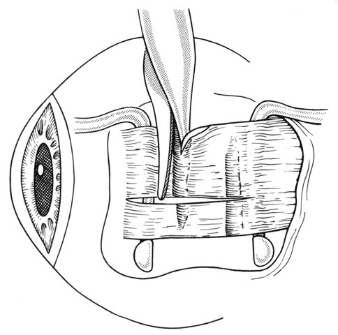
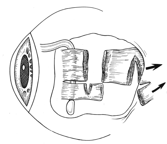
If a muscle tendon has been split during mobilization of the tendon or by passing a muscle hook under a scarred tendon, the marginal myotomy should be avoided. Placing cuts in the muscle may cause the muscle to be transsected (Figs. 37 through 40).

Fig. 37. When a muscle is split, a marginal myotomy is contraindicated.

Fig. 38. The split muscle is clamped.

Fig. 39. The cuts in the muscle divide it.

Fig. 40. The muscle can retract into the orbit.

If a muscle has been lost, or has slipped from its original insertion, or when only the muscle capsule is attached to the globe and the actual muscle tissue has slid back through the muscle sleeve, the force generated by the muscle will be greatly diminished. Effort must be directed to finding and recovering the muscle tissue and reattaching it to the globe, thus restoring the contractile force of the muscle. Once disinserted, the most difficult muscle to find is the medial rectus muscle. This is because once the intermuscular membranes are cut, it has no attachments to other muscles and it can retract into the orbit. The other rectus muscles have attachments to other muscles that facilitate recovery. The superior rectus is attached to the superior oblique, the inferior rectus is attached to the inferior oblique, and the lateral rectus has attachments between the undersurface of the lateral rectus and the orbital surface of the inferior oblique. Once these attachments are identified, the small operculum in Tenon's capsule, through which the muscle sleeve passes, can usually be identified. When this operculum has been found, a forceps is used to grasp muscle tissue. If the patient has not been given atropine, bradycardia may occur when traction is applied to muscle tissue. It is important to avoid pulling Tenon's fascia forward in a blind attempt to find the muscle. This may cause the muscle to further retract from the opening in Tenon's capsule. It is preferable to gently displace the globe away from the area of surgical exploration with blunt retraction; vigorous rotation of the globe may cause further posterior slippage of the lost muscle. Once the muscle has been identified, it is helpful to place a locking Castroviejo forcep on the muscle or its tendon. This will reduce the chance that the recovered muscle will inadvertently slip out of the instrument and retract into the orbit. A hand-over-hand technique can be used to bring the remainder of the muscle tissue anterior. Once the muscle is brought anterior, a Jameson resection clamp can be applied to the muscle tendon. When a suture has been placed through the muscle, the clamp is removed (Fig. 41). The muscle can then be reattached to the globe in a position that, in the surgeon's best judgment, will provide the best alignment.

Fig. 41. When a muscle slips out of a clamp, becomes disinserted from its tendon, or is “lost,” it can be found by searching along the scleral surface to find an operculum in Tenon's capsule. Once muscle tissue is identified, it is brought handover-hand through the operculum and placed on a secure clamp such as a Jameson resection clamp.

Reattachment of the muscle to the globe may be difficult in the presence of retinal appliances, and in patients with high myopia or ectatic sclera. For these situations, a hang-back technique to reattach the muscle to sclera can be used.

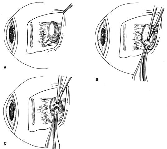
Conjunctival inclusion cysts may overlie the muscle insertion, be located more posterior, or be located on the underside of the rectus muscle (Figs. 42 and 43). These are dissected free and removed. If the epithelial cells are removed, they will not recur. Some epithelial cysts have been shown to be intimately attached to the rectus muscle, as part of a pseudotendon. Caution must be used not to disturb the muscle attachment to the globe when removing these cysts.

Fig. 42. A large conjunctival cyst is present over the left medial rectus.

Fig. 43. A. When a cyst is located near the anterior portion of the insertion, the conjunctiva is carefully reflected over the cyst to gain exposure (B). C. A Vannas scissors may assist this dissection.

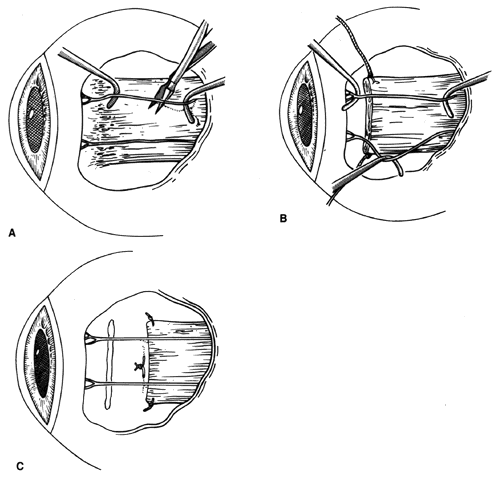
Circulation to the anterior segment comes from the anterior ciliary arteries that course with the rectus muscles and, to a lesser degree, from the conjunctival anastomoses at the limbus.36,41,42 McKeown and Lambert42 have devised a method to recess a rectus muscle while preserving this circulation. This technique, or one of its modifications, can be used to recess or resect a rectus muscle when preservation of the circulation to the anterior segment is important (Fig. 44).

Fig. 44. A. The anterior ciliary arteries are identified under loupe or microscope magnification. They are lifted from the muscle capsule with Stevens muscle hooks. B. A suture is placed through the tendon. C. The muscle can then be recessed or resected.

We have found that upon completion of the procedure, the injection of Decadron, 4 mg/mL, into the surrounding tissue tends to suppress the formation of scar and reduces the inflammation associated with the healing process.43 Others have also made this observation.44 Usually, cul-de-sac incisions are not closed. If the conjunctival incision gapes, it can be closed with an interrupted suture using 7.0 mild chromic or a 6.0 plain gut suture.

If a limbal incision requires closure, care should be taken to ensure that the conjunctival surface adjacent to the cornea is not elevated. Elevated conjunctiva at the limbus may cause a focal point of drying of the cornea (dell or dellen).

Many authors use the limbal approach for reoperations because it gives them the capability of performing a bare sclera closure. This reduces or eliminates the restrictive effects of contracted or scarred conjunctiva.8,10,45

Antibiotic-steroid ointment may be applied to the eye if the procedure is lengthy. If there has been a moderate amount of hemorrhage during the procedure, the surgeon may elect to apply an occlusive dressing on the eye.

Back to Top
COMPLICATIONS
If procedures are performed frequently enough, complications will occur. The surgeon's goal should be to produce no complications; however, this goal cannot be guaranteed.

This section will not cover all the complications that are associated with strabismus surgery. Many of these are reported elsewhere in these volumes.

Complications for the original strabismus operation are also potential complications for reoperation. Certain complications are more likely to occur during reoperation, and these will be discussed in this section with emphasis on prevention. Complications are more frequent after reoperations because of altered anatomy, the impact of repeat surgical trauma, changes in vascular supply, and the presence of scar tissue, which can distort the surface of the globe.

ANTERIOR SEGMENT ISCHEMIA

The anterior segment of the eye receives its vascular needs from the anterior ciliary vessels that course through the rectus muscles. If more than two rectus muscles are detached from the surface of the globe, and the anterior ciliary arteries are severed, the risk of ischemia of the anterior segment rises. This is especially true in patients with thyroid orbitopathy or sickle-cell disease, and in older patients who have compromised hematologic or cardiovascular status. Careful review of what rectus muscles were removed from the sclera during previous operations, as well as careful physical examination, will alert the surgeon to these risks. If, despite the recognition of a significant risk of anterior segment ischemia, the surgeon has no other option but to operate on the remaining untouched rectus muscle, surgical techniques exist that attempt to preserve the anterior ciliary arteries (see Fig. 44).42 In addition to these measures, we recommend pretreating the patient with aspirin, ten grains per day for 5 days prior to the procedure, to reduce platelet adhesiveness. Injection of corticosteroids such as Decadron Phosphate, 4 mg/mL, into the Tenon's tissue after the procedure will reduce tissue swelling and inflammation.

The emphasis for preventing anterior segment ischemia is on taking precautionary measures in surgical planning and execution of the procedure with meticulous technique. Fortunately, most cases of anterior segment ischemia will resolve without serious sequelae.

GLOBE PERFORATION

Globe perforation is more likely to occur with strabismus reoperations. This may be due in some cases to adherent bands of tissue that lie on the scleral surface and cause the sclera to be distorted. If an extraocular muscle is physically contracted, and a significant force is needed to elevate it near its insertion, distortion of the sclera may occur. The surgeon must be particularly cautious when passing needles through the muscle near its altered insertion. Meticulous care must be taken to achieve optimum exposure and to release or remove abnormal scar tissue. A surgical head lamp may facilitate this exposure. The surgeon should also use loupes or microscope magnification during these procedures. If scleral perforation is a concern, reattachment of the muscle to the sclera can be achieved with a “hang-back” technique. The hang-back reattachment of the rectus muscle is also helpful in reoperating on patients who have encircling bands or appliances on the sclera that rest where the new insertion is to be.

VORTEX VEIN BLEEDING

The vortex veins penetrate the sclera posterior to the equator of the globe. They are generally located above and below the horizontal recti and 1 or 2 mm medial and lateral to the vertical rectus muscles. Vortex veins are seldom disrupted during the initial strabismus surgery unless muscle hooks are passed excessively deep into the orbit to secure the rectus muscle. Deep, blind passage of hooks during strabismus operations is to be discouraged. However, after recession or resection, the surrounding tissue response may incorporate the vortex vein (see Fig. 33). When reoperating on a horizontal rectus muscle, the vortex vein may be contained in the fibrotic tissue produced by the first procedure. The vortex veins may be avulsed or cut during efforts to free a rectus muscle from surrounding tissue. When this happens, copious bleeding will occur and visualization will be compromised. Attempts to visualize the bleeding site are usually not effective and may cause further hemorrhage. Vortex vein bleeding is best managed by removing the lid speculum, closing the eyelids, and applying firm pressure to the orbit for about 5 minutes. Pressure should be released about every 30 seconds to permit the central retinal artery to perfuse the retina. After this, reinsertion of the speculum and removal of coagulated blood will re-establish visualization. The vortex vein rent will usually seal and will not bleed unless disturbed again.

Inclusion cysts and pyogenic granulomas are more common after reoperations. Inclusion cysts originate from the transfer of epithelial cells underneath the conjunctiva. These implanted epithelial cells will divide and create fluid-filled cysts on or around the muscle insertion. These cysts may enlarge, and when this occurs, excision of the cyst, including the cyst wall, is recommended.

Pyogenic granuloma occurs when Tenon's tissue prolapses through an incision. When this happens, exuberant granulation tissue will form. This tissue will frequently form a slender pedicle. Treatment with topical corticosteroids will cause the granulation tissue to shrink in size. The granuloma may twist on its vascular pedicle and will usually drop off. If the lesion persists for 3 to 4 weeks, simple excision with a Wescott scissor will correct this problem.

DELL OR DELLEN

Dell or dellen are caused by interference with the normal wetting of the cornea or sclera due to tissue elevations resulting from surgical trauma or faulty conjunctival closure. A dell is a dehydrated portion of the cornea. Typically, the defect is covered with epithelium, and removal of the elevated tissue adjacent to the dellen, or simple patching to rehydrate the cornea, will correct this problem.

LOSS OF VISION

Loss of vision is uncommon after strabismus surgery. Excessive traction on rectus muscles may compromise blood supply to the globe. Patients who have had radiation to the orbit or extensive amounts of orbital surgery, from either repair of retinal detachment or multiple strabismus operations, are at greatest risk. If a procedure is performed under intravenous sedation combined with retrobulbar anesthesia, the restriction of the orbital contents due to excessive scarring may fixate the optic nerve, and the anesthetic may be injected into the optic nerve, causing optic nerve trauma (see Fig. 6). This can compromise the circulation of the central retinal artery. The use of a general anesthetic or performing the surgery with a perimuscular block will decrease the frequency of this very rare complication.

PERSISTENT UNSATISFACTORY ALIGNMENT

Persistent unsatisfactory alignment may be a complication. Use of adjustable sutures reduces the frequency of this complication. We prefer to perform suture adjustment in the operating room. To do this, we use a perimuscular anesthetic combined with short-acting intravenous sedation (Fig. 45). After the muscles have been temporarily tied into position, sedation is withdrawn. The patient can sit up and assist the surgeon in determining the position of the muscles before permanent knots are tied. Short-acting general anesthesia with agents such as propofol may also be used to achieve rapid recovery of consciousness so that adjustment may be performed in the recovery area.

Fig. 45. Suture adjustment can be accomplished intraoperatively if perimuscular anesthesia is combined with short-acting intravenous sedation. After sedation is withdrawn, the patient will become alert. The patient is seated, and glasses are used when needed. Cover testing is performed with use of an eye chart for fixation. The position of the muscle is adjusted until the ocular alignment is satisfactory.

SLIPPED OR LOST MUSCLE

In reoperations involving muscles that have had previous surgery, the anatomy of the muscle as well as surrounding tissues may be disturbed, and the accuracy of suture placement in muscle tissue and the sclera may be compromised. This may cause failure of the suture to hold the muscle to the sclera. Good illumination (fiberoptic headlight), magnification, and adequate exposure will reduce the occurrence of insufficiently secured eye muscles. To avoid losing the muscle or damaging it at the time of surgery, great care must be used when freeing up restrictive tissues and removing scar tissue in the vicinity of the muscle insertion. Care should be taken to incorporate the entire muscle with the suture. Tight, restricted muscles may be particularly prone to slippage.

RESTRICTION OF GLOBE MOVEMENT

Surgical manipulation and excessive cautery of conjunctiva, Tenon's fascia, and extraocular muscles, combined with bleeding and prolapse of orbital fat, may produce a disruption of tissue planes and produce a fibrotic healing process that results in restrictive bands or the fat adherence syndrome. The adherence syndrome is characterized by fibrous adhesions developing between the sclera, the muscle, the surrounding Tenon's tissue, and the fat pad. These adhesions may tether the globe and produce a leash effect (limitation of movement of the globe beyond a certain position, either in the field of action of the affected muscle or away from its field of action). When a muscle has been repositioned one or more times, production of scar tissue is enhanced. If restriction is present and not released at the time of the procedure, it will reduce the success of the procedure. To avoid this problem, measures should be taken to remove all restrictive components, to limit the dissection during the procedure to a circumscribed area as small as possible, and to limit the use of techniques that disrupt tissues or have the potential for leaving foreign material in the wound. Blunt and sharp dissection techniques with blunt-tip Wescott scissors, forceps, and muscle hooks should be used to manipulate tissue. Corticosteroids are frequently used in reoperations to suppress fibroblastic proliferation and decrease the amount of mechanical restriction.

Some authors have placed sleeves or have used viscoelastic material around the rectus muscles to reduce restrictive forces.46–49 We have not been convinced that these measures are beneficial.

Restriction due to internal noncompliance of the extraocular muscle may also occur. This happens when inflammation is present or when swelling of the muscle is present (i.e., in conditions such as Graves' orbitopathy). Noncompliant muscles are also due to excessive scar tissue deposited on and around the muscle capsule. To achieve surgical success, tight muscles must be weakened and their restrictive effect must be neutralized.

RESIDUAL DIPLOPIA

After a repeat procedure to correct strabismus, diplopia may persist. In some situations, this is due to inadequate understanding of the problem resulting in a faulty strategy for correction or failure to achieve the desired eye position. Other times, it is due to restrictive forces that produce incomitance. Adjustable suture techniques have proved valuable in allowing postoperative adjustment of ocular alignment to reduce the frequency of this problem.

EYELID ABNORMALITIES

Reoperations on vertical rectus muscles may alter the position of the eyelid or its relationship to the globe. Recessions may cause the upper or lower eyelid to retract, and resection may cause lid ptosis, especially if dissection of surrounding fascial attachments to the muscle are inadequate.

Lid effects may also be encountered when large horizontal muscle resections that cause enophthalmos are performed. This may occur in primary gaze but more commonly occurs as the eye rotates away from the field of action of a resected muscle, producing a pseudo-Duane's syndrome. Similarly, large recessions may allow the globe to become slightly proptotic compared with the fellow eye.

Back to Top
RESULTS OF SURGERY
Prior to commencing a reoperation, there must be an understanding and acceptance between the surgeon and the patient that surgery can improve the problem(s) but that it may be difficult to eliminate the problem(s) entirely. It is important to emphasize realistic expectations of what the operation can and cannot do. In children, the eyes should be placed in a position that promotes development of the highest level of binocularity possible. This may not be orthophoric in all fields of gaze. In patients who have had binocularity and who now have diplopia, the goal of surgery should be to re-establish single binocular vision in useful gaze positions. In patients with suppression, achievement of a small-angle stable deviation with motor fusion is a realistic goal. The result for any given patient, therefore, will depend on the underlying sensory capabilities and adaptations, including the presence of fusion or suppression as well as other factors, such as the number of procedures, abnormalities in innervation, mechanical restriction, and visual acuity.

Surgery can be classified as an unqualified or qualified success, or as a failure. An unqualified success is one in which the desired goal is achieved without the use of adjunctive treatment, such as bifocals, prisms, botulinum A toxin, and exercises. A qualified success is the same, except that adjunctive measures are needed to achieve the outcome. Failure occurs when the goal of surgery, which is agreed upon prior to surgery, is not achieved with or without adjunctive measures. In the case of children in whom the goal is subnormal binocular vision with motor fusion, success is usually defined as a horizontal deviation of 8 Δ of heterotropia or less. In patients who have no suppression and constant diplopia, functional benefit is defined as elimination of diplopia in primary gaze positions and in the reading position, and it corresponds to an area of single binocular vision approximately 10° to 20° to each side of primary gaze.

Surgical success can be measured both subjectively (e.g., presence or absence of diplopia) and objectively. Objective measures include restoration of stereopsis, enhanced area of single binocular vision measured clinically or with binocular perimetry, and angle of deviation less than or equal to 8 Δ of heterotropia.

Surgical success may also be classified as early and late. The procedure cannot be considered successful if the benefit is not lasting. Techniques to improve the outcome of early alignment include use of adjustable suture surgery and botulinum toxin injections. Even with these modifications, however, the success rate may decrease with time because of the presence of residual or new mechanical restriction, abnormal innervation, decreased visual acuity, and a poor fusion mechanism.

Although no two strabismus problems are identical, there are factors that favor success and others that reduce chances of producing a lasting successful outcome. Favorable preoperative characteristics include comitancy of the deviation, potential for high levels of fusion, horizontal deviations, normal innervation of all extraocular muscles, absence of a restrictive component, and stability. Unfavorable factors include mechanical restriction of eye movement, history of multiple operations, limitations of surgical strategy due to risk of or previous episodes of anterior segment ischemia, poor vision in one or both eyes, instability of the deviation, abnormal innervation, abnormal anatomy (such as anomalous muscle insertions described in some craniosynostoses), orbital disease, orbital defects, trauma, disease of the central nervous system, abnormal size of the globe, more than one etiologic factor causing the strabismus, reduced visual field (e.g., hemianopia), and nystagmus.

Back to Top
SUMMARY
With careful planning and meticulous attention to findings, in the examination lane and in the operating room, a strategy to correct strabismus, after other failed attempts, can successfully align the eyes and produce a very grateful and satisfied patient and family.
Back to Top
REFERENCES

1. Helveston EM: Reoperations in strabismus. Ophthalmology 86:1379, 1979

2. Cooper EL: The surgical management of secondary exotropia. Trans Am Acad Ophthalmol Otolaryngol 65:598, 1961

3. Parks MM: Causes of the adhesive syndrome. In: Symposium on Strabismus. Transactions of the New Orleans Academy of Ophthalmology, p 269. St. Louis, CV Mosby, 1978

4. Jampolsky A: Strabismus reoperation techniques. Trans Am Acad Ophthalmol Otolaryngol 79:704, 1971

5. Jampolsky A: Surgical leashes and reverse leashes in strabismus surgical management. In: Symposium on Strabismus. Transactions of the New Orleans Academy of Ophthalmology, p 244. St. Louis, CV Mosby, 1978

6. Hermann JS: Masked bilateral superior oblique paresis. J Pediatr Ophthalmol Strabismus 18:43, 1981

7. Raab EL, Parks MM: Recession of the lateral recti: early and late postoperative alignment. Arch Ophthalmol 82:203, 1969

8. Knapp PK: The surgical treatment of persistent horizontal strabismus. Trans Am Ophthalmol Soc 63:75, 1965

9. Metz HS: Restrictive factors in strabismus. Surv Ophthalmol 28:71, 1983

10. Eggers HM, Knapp P: Management of reoperations in strabismus surgery. Int Ophthalmol Clin 25:161, 1985

11. Knapp P: The treatment of persistent horizontal strabismus. Trans Am Ophthalmol Soc 63:75, 1965

12. Pratt-Johnson JA: Fusion and suppression: Development and loss. J Pediatr Ophthalmol Strabismus 29:4, 1992

13. Roper-Hall G, Feibel RM: Measurement of the field of binocular single vision in the evaluation of incomitant paralytic strabismus. Am Orthoptic J 24:77, 1994

14. Sullivan TJ, Kraft SP, Burach C et al: A functional scoring method for the field of binocular single vision. Ophthalmology 99:575, 1992

15. Repka MX, Arnoldi KA: Lateral incomitance in exotropia: Fact or artifact. J Pediatr Ophthalmol Strabismus 28:125, 1991

16. Guyton DL: Clinical assessment of ocular torsion. Am Orthoptic J 33:7, 1983

17. Locastro AJ, Kim SJ, Biglan AW: Treatment of cyclodiplopia with the Harada-Ito operation. Am Orthoptic J 42:111, 1992

18. Biglan AW, Burnstine RA, Rogers GL et al: Management of strabismus with botulinum A toxin. Ophthalmology 96:935, 1989

19. Rosenbaum AC, Kushner BJ, Kirschen D: Vertical rectus muscle transposition and botulinum toxin (Oculinum) to medial rectus for abducens palsy. Arch Ophthalmol 107:820, 1989

20. Kittleman WT, Mazow ML: Reoperation in esotropia surgery. Am J Ophthalmol 18:174, 1986

21. King RA, Calhoun JH, Nelson LB: Reoperations for esotropia. J Pediatr Ophthalmol Strabismus 24:136, 1987

22. Biedner B, Yassur Y, David R: Medial rectus re-recession in undercorrected esotropia. J Pediatr Ophthalmol Strabismus 29:89, 1992

23. Lockhart LB, Biglan AW, Hiles DA: A comparison of the effectiveness of bilateral resection to unilateral marginal myotomy and resection for correcting residual exodeviations. Am Orthoptic J 36:49, 1986

24. Yassur BB: Effect of resection of lateral rectus muscle in undercorrected esotropia. Ophthalmologica 195:45, 1987

25. von Noorden GK: An alternative to marginal myotomy. Am J Ophthalmol 94:285, 1992

26. de Faber JT, von Noorden GK: Medial rectus muscle marginal myotomy for persistent esotropia. Am J Ophthalmol 112:702, 1991

27. Repka MX, Connett JE, Baker JD et al: Surgery in the Prism Adaptation Study: Accuracy and dose response. J Pediatr Ophthalmol Strabismus 29:150, 1992

28. Hiles DA, Watson BA, Biglan AW: Characteristics of infantile esotropia following early bimedial rectus recession. Arch Ophthalmol 98:676, 1980

29. Parks MM: The monofixation syndrome. Trans Am Ophthalmol Soc 67:609, 1969

30. Arthur BW, Smith JT, Scott WE: Long-term stability of alignment in the monofixation syndrome. J Pediatr Ophthalmol Strabismus 26:224, 1989

31. Prism Adaptation Study Research Group: Efficacy of prism adaptation in the surgical management of acquired esotropia. Arch Ophthalmol 108:1248, 1990

32. Shuchett EP, Hiles DA, Biglan AW et al: Posterior fixation suture operation (Faden operation). Ophthalmic Surg 12:578, 1981

33. Ohtsuki H, Hasebe S, Tadokow Y et al: Advancement of medial rectus muscle to the original insertion for consecutive exotropia. J Pediatr Ophthalmol Strabismus 30:301, 1993

34. Wilson ME, Hoxie J: Facial asymmetry in superior oblique muscle palsy. J Pediatr Ophthalmol Strabismus 30:315, 1993

35. Guyton DL: Exaggerated traction test for the oblique muscles. Ophthalmology 88:1035, 1981

36. Fishman PH, Repka MX, Green WR et al: A primate model of anterior segment ischemia after strabismus surgery. Ophthalmology 97:456, 1990

37. Helveston EM, Alcorn DM, Ellis FD: Inferior oblique inclusion after lateral rectus surgery. Graefes Arch Clin Exp Ophthalmol 226:102, 1988

38. Schlossman A: Personal communication

39. Kennedy JA: Marginal myotomy of the medial rectus. Arch Ophthalmol 84:625, 1970

40. Krocek SE, Heyde EL, Helveston EM: Quantifying the marginal myotomy. Am J Ophthalmol 70:204, 1970

41. Virdi PS, Hayreh SS: Anterior segment ischemia after recession of various recti. Ophthalmology 94:1258, 1987

42. McKeown CA, Lambert HM, Shore JW: Preservation of the anterior ciliary vessels during extraocular muscle surgery. Ophthalmology 96:498, 1989

43. Davis JS, Biglan AW: Prospective Investigation of the Effectiveness of Intraoperative Adjustable Sutures for Correction of Strabismus. Trans Am Ophthalmol Soc XCII:325, 1994

44. Yaacobi Y, Hamed LM, Koul KS et al: Reduction of postoperative adhesions secondary to strabismus surgery in rabbits. Ophthalmic Surg 23:123, 1992

45. Cole JG, Cole HG: Recession of the conjunctiva in complicated eye muscle operations. Am J Ophthalmol 53:618, 1962

46. Dunlap EA: Plastic implants in muscle surgery: A study of the possible use of plastic materials in the management of extraocular motility restrictions. Trans Am Ophthalmol Soc 65:393, 1967

47. Searl SS, Metz HS, Lindahl KJ: The use of sodium hyaluronate as a biologic sleeve in strabismus. Am J Ophthalmol 19:259, 1987

48. Elsas FJ, Gowda C, Urry DW: Synthetic polypeptide sleeve for strabismus surgery. J Pediatr Ophthalmol Strabismus 29:284, 1992

49. Scott AD, Miller JM, Collins CC: Eye muscle prosthesis. J Pediatr Ophthalmol Strabismus 29:216, 1992

Back to Top