Chapter 108
Treatment of Advanced Stages of Retinopathy of Prematurity
MATTHEW T. S. TENNANT and J. ARCH MCNAMARA
Main Menu   Table Of Contents

Search

INTERNATIONAL CLASSIFICATION
CRYOTHERAPY
LASER PHOTOCOAGULATION
SCLERAL BUCKLE
VITRECTOMY
FUTURE DIRECTIONS
CONCLUSION
REFERENCES

The incidence of retinopathy of prematurity (ROP) for early gestational age and low-birth-weight infants has decreased over the past decade despite the fact that medical advances continue to improve the survival rates in very premature infants.1–4 The use of surfactants and antenatal steroids coupled with improved neonatal nutrition, oxygen monitoring, and ventilation is believed to underlie this improvement.1,3

Infant mortality rates for infants treated in neonatal intensive care units correlate directly with weight and gestational age at birth. Infants who weigh between 500 and 749 g or are less than or equal to 23 weeks gestational age (GA) at birth have less than a 50% chance of survival.5 It is these very small infants who are most susceptible to ROP. ROP is identified in greater than 60% of infants of less than 750 g birth weight and less than 20% of infants of between 1000 and 1250 g.3,4 The incidence of ROP is less than 10% for infants between 1250 and 1500 g birth weight.6,7 The incidence of ROP declines from greater than 70% among infants of less than or equal to 23 weeks GA to less than 10% at 32 weeks GA.4 Fortunately, most cases of acute ROP undergo regression.8 In the minority of cases that progress, various treatments can lead to regression or stabilization of the disease.

Back to Top
INTERNATIONAL CLASSIFICATION
In 1984, the committee for the classification of ROP, a group of 23 ophthalmologists from 11 countries, all with experience in the treatment of ROP, published the international classification of ROP. 9 This classification system provided an objective, reproducible method for ophthalmologists to grade the location, stage and extent of ROP. It enabled the establishment of multicenter, randomized clinical trial for the treatment of ROP (CryoROP study).10–13

LOCATION

The retina is divided into three zones when identifying the location of ROP (Fig. 1). Zone I is a circle centered at the optic nerve with a radius of twice the distance from the optic nerve to the center of the macula. Clinically, this radius of 30 degrees approximates the diameter of field visualized with a 25D indirect lens.(14) Zone II extends from the outer edge of zone I to the nasal ora serata then forms a circle with the optic nerve at the center. Zone III is composed of the remaining crescent of retina temporal to zone II.

Fig. 1. Scheme of the retina of right eye (RE) and left eye (LE) showing zone borders and clock hours employed to describe location and extents of ROP. (From An international classification of retinopathy of prematurity. The Committee for the Classification of Retinopathy of Prematurity. Arch Ophthalmol 102:1130, 1984.)

EXTENT

Extent is quantified by clock hours of retinal involvement.

STAGING

ROP is divided into stages of severity from stage I or demarcation line denoting minimal ROP to stage 5 or total retinal detachment (Table 1).


Table 1. Stages of ROP as Defined by the International Classification of ROP(15)


StageCharacteristics
1Demarcation line
2Ridge
3Ridge with extraretinal fibrovascular proliferation
4Subtotal retinal detachment
  A. Extrafoveal
  B. Foveal involvement
5Total retinal detachment
Funnel
AnteriorPosterior
OpenOpen
OpenNarrow
NarrowOpen
NarrowNarrow


“PLUS” DISEASE

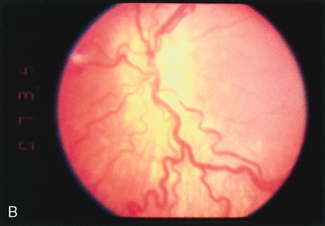
Vascular changes associated with ROP include tortuosity and dilation of the peripheral retina and iris. When these changes progress to include the posterior pole, then a plus sign is added to the ROP stage number.

THRESHOLD DISEASE

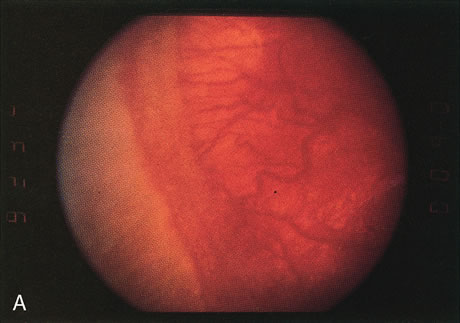
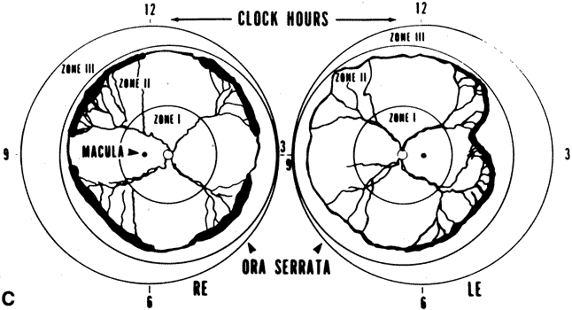
Threshold disease is defined as five contiguous or eight accumulated clock hours of stage 3 ROP with plus disease (Fig. 2). At threshold, the risk of 20/200 or worse final visual acuity is approximately 60% without treatment,10,11 and it is the level of disease at which treatment should be applied. When threshold disease is present, treatment should be performed within 72 hours of detection.13

Fig. 2. A. Clinical appearance of stage 3 ROP. This stage has growth of vessels with fibrous tissue out of the plane of the retina (extraretinal fibrovascular proliferation). B. Clinical appearance of plus disease. In the posterior pole, the retinal veins are engorged and tortuous. C. Two representative eyes that have reached threshold for treatment. The right eye (RE) has at least eight accumulative clock hours of stage 3 ROP. The left eye (LE) has at least five contiguous clock hours of stage 3 ROP. The thin line of ROP represents stage 1 or stage 2 disease, the broader sketched line signifies stage 3 disease. (From Cryotherapy for Retinopathy of Prematurity Cooperative Group: Multicenter trial of cryotherapy for retinopathy of prematurity. Preliminary results. Arch Ophthalmol 106:471–479, 1988. Copyright, Archives of Ophthalmology.)

PRETHRESHOLD DISEASE

Prethreshold disease is ROP that does not meet the definition for threshold disease but is likely to progress and require treatment. It is defined as ROP of any stage in zone I, ROP of stage 2 in zone 2 with plus disease or ROP of stage 3 in zone II of less than five contiguous clock hours or eight cumulative clock hours. Eyes with prethreshold disease should be examined on a weekly basis.13

Back to Top
CRYOTHERAPY

INDICATIONS

Cryotherapy was the first treatment for ROP proven to be effective by multicenter randomized clinical trial. The multicenter trial of cryotherapy for retinopathy of prematurity (CryoROP) study assessed structural and functional outcomes in eyes randomized to either cryotherapy or observation for infants of less than 1251 g birth weight with threshold disease.13 The presence of a retinal fold involving the fovea, a retinal detachment involving zone I, retrolental tissue, or mass was deemed an unfavorable structural outcome, whereas functional outcome was deemed unfavorable if final best corrected visual acuity was 20/200 or worse.10,11 Preliminary study results at 3 months indicated a strong structural benefit for eyes with threshold disease treated with cryotherapy; therefore, patient enrollment was halted before the planned completion date.13 Threshold disease continues to be the primary indication for treatment in eyes with ROP.

CONTRAINDICATIONS

If the fundus view is obscured by anterior segment changes or vitreous hemorrhage, cryotherapy should not be applied. Ultrasonography should be performed when there is no fundus view. If obscuring vitreous hemorrhage is present during the active stage of ROP, vitrectomy should be considered to clear the view for treatment. If anterior segment changes, such as corneal opacity or cataract, are present in both eyes, or in a fellow eye when the other eye is blind, and if retinal detachment is seen on ultrasound, combined anterior segment and vitreous surgery should be considered. The infant's systemic status, if unstable, may preclude treatment. Treatment should be deferred if the neonatologist believes that treatment may be too stressful for the infant.

PROCEDURE

Cryotherapy can be applied with the use of either topical, local infiltration, or general anesthesia. If topical or local infiltration anesthesia is used, intravenous analgesic and sedative medication should be given.16 With topical anesthesia, the ballooning of the conjunctiva associated with subconjunctival infiltration does not occur. Sometimes, the swelling of the conjunctiva from injection of local anesthesia in addition to the edema that occurs during cryotherapy obscures the cornea, necessitating termination of the procedure. For topical anesthesia, proparacaine hydrochloride 0.5% or tetracaine hydrochloride 0.5% can be applied to the cornea every 20 minutes during treatment. Local infiltration anesthesia can be given by injecting 0.5 mL of lidocaine hydrochloride 1% into the subconjunctival space. To avoid possible cardiopulmonary complications, no more than 0.5 mL should be injected.17 General anesthesia can be used and was given to 28% of the patients in the CryoROP study.18 For general anesthesia, the infant usually must be removed from the nursery and be transported to the operating room. The infant is separated from the staff familiar with the case, and faces all of the risks associated with general anesthesia.

Before cryotherapy, the pupils are dilated. Homatropine hydrobromide 2% and phenylephrine hydrochloride 2.5% drops instilled 3, 2, and 1 hours preoperatively provide adequate dilation. After a lid speculum is inserted, cryotherapy may proceed under indirect ophthalmoscopic viewing. A standard retinal or neonatal cryoprobe may be used. It is best to start treatment nasally because pressure from the cryoprobe will soften the globe and thus facilitate treatment temporally, when the avascular zone usually is more posterior. Contiguous spots of cryotherapy should be applied throughout the entire avascular retina anterior to the ridge and extraretinal fibrovascular proliferation. Usually, it takes just a few seconds for the avascular retina to whiten from the cryotherapy. This whitening is the endpoint for treatment (Fig. 3). Typically, depending on the extent of avascular retina, 30 to 50 applications of cryotherapy are necessary to complete treatment. Care should be taken to avoid prolonged scleral depression, which increases the intraocular pressure and risk for central retinal artery occlusion.

Fig. 3. Appearance of an avascular zone immediately after several applications of cryotherapy. This diagram shows the ideal spacing of freeze spots and their relationship to the ora serrata anteriorly and to the stage 3 ridge of ROP posteriorly.

Most often, treatment can be accomplished without making conjunctival incisions. When threshold disease is present in zone I or posterior zone II, it may not be possible to treat the entire avascular zone without a conjunctival incision. A small incision 4 to 6 mm posterior to the limbus within the center of the quadrant is adequate to allow the cryoprobe tip to fit. Depending on the extent of disease, one to four incisions may be made. These incisions need not be sutured if they are less than 3 to 4 mm long.

Occasionally, corneal clouding occurs during treatment, obscuring the view. Removing the lid speculum and waiting several minutes often results in clearing. If waiting fails, the epithelium may be removed with a cotton-tipped swab soaked in cocaine hydrochloride 4% solution. Use of the swab provides excellent topical anesthesia as well. If removing the corneal epithelium with cocaine does not result in clearing of the cornea, the procedure can be completed later.

Vitreous hemorrhage also may occur during treatment, necessitating termination of the procedure. The hemorrhage usually results from bleeding from a florid area of extraretinal fibrovascular proliferation caused by pressure on the globe from the cryoprobe. Treatment should be completed when the vitreous hemorrhage is clear enough to permit further photocoagulation.

EXPECTED RESULTS

The desired outcome after cryotherapy is regression of both plus disease and extraretinal fibrovascular proliferation (Fig. 4). Without regression, features of unfavorable outcome, both anatomic and functional, may develop. The structural and functional benefits of cryotherapy in eyes with threshold disease have been demonstrated by the CryoROP study at 1 year, 3 ½, and 5 ½ years post enrollment.11,19 At 10 years post enrollment structural outcome was unfavorable in 47.9% of control eyes versus 27.2% of treated eyes. Unfavorable functional outcome (Snellen visual acuity of 20/200 or worse) was identified in 62.1% of control eyes versus 44.4% of treated eyes at 10 years. The percentage of eyes with 20/40 or better visual acuity was similar for both treated (25.2%) and untreated (23.7%) eyes at 10 years with no statistical difference between the two groups.10

Fig. 4. Preoperative (A) and postoperative (B) appearance of the posterior pole of an infant treated for threshold ROP with cryotherapy. Note the regression of plus disease after treatment. Preoperative (C) and postoperative (D) appearance of the retinal periphery of the same infant. Avascular zone (arrow). Note the regression of the extraretinal fibrovascular proliferation and the cryotherapy scarring.

A reduction in both plus disease and extraretinal fibrovascular proliferation should be seen by 1 week after treatment.

COMPLICATIONS AND THEIR MANAGEMENT

Both ocular and systemic complications may occur during cryotherapy for ROP. Systemic complications may be life threatening. All patients treated with cryotherapy have periorbital edema, conjunctival injection, and chemosis. The periorbital edema usually subsides in a few days, whereas the conjunctival injection and chemosis take 1 to 2 weeks to regress. In the CryoROP study, conjunctival or subconjunctival hematoma occurred in 11.7% of treated eyes, and unintended conjunctival laceration occurred in 5.3% of eyes.18 These complications are self-limited and do not lead to long-term problems.

The most frequent and most serious ocular complication in the CryoROP study was intraocular hemorrhage. Retinal, preretinal, and vitreous hemorrhage occurred in 22.3% of treated eyes.18 At times intraocular hemorrhage may prevent the completion of treatment. In such cases, treatment is aborted and the eye is carefully followed for clearing of the hemorrhage so that treatment may be completed.

Because treatment is delivered with a probe that must be pressed onto the globe, there is the potential for severe injury, such as perforation of the globe, eye muscle laceration or avulsion, and orbital wall injury. Central retinal artery occlusion may occur as a result of excessive pressure placed on the globe during cryotherapy. In the CryoROP study, transient closure of the central retinal artery occurred in one case.18 Care should be taken to limit the pressure that is placed on the globe and adnexal structures.

In the CryoROP study,18 9.4% of infants had bradycardia, arrhythmia, or significant apnea during the administration of anesthesia and cryotherapy. An additional 1.1% of infants had acquired or increased cyanosis. One infant had a seizure during treatment. Brown and others17 reported three cases of respiratory arrest and one of cardiopulmonary arrest in 80 consecutive infants treated with cryotherapy for ROP. These severe systemic complications underscore the need for careful monitoring of infants by a neonatologist or anesthesiologist during treatment. Evidence has shown that the addition of appropriate systemic anesthesia to topical anesthesia for cryotherapy can reduce the rate of systemic complications.16

Late-onset retinal detachment after cryotherapy has been reported.20 Breaks are often found at the border of the cryotherapy-treated and untreated retina. The retinal breaks may occur as a result of the inability of treated retina to stretch as the eye grows or as a result of vitreous traction.21 Surgical treatment includes standard buckling, vitrectomy, or combination techniques although the need for subsequent surgery is as high as 30%.22 An increased risk of rhegmatogenous retinal detachment is present for life.

The incidence of myopia is increased in children with a history of ROP with or without treatment. This increased risk of myopia correlates directly with severity of eye disease. Severe ROP leads more often to high myopia.23 When compared with observed control eyes with threshold ROP, eyes treated with cryotherapy have an increased risk of myopia greater than 8.00 Diopters (D).24

The visual field in eyes with a history of threshold ROP is decreased when compared with normal controls. Results from the CryoROP study at 10 years demonstrated a reduction in visual field area from 30% to 37% in eyes treated with cryotherapy when compared with healthy controls.25 This reduction in visual field was 5% greater for eyes that underwent cryotherapy than in eyes with threshold ROP that did not undergo cryotherapy.

There does not appear to be an increased incidence of amblyopia in eyes with threshold ROP treated with cryotherapy when compared to observed threshold ROP. In the CryoROP study, strabismus surgery was required for 10% of eyes treated with cryotherapy and 7% of control eyes with threshold ROP.26

Back to Top
LASER PHOTOCOAGULATION

INDICATIONS

Photocoagulation was the first modality of treatment of the anterior avascular zone for acute ROP. In 1967, Nagata and colleagues27,28 used xenon arc photocoagulation to induce regression of acute ROP. This method of treatment was difficult to deliver, and it was supplanted by cryotherapy, which was first reported to induce regression by Yamashita29 in 1973.

In the late 1980s, indirect ophthalmoscopic delivery systems for laser photocoagulation became available. Shortly thereafter, McNamara and colleagues,30 Iverson and colleagues,31 (Table 2) Hunter and Repka32 (Table 3) compared transpupillary laser photocoagulation with cryotherapy for threshold ROP in prospective, randomized trials. An argon laser was used to treat in the McNamara30 and Iverson31 studies, whereas a diode laser was used in the Hunter32 study. Similar favorable regression rates were reported with either treatment in each of these studies. McGregor and colleagues33 and Pearce and colleagues34 retrospectively compared laser photocoagulation with cryotherapy for threshold ROP. No statistical difference was found between the two treatment groups.


Table 2. Prospective, Randomized, Comparison of Indirect Ophthalmoscopic Delivery of Diode Laser to Cryotherapy for Threshold ROP


StudyDiode LaserCryotherapy
FavorableUnfavorableFavorableUnfavorable
McNamara and associates4125/28 (89%)3/28 (11%)20/24 (83%)4/24 (17%)
Hunter and Repka3216/17 (94%)1/17 (6%)14/15 (93%)1/15 (7%)


Table 3. Prospective, Randomized, Comparison of Indirect Ophthalmoscopic Delivery of Argon Laser to Cryotherapy for Treatment of Threshold ROP


StudyArgon LaserCryotherapy
FavorableUnfavorableFavorableUnfavorable
McNamara and associates3015/16 (94%)1/16 (7%)9/12 (75%)3/12 (25%)
Iverson and Associates316/6 (100%)0/6 (0%)5/6 (83%)1/6 (17%)


Seiberth and colleagues35 compared transscleral with transpupillary diode laser photocoagulation for the treatment of threshold ROP. They found transscleral diode laser to be as effective as transpupillary diode laser in causing regression of threshold ROP.

CONTRAINDICATIONS

Treatment of threshold ROP with laser photocoagulation requires clear media. Anterior segment changes, such as cataract and corneal clouding, and vitreous hemorrhage may preclude laser treatment, yet the media may be clear enough for cryotherapy to be performed. A persistent tunica vasculosa lentis may lead to cataract formation with argon laser photocoagulation.36 This risk may be decreased when using a diode laser.37

Systemic contraindications may be fewer with laser photocoagulation than with cryotherapy, perhaps due to the less invasive nature of the procedure.32

PROCEDURE

Indirect laser photocoagulation for ROP should be performed with either topical anesthetic supplemented with intravenous analgesia and sedation or with general anesthesia.16 There is no need for local infiltration anesthesia because only gentle manipulation of the globe with a scleral depressor is necessary. McNamara and others30 used topical anesthesia alone, Hunter and Repka32 used topical anesthesia supplemented with systemic analgesia, whereas all infants treated by Iverson and colleagues31 and Landers and associates40 were given general anesthesia. Transient bradycardia occurred in three patients (19%) treated with laser and three (25%) treated with cryotherapy in the McNamara study. The bradycardia resolved when the scleral depressor or cryoprobe was removed for several seconds. Intubation following apnea and bradycardia was required for two patients (13%) treated with cryotherapy and none with laser in the Hunter and Repka32 study. No other systemic complications occurred.

Before laser photocoagulation, the pupils are dilated. Homatropine hydrobromide 2% and phenylephrine hydrochloride 2.5% drops instilled 3, 2, and 1 hours preoperatively provide adequate dilation. If topical anesthesia supplemented with systemic analgesia and sedation is to be used, treatment can be performed in the operating room or in the nursery. Proparacaine hydrochloride 0.5% is applied to the globe. A lid speculum is placed. Gentle manipulation with a scleral depressor to position the globe for viewing of the peripheral retina is performed. Although the peripheral retina is viewed through the indirect ophthalmoscope, the laser aiming beam can be seen. When the aiming beam is in focus on the peripheral avascular retina, laser discharge is accomplished by pressing a foot pedal. Care should be taken to assure that the aiming beam is focused before discharge to avoid laser burns in unwanted areas, such as the posterior vascularized retina, cornea, iris, and lens. The entire anterior avascular retina should be treated with avoidance of laser to the active ridge. Steinmetz and Brooks38 have suggested a possible benefit to treating the ridge in addition to the avascular retina but to date no statistical benefit has been shown. Laser spots should be placed one-half burn width apart, with a dull white laser photocoagulation mark used as the endpoint. Banach and colleagues have demonstrated an increased risk of disease progression with a less dense laser photocoagulation pattern.39 For argon laser, the initial power setting should be 200 mW, with a duration of 0.1 second. The power can be increased gradually to achieve the appropriate laser mark in the fundus. For diode laser, the initial power setting should be slightly lower (150 mW), with a longer duration (0.2 second), to avoid causing choroidal hemorrhage and rupture of Bruch's membrane. A neonatologist or anesthesiologist should monitor the infant during treatment.

The advent of portable diode (Fig. 5) and argon lasers has simplified the treatment of ROP and has largely supplanted cryotherapy as the treatment of choice for ROP at most centers. The portable laser can be brought directly into the nursery, which avoids the need for costly and sometimes hazardous transfer of the infant to a facility with a freestanding laser. For safety reasons, treatment should be performed in an isolated room in the nursery.

Fig. 5. Oculight SLx (Iris Medical, Mountain View, CA) portable diode laser with indirect ophthalmoscope delivery unit.

EXPECTED RESULTS

Several small prospective randomized trials have compared the efficacy of laser photocoagulation to cryotherapy for threshold ROP30,31,40,41 No statistically significant study has proven its efficacy, but they have suggested that laser treatment is as effective as cryotherapy in the treatment of threshold ROP (Fig. 6). A number of other studies have assessed the structural and functional outcomes following both argon and laser treatment of threshold ROP, with good results (Table 4).


Table 4. Structural and Visual Outcomes After Laser for Threshold ROP: Seven Studies.


StudyLaserStructural outcomeVisual Outcome
Landers and Associates40Argon11/15 – Favorable* No information given
4/15 – (2 stage 4B, 2 stage 5)
Dejonge and Associates42Diode57/61 – FavorableNo information given
4/61 – (2 stage 4A, 1 stage 4B)
Ling and Associates43Diode13/13 – Favorable13/13 Favorable*
Axer-Siegel and Associates44Diode41/48 – FavorableNo information given
7/48 – (4 stage 4A, 1 stage 4B, 2 stage 5)
Sieberth and Associates45Diode41/42 – FavorableNo information given
1/42 – (Stage 5)
Foroozan and Associates46Diode or Argon109/120 – FavorableNo information given
11/120 (1 stage 4A, 3 stage 4B, 7 stage 5)
Connolly and Associates47DiodeNo information given20/60 – 20/50 or better Snellen visual acuity

*As defined by the Cryo-ROP Study Group


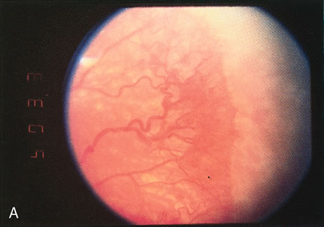
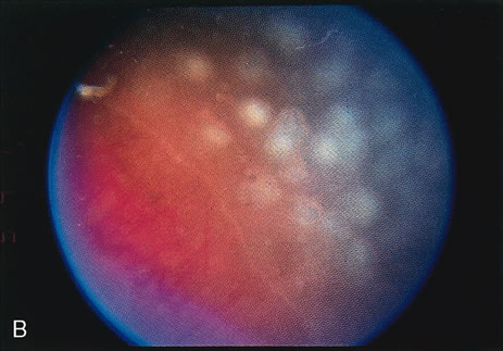
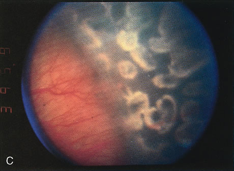
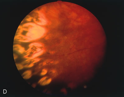
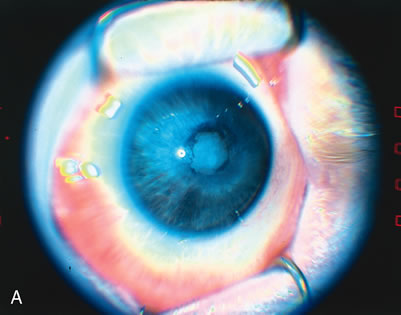
Fig. 6. A. Peripheral neovascularization in zone I. B. Peripheral retina immediately after argon laser photo-coagulation. C. Peripheral retina 1 week after argon laser photocoagulation. D. Peripheral retina 1 month after argon laser photocoagulation. Note the complete regression of neovascularization.

Connolly and colleagues48 assessed visual function and structural outcomes between photocoagulation and cryotherapy for ROP at 10 years post-treatment. They found that the laser treated group had a mean best-corrected ETDRS (Early Treatment Diabetic Retinopathy Study) visual acuity (BCVA) of 20/66 compared with a BCVA of 20/182 in the cryotherapy group. Four of the 21 eyes treated with cryotherapy had unfavorable structural outcomes, and two of the 23 eyes treated with laser had unfavorable structural outcomes. Despite unintended sample bias, this study compares well with that of Shalev and colleagues49 who reviewed their results comparing cryotherapy with laser at 7 years post-treatment for threshold ROP. They found that mean BCVA was 20/33 for laser-treated eyes and 20/133 for cryotherapy-treated eyes. These studies suggest that laser treatment for threshold ROP may maximize visual potential when compared with cryotherapy.

COMPLICATIONS AND THEIR MANAGEMENT

As with cryotherapy, both systemic and ocular side effects may occur with laser photocoagulation for ROP. As mentioned earlier, bradycardia during treatment, presumably due to scleral depression, can occur. Releasing the depressor usually results in resolution of the arrhythmia.

Mild conjunctival injection from scleral depression may be present for a day or so after laser photocoagulation. This effect is much less than the anterior segment changes that typically occur after cryotherapy. Additionally, conjunctival incisions are never needed for laser photocoagulation because posterior treatment is delivered easily. Conjunctival incisions frequently are necessary for the treatment of zone I disease with cryotherapy.

Anterior segment burns to the cornea, iris, or lens may occur with laser treatment.50 Landers and colleagues40 noted small burns to the pupil margin in 5 of 15 laser-treated patients. More powerful laser damage to the iris may rupture iris blood vessels and lead to a hyphema.51,52 Lens changes following laser photocoagulation for ROP may range from transient lenticular opacities that resolve over a few weeks to permanent cataracts. 53–56 Ensuring that the aiming beam is focused on the retina is essential. Diode laser may lessen the likelihood of lens burns in premature infants with persistent tunica vasculosa lentis.37 The diode laser, emitting at 810 nm, is not absorbed by the hemoglobin in those vessels, whereas argon green, emitting at 514 nm, is absorbed by hemoglobin.

Vitreous hemorrhage may occur with laser photocoagulation. There were three instances of mild vitreous hemorrhage in each group (laser and cryotherapy) in the McNamara study.30 These hemorrhages resolved within 1 week. Choroidal hemorrhages with associated ruptures in Bruch's membrane are a known complication of laser treatment. Appropriate burn intensity decreases the likelihood of this complication.

Late-onset retinal detachment has been reported after cryotherapy,20 and it is a potential complication after laser treatment. The risk may be less with laser treatment because the laser is applied in a scatter pattern, with intervening areas of untreated retina. This approach may allow the retina to grow and stretch better, thus decreasing the likelihood of breaks at the border between treated and untreated retina. A single case of an exudative retinal detachment, which resolved spontaneously, has been reported following laser treatment for threshold ROP.57

Increased myopia is a known side effect of cryotherapy for threshold ROP.24 Laser therapy for threshold ROP causes significantly less myopia than cryotherapy.58 Connolly and colleagues59 noted a mean spherical equivalent (SE) of –4.48 D following laser treatment compared with a mean SE of –7.65 D for eyes treated with cryotherapy. The crystalline lens power and not axial length was found to account for the difference in refractive power between the two treatment modalities.

Back to Top
SCLERAL BUCKLE

INDICATIONS

Cryotherapy and laser photocoagulation do not cure all cases of threshold ROP. Progression to stage 4 or 5 disease may occur, despite appropriate treatment. These more advanced stages represent retinal detachments that range from small and localized to closed anterior and posterior funnel detachtments.60 Retinal detachments in ROP may have a serous and tractional component. Exuberant extraretinal fibrovascular proliferation may lead to subretinal serous fluid accumulation. With progressive fibrous proliferation, tractional detachment of the retina may occur. This proliferation is often circumferential, and the retina may detach in a purse string configuration. Rhegmatogenous retinal detachments do occur but are rare, often occurring years later as a delayed complication of vitreoretinal traction to an abnormal retinal periphery.20,22

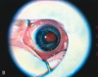
The indications for scleral buckling surgery are stage 4A with progression, stage 4B or open funnel stage 5 retinal detachment. Stage 4A retinal detachment (Fig. 7A) is subtotal, and spares the fovea. The CryoROP study demonstrated resolution in 46.1% of eyes with stage 4A retinal detachments at 4½ years. 61 Unfortunately, the study also showed a 17.9% risk of progression for stage 4A eyes to total retinal detachment. Stage 4B retinal detachment (Fig. 7B), which is due to progressive exudation or traction, involves detachment of the fovea. Stage 5 retinal detachment (Fig. 7C) is total; it may have an open or closed funnel configuration. When the detachment is funnel shaped, the funnel is divided into anterior and posterior parts, and may be open or closed in either location.

Fig. 7. A. Stage 4A ROP. Subtotal retinal detachment does not involve the fovea. B. Stage 4B ROP. Subtotal retinal detachment involves the fovea. C. Stage 5 ROP. Total retinal detachment. The detachment may be shallow, with an open funnel configuration, as in this example. Note the loss of the choroidal vascular pattern because of subretinal fluid.

Since the last edition, lens-sparing vitrectomy for Stage 4A has been advocated by Trese and Capone.60A They report excellent visual results. However, it is still sometimes difficult to determine when spontaneous regression might occur. Once the fovea detaches, progressive damage to photoreceptors may occur, precluding visual development.

CONTRAINDICATIONS

Stage 4A ROP may regress spontaneously or as a result of cryotherapy or laser photocoagulation.61 These detachments should be followed closely, but repair is not necessary unless progression is observed. Once the fovea is threatened (stage 4B), scleral buckling or lens sparing vitrectomy should be considered.

A closed or partially closed funnel stage 5 retinal detachment is not amenable to scleral buckling. Apposition of the choroid and retinal pigment epithelium to the retina cannot occur because of fibrous tissue elevating the retina. These more severe forms of retinal detachment may be treated with vitreous surgery.

Vitreous hemorrhage is a relative contraindication to scleral buckling surgery. If the hemorrhage is mild and an adequate view is still present, allowing staging and appreciation of the amount of fibrous proliferation, then a scleral buckling operation may be performed. If the hemorrhage is too dense, or if there is any concern that a funnel-shaped retinal detachment is present, vitrectomy should be considered.

The infant's systemic condition must be considered. Scleral buckling is performed under general anesthesia, and any contraindications to this form of anesthesia will necessitate delaying the surgery.

PROCEDURE

Scleral buckling for ROP is performed under general anesthesia in the operating room. An anesthesiologist skilled in pediatric anesthesia and a neonatologist familiar with the patient should be in attendance.

Before surgery, the pupils are dilated. Homatropine hydrobromide 2% and phenylephrine hydrochloride 2.5% drops instilled 3, 2, and 1 hours preoperatively provide adequate dilation. After a pediatric lid speculum is inserted, a 360-degree conjunctival peritomy is performed. The rectus muscles are bridled with 4-0 silk sutures. The sclera should be inspected for thin areas. The standard instruments for adult scleral buckling procedures are too large, so a Halsted malleable retractor can be used to expose the sclera in each quadrant.

If cryotherapy was not performed on the avascular peripheral retina previously, or if inadequate cryotherapy is present, cryotherapy should be performed as described earlier.

Scleral buckling can be accomplished with either an implant or an exoplant. If an implant procedure is performed, the surgeon should be highly skilled in preparing a scleral bed; the sclera in these infants is extremely thin, and perforations may occur during preparation of the scleral flaps.

The globe should be encircled with a 2-mm-wide no. 40 band or a 2.5-mm-wide no. 240 band. The latter band provides a broader area of indentation, but it may be too large for smaller eyes. If a localized area of greater height is desired, such as over a broader area of extraretinal fibrovascular proliferation that has greater elevation, then a segment of a 4.5-mm-wide no. 219 strip can be placed (Fig. 8). The silicone element or elements are secured in place with a single 5-0 nylon mattress suture in each quadrant. The ends of the band are secured to each other with a Watzke sleeve or a 5-0 nylon suture.

Fig. 8. Scleral buckle for ROP. Note that the buckle is placed over the extraretinal fibrovascular proliferation, where traction is the greatest.

External drainage of subretinal fluid remains controversial.62–68 The risks of retinal penetration, incarceration, subretinal hemorrhage, and infection must be balanced against the prolonged time that subretinal fluid may remain if no drainage is performed. In cases of shallow retinal detachment, an endolaser probe can be used to assist with external drainage of fluid.69 A scleral cut down is performed, and the endolaser is positioned adjacent to the bare choroid. Set at 200 mw and 0.2 of a second one burn is usually effective at creating a choroidotomy with subsequent drainage. If external drainage is not performed, an anterior chamber paracentesis can be done to allow increased buckle height.

Once the buckle is cinched, the intraocular pressure should be assessed. If the pressure is too high, anterior chamber paracentesis can be performed. Alternatively, or in addition to paracentesis, acetazolamide (5 mg/kg) can be given intravenously to lower the intraocular pressure. The central retinal artery should be checked for lack of pulsation by indirect ophthalmoscopy to ensure that the pressure is not too high.

At the end of the procedure, the bridle sutures are removed, the conjunctiva is closed with absorbable sutures, and antibiotic and steroid medications are applied.

The eyes often are very small when an infant has scleral buckling surgery. To promote normal growth of the eye, transection of the scleral buckle can be performed from 3 to 12 months after the original procedure.62,68 Choi and Yu65 have shown that postoperative removal of the scleral buckle in infants with ROP is associated with reduced refractive myopia.

EXPECTED RESULTS

Anatomic success after scleral buckling for retinal detachment in ROP is defined as macular reattachment (full attachment of the retina between the temporal vascular arcades).

The success rate of scleral buckling varies with the severity of ROP. Analysis of the results from seven studies on scleral buckling for ROP demonstrates a wide range in reattachment success (Table 5). Greater reattachment rates were achieved in all studies when additional vitreous surgery was performed.


Table 5. Data from Seven Studies That Used Scleral Buckling Surgery to Treat Stage 4 and 5 ROP


StudyStageNo Reattached/
total number
Vision in eyes with
reattached retina
Chuang and Yang644A9/14 (64%)No information given
4B6/9 (67%)
Greven and Tasman684B3/3 (100%) 20/200 to fix/follow
5  10/19 (52%)20/100 to NLP
Hinz and associates634A6/8 (75%)20/180 to LP
McPherson and associates704  19/21 (90%)No information given
5  5/11 (45%)
Noorily and associates664B10/15 (67%)Fix/follow to NLP
Trese624A12/17 (70%)No information given
4B29/43 (67%)
5  4/10 (40%)
Ricci and associates674A11/18 (61%)20/30 to NLP
4B2/10 (20%)20/50 to NLP


Although the anatomic success rates in these studies are encouraging, the visual results in general are disappointing. The reasons for poor visual results in these infants remain speculative. Some infants may have cerebral dysfunction as a result of complications of prematurity, such as intraventricular hemorrhage. In these infants, the macula is immature, and detachment of this immature retina may have a more devastating effect on photoreceptors and their development than in the adult macula. Amblyopia may play a role as well.

COMPLICATIONS AND THEIR MANAGEMENT

The complications of scleral buckling surgery for ROP are similar to those that can occur with adult scleral buckling surgery, with some special considerations for the infant eye. The premature infant's eye is significantly smaller than an adult eye. Full adult size of the globe is not attained until 3 years of age. An infant at 34-weeks' gestation has an eye that is 15 mm in diameter (Fig. 9). The sclera of the infant eye is thin, and the surgeon must be especially careful when depressing, cutting a bed, and passing sutures.

Fig. 9. Gross specimens of an adult eye (A) and a 34-week-gestation infant eye (B). (Courtesy of James P. Bolling, MD, Mayo Clinic, Jacksonville, FL)

Cryotherapy done during a scleral buckling procedure carries the risks of complications mentioned earlier. Because of the risk of corneal clouding and central retinal artery occlusion, prolonged depression during cryotherapy should be avoided.

Perforation of the thin infant globe can occur while a scleral bed is being prepared and while sutures are passed to secure the buckling element. Dissection of scleral beds is a technically difficult procedure that should be attempted only by highly experienced surgeons. Similarly, great care should be taken when passing sutures through the sclera. If perforation occurs during passage of a suture, the suture should be removed and replaced so that the buckling element will cover the perforation. Indirect ophthalmoscopy should be done immediately to determine whether there has been retinal damage. If the retina became detached in the area of perforation, fortuitous drainage may occur. If retinal perforation has occurred, cryotherapy should be applied.

A decision to drain subretinal fluid should be made carefully because the risks are significant. If there is only shallow fluid, and if the buckle provides adequate height, drainage may not be necessary. The risks of drainage are perforation, incarceration, hemorrhage, and infection. Choosing a drainage site over bullous retina can minimize the risk of perforation. If perforation occurs, it should be managed by buckling and cryotherapy, as mentioned earlier. Drainage under the buckle obviates the need to add an additional buckling element over a perforation at a drainage site. Incarceration is less likely with tractional retinal detachment than with the rhegmatogenous variety because the retina is pulled inward by tractional forces. Nonetheless, if incarceration occurs, it should be treated with cryotherapy and buckled. Diathermizing the choroid prior to drainage will decrease the risk of hemorrhage. Once the globe is entered, the possibility of intraocular infection increases. Endophthalmitis after scleral buckling is rare. It should be managed with standard techniques, including injection of intravitreal antibiotics.

Careful attention to the intraocular pressure at the end of the procedure is important. If the pressure is too high, central retinal artery occlusion may occur. As mentioned earlier, the pressure should be normalized, and the central retinal artery checked for the desired absence of pulsations.

In the small infant eye, the buckle may induce angle-closure glaucoma in the postoperative period. The cornea should be assessed for clarity, and the intraocular pressure can be checked with an electronic tonometer. If angle-closure glaucoma develops, the scleral buckle must be loosened.

A tight buckle also may induce anterior segment necrosis. This condition is manifested by corneal edema without high intraocular pressure, chemosis, and a cellular reaction in the anterior chamber. The buckle should be loosened or removed.

Scleral buckle infection is manifested by conjunctivitis overlying the buckle and a clear cornea in mild cases. Intraocular inflammation is seen in more severe cases. This condition is managed by removal of the buckle and antibiotics.

Extrusion of the scleral buckle as a result of release of the sutures is managed by resecuring the buckle, or removal of the buckle, if it is no longer necessary.

Although not technically a complication, persistent detachment of the retina is an unsatisfactory result. Nonattachment may be due to the disease process itself (persistent or progressive tractional detachment), or it may be due to a loose buckle. If the height of the buckle seems inadequate, revision of the scleral buckling procedure should be done.

As mentioned earlier, the scleral buckle may cause erosion or may interfere with growth of the eye as the infant matures. The scleral buckle should be transected or removed after 6 to 12 months to promote normal eye growth.

Back to Top
VITRECTOMY

INDICATIONS

Vitreous surgery may be performed for stage 4 and 5 ROP. Controversy remains as to whether or not vitrectomy surgery should be offered for stage 4A disease. This is in large part because of two different findings from the CryoROP study. First, visual acuity was worse than 20/200 in 82% of eyes at 4½ years with stage 4A retinal detachments at the 3 months examination.61 Second, the CryoROP study71 demonstrated a uniformly poor anatomic and visual outcome following vitrectomy for stage 4 and 5 ROP. Fortunately, improvements in closed vitrectomy surgical techniques over the past two decades now offer hope for many eyes with advanced disease.72 The surgery is complicated and the visual results may be poor, so patients should be selected carefully.

CONTRAINDICATIONS

A fellow eye with minimal or regressed ROP is a relative contraindication to vitreous surgery for advanced disease. Corneal opacification may also preclude surgical repair, although an open-sky vitrectomy with corneal transplantation can be performed if ultrasound demonstrates an open funnel retinal configuration.73,74 Eyes with obvious buphthalmos and glaucoma have a poor visual result, and should not have surgery. High intraocular pressure at the time of immediate preoperative evaluation is a relative contraindication to surgery. Permanent optic nerve and retinal damage may have occurred, and will result in a poor visual prognosis, even if the retina is reattached. No light perception vision (no response to a bright light) is a relative contraindication to surgery. Bound-down iris prevents the pupil from reacting to light, and a completely detached retina with epiretinal membrane formation may prevent the child from wincing when exposed to a bright light. Testing by visual evoked potential may objectively confirm a clinical suspicion of no light perception.75

As with other forms of surgery, the infant's systemic condition should be considered. As with scleral buckling, vitreous surgery is performed under general anesthesia, and any contraindications to this form of anesthesia will necessitate delaying the surgery.

PROCEDURE

Vitreous surgery for advanced ROP continues to evolve with improvements in surgical technique and equipment. The advent of wide-field viewing systems, the infusion light pipe, and improved surgical instrumentation have facilitated surgical repair.

The timing of surgery for advanced ROP is important. If plus disease with engorgement of the iris vasculature and tortuosity and dilation of the retinal vessels are present, vitreous surgery should be postponed until ablative therapy has been performed to the avascular retina. Ablation of avascular retina does not reduce traction from the vitreous, but it does induce regression of plus disease thereby reducing the risk of intraoperative hemorrhage. If there is persistent, severe plus disease, Trese76 recommends a two-step approach. Patients initially are treated with ablative therapy to the peripheral attached avascular retina. Within 3 weeks, in most cases, the plus disease has regressed to a point that vitreous surgery may proceed.

There are two approaches to vitreous surgery in infants with advanced ROP; open-sky vitrectomy and closed vitrectomy. Both have advantages and disadvantages. In the past, the main advantage to open-sky vitrectomy (vitreous surgery through a trephined opening in the cornea) was easy access and visualization of the anterior retina and epiretinal membranes. Today, improved closed vitrectomy surgical techniques have negated these advantages. The disadvantages of open-sky vitrectomy include lack of intraocular pressure control and difficulty in posterior manipulation. Closed vitrectomy has the advantages of maintenance of intraocular pressure, minimal distortion of tissue, and good visualization in the posterior pole. There are very few if any disadvantages of closed vitrectomy now that bimanual manipulation is a reality. The goal of vitreous surgery for severe stage 4 and 5 ROP is mechanical release of vitreoretinal traction, which can be accomplished with either form of surgery.

Before undergoing vitrectomy, the patient should be examined under anesthesia. This assessment should include biomicroscopy, measurement of corneal diameter, tonometry, indirect ophthalmoscopy, fundus photography, A-scan and B-scan ultrasonography, visual evoked potential if necessary, and examination under the operating microscope.

The technique of open-sky vitrectomy was pioneered by Schepens77 and Hirose and others,78and was modified by Hirose and associates73 and Tasman and colleagues (Fig. 10).74 Improvements in closed vitrectomy techniques have now almost eliminated the need for open-sky vitrectomies in the treatment of advanced ROP. If corneal opacification is present, corneal button removal followed by placement of a temporary keratoprothesis can be used to provide a closed working environment. This combined procedure allows excellent visualization of the anterior retina during bimanual surgery while providing a controlled intraocular pressure.

Fig. 10. A. Preoperative ROP stage V. B. Postoperative after open sky technique (Courtesy of William Tasman, MD, Philadelphia, PA)

Treister and Machemer79 first described closed vitrectomy for the retinal detachment associated with ROP. Charles,80 Trese,76,81 and DeJuan and colleagues82,83 refined the indications and technique. Innovations in surgical technique now allow lens-sparing vitrectomy in those instances in which anterior loop traction has not pulled the retina up to the lens (Fig. 11).84

Fig. 11. Diagram of two-port lens sparing vitrectomy for open funnel stage 5 ROP.

The entry site for closed vitrectomy is through the pars plicata 1 to 2 mm posterior to the limbus, because no pars plana is present in the infant. Two or three ports may be used, with most surgeons preferring the less invasive two-port technique. In the three-port system, a blunt-tipped 2-mm infusion cannula is sewn into the anterior chamber at the limbus inferotemporally. The remaining two ports are created in the superior quadrants and are used for a fiberoptic light pipe and various vitrectomy instruments (e.g., suction/cutter, membrane peeler/cutter, scissors, diathermy). In a two-port system, a 20-gauge end-irrigating light pipe is used through one of the superior sclerostomy sites to provide both irrigation and illumination. If iris dilation is poor, iris retractors can be used. Once in the anterior chamber with the vitreous cutter, the soft infant lens is removed by suction and cutting. The retrolental tissue is grasped in the center with tissue forceps, lifted, and incised with a microvitreoretinal blade or scissors. When a plane has been created, further dissection with forceps and a membrane peeler/cutter can be accomplished. Flaps of cut membrane are removed with the vitreous cutter. Care should be taken not to create a retinal break in the thin periphery. Replacement of balanced saline solution with viscoelastic is used by some to stabilize the retina, as well as assist with membrane dissection.85 It is not necessary to remove all of the fibrous tissue; the goal of surgery is zone I reattachment. A retinal break in the periphery will likely result in nonattachment. With the hole in the retrolental tissue enlarged as much as possible, the dissection is carried further posteriorly. A narrow configuration posteriorly is more difficult to dissect because of greater accumulation of fibrous proliferation. Dissection must be performed because peeling can cause retinal breaks. As the dissection is carried posteriorly, a peripheral anterior loop configuration may become apparent. If the loop is not too advanced, opening the trough with pic forceps and the membrane peeler/cutter may be possible. Dissection of membranes with removal of vitreous traction from the trough is essential if the retina is to flatten in the postoperative period. Wide-field viewing with scleral depression by an assistant frequently aids this dissection. When as much membrane as possible has been removed, the operation is finished. Drainage of subretinal fluid is not necessary. At the end of the operation, air may be injected into the vitreous cavity to help maintain retinal separation while the subretinal fluid is reabsorbing. The entry sites are closed with 9-0 absorbable polyglactin sutures (Vicryl–Ethicon, Somerville, NJ).

In selected patients with posterior proliferation and an attached peripheral retina, the lens may be spared.84,86,87 A two-port system is used. Incisions should be directed parallel to the visual axis, and great care should be taken to avoid moving the instruments across the midline because the lens may be struck. This age group is highly susceptible to amblyopia, so saving the lens is desirable.

EXPECTED RESULTS

Vitreous surgery for advanced ROP is performed for eyes with stage 4A or greater retinal detachments. The anatomic results following vitrectomy have improved with improved surgical techniques, however surgical failure is not uncommon. The complicated pathoanatomy and the high likelihood of inducing a retinal break during surgery combine to produce this encouraging, but low, rate of success.

The visual results are even more disappointing (Table 6). In a standardized assessment of visual acuity for eyes with retinal detachments that underwent vitrectomy as part of the CryoROP study, only two eyes of one infant had pattern vision at the lowest measurable threshold.88 None of the 58 control eyes with retinal detachment that did not undergo vitrectomy had pattern vision. These results underscore the suggestion that efforts are well spent in attempting to prevent retinal detachment in ROP.


Table 6. Data from Six Studies That Used Vitrectomy to Treat Stage 4 and 5 ROP


StudyStageAnatomic Success
Total attachmentPartial attachmentVision in eyes with reattached retina
CryoROP71 (Closed + Open Vitrectomy)51/72 (1%)2/72 (3%)Fix/Follow (1) LP (2)
Capone and Trese84 (Closed vitrectomy)4A36/40 (90%) Central, steady and maintained fixation in all eyes
Choi and Suk89 (Closed vitrectomy)4–520/87 (23%)24/87 (28%)LP (17), Fix/Follow (15), identify form (5)
Chong and associates90 (Closed Vitrectomy)4B0/1210/12 (83%)Fix/Follow (6), LP (2)
50/4615/46 (33%)20/800 (1), Fix/Follow (4)
Fuchino and associates91 (Closed vitrectomy)50/4929/49 (59%)LP (4), HM (3), 20/2000–20/200 (7), 20/200–20/25 (5), 20/25 (1)
Tasman and associates7450/238/23 (35%)Fix/Follow (1) LP (4)
Trese72 (Closed vitrectomy + Buckle)518/40 (45%)* 22/40 (55%)Fix/Follow (11)
Trese and Droste92 (Closed Vitrectomy)4A 1/1 (100%)LP or better (24)
4B 20/24 (83%) 
5 4/8 (50%) 
Zilis and associates82(Closed Vitretomy)4A/B4/14 (29%)9/14 (64%)Fix/Follow (6), LP (5)
511/21 (9%)38/121 (31%)Fix/Follow (13)

*(total attachment posterior to the scleral buckle)>CryoROP71 (Closed 1 Open Vitrectomy)


COMPLICATIONS AND THEIR MANAGEMENT

Other than massive hemorrhage and infection, the most serious complication in eyes undergoing vitrectomy for advanced ROP is the formation of retinal breaks. Most retinal breaks occur anteriorly in the region of the pars plana during dissection of the tenacious epiretinal membrane. Once a retinal break is created, the case is almost certainly lost because flattening of the retina to allow for adequate cryotherapy to the break and scleral buckling is almost impossible. Reattachment did not occur in any of the cases reported by Zilis and others82 in which a retinal break was noted during surgery. Sometimes, a retinal break is not seen, but Schlieren (optical phenomenon occurring when two clear fluids of different optical densities mix) occurs. A retinal break must be assumed in those cases.

Bleeding is a frequent problem during both open-sky and closed vitrectomy. Most often, eyes are operated on when the active phase of ROP has subsided. If some degree of plus disease is present at surgery, however, hemorrhage is much more likely. Intraocular diathermy usually is sufficient to stop bleeding. Blacharski and Charles93 used intravitreal thrombin to control bleeding during vitrectomy for stage 5 ROP.

Back to Top
FUTURE DIRECTIONS
Despite improvements in early identification and treatment over the past three decades, ROP continues to be a cause of blindness. Eyes with posterior ROP (ROP within zone I or posterior zone II are at particular risk of visual impairment, often progressing to total retinal detachment even with appropriate ablative treatment when threshold is reached.19 Research to improve outcomes is advancing on several fronts.

In an effort to improve structural and functional outcomes for eyes with posterior ROP, the National Eye Institute is currently enrolling patients at high risk for unfavorable outcomes. The Early Treatment ROP study (ETROP) is randomizing patients to laser treatment of high risk prethreshold disease versus observation until threshold is reached.94 Vander and associates14 had previously tested this hypothesis in a small, multicentered, randomized trial of 19 infants with posterior ROP. They found no statistical difference between outcomes for those eyes treated at prethreshold levels compared to waiting until threshold had been reached; however without treatment, 88% of control prethreshold eyes progressed to threshold disease.

Surgical techniques in the treatment of advanced ROP with retinal detachment continue to be refined and improved. The introduction of wide-field microscope viewing systems coupled with lighted instruments now allows true bimanual techniques within a closed vitrectomy system. New sutureless technologies such as the 25-gauge vitrectomy system are proving beneficial in reducing iatrogenic tissue damage during vitrectomy in these very small eyes.95

Experimental chemical inhibition of animal model ROP is beginning to show promise. Anacortave acetate has been shown to reduce the severity of abnormal blood vessels associated with ROP while allowing normal retinal vasculature to remain unchanged.96 In the future, eyes with posterior ROP may undergo intravitreal injection of agents that inhibit new vessel growth with or without laser ablation in an effort to reduce the risk of progression to retinal detachment.

Back to Top
CONCLUSION
Early identification and treatment of threshold ROP is of utmost importance to reduce the risk of progression to stage 4 and 5 ROP. In those cases in which ablative therapy in unsuccessful in preventing progression, scleral buckling should be reserved for progressive exudative stage 4A ROP while more tractional stage 4 or Stage 5 ROP should be treated with closed vitrectomy with or without lensectomy.
Back to Top
REFERENCES

1. Rowlands E, Ionides AC, Chinn S et al: Reduced incidence of retinopathy of prematurity. Br J Ophthalmol 85:933-935, 2001

2. Fledelius HC, Dahl H: Retinopathy of prematurity, a decrease in frequency and severity. Trends over 16 years in a Danish county. Acta Ophthalmol Scand 78:359, 2000

3. Bullard SR, Donahue SP, Feman SS et al: The decreasing incidence and severity of retinopathy of prematurity. J Aapos 3:46, 1999

4. Hussain N, Clive J, Bhandari V: Current incidence of retinopathy of prematurity, 1989–1997. Pediatrics 104:e26, 1999.

5. Gould JB, Benitz WE, Liu H: Mortality and time to death in very low birth weight infants: California, 1987 and 1993. Pediatrics 105:E37, 2000.

6. Campbell PB, Bull MJ, Ellis FD et al: Incidence of retinopathy of prematurity in a tertiary newborn intensive care unit. Arch Ophthalmol 101:1686, 1983

7. Kalina RE, Karr DJ: Retrolental fibroplasia. Experience over two decades in one institution. Ophthalmology 89:91, 103, 1982.

8. Repka MX, Palmer EA, Tung B: Involution of retinopathy of prematurity. Cryotherapy for Retinopathy of Prematurity Cooperative Group. Arch Ophthalmol 118:645, 2000

9. An international classification of retinopathy of prematurity. The Committee for the Classification of Retinopathy of Prematurity. Arch Ophthalmol 102:1130, 1984

10. Multicenter trial of cryotherapy for retinopathy of prematurity: Ophthalmological outcomes at 10 years. Arch Ophthalmol 119:1110, 2001

11. Multicenter trial of cryotherapy for retinopathy of prematurity: Snellen visual acuity and structural outcome at 5 ½ years after randomization. Cryotherapy for Retinopathy of Prematurity Cooperative Group. Arch Ophthalmol 114:417, 1996

12. Multicenter trial of cryotherapy for retinopathy of prematurity: 3 ½-year outcome—structure and function. Cryotherapy for Retinopathy of Prematurity Cooperative Group. Arch Ophthalmol 111:339, 1993

13. Multicenter trial of cryotherapy for retinopathy of prematurity: Preliminary results. Cryotherapy for Retinopathy of Prematurity Cooperative Group. Pediatrics 81:697, 1988

14. Vander JF, Handa J, McNamara JA et al: Early treatment of posterior retinopathy of prematurity: a controlled trial. Ophthalmology 104:1731; discussion 1735, 1997

15. Flynn JT: An International Classification of Retinopathy of Prematurity: development of the classification of the late stages of retinopathy of prematurity. Birth Defects Orig Artic Ser 24:175, C172, 1988

16. Haigh PM, Chiswick ML, O'Donoghue EP: Retinopathy of prematurity: systemic complications associated with different anaesthetic techniques at treatment. Br J Ophthalmol 81:283, 1997

17. Brown GC, Tasman WS, Naidoff M et al: Systemic complications associated with retinal cryoablation for retinopathy of prematurity. Ophthalmology 97:855, 1990

18. Multicenter trial of cryotherapy for retinopathy of prematurity: Three-month outcome. Cryotherapy for Retinopathy of Prematurity Cooperative Group. Arch Ophthalmol 108:195, 1990

19. Multicenter trial of cryotherapy for retinopathy of prematurity. One-year outcome—structure and function. Cryotherapy for Retinopathy of Prematurity Cooperative Group. Arch Ophthalmol 108:1408, 1990

20. Greven CM, Tasman W: Rhegmatogenous retinal detachment following cryotherapy in retinopathy of prematurity. Arch Ophthalmol 107:1017,1989

21. Topilow HW, Ackerman AL: Cryotherapy for stage 3+ retinopathy of prematurity: visual and anatomic results. Ophthalmic Surg 20:864, 1989

22. Kaiser RS, Trese MT, Williams GA, Cox MS Jr: Adult retinopathy of prematurity: outcomes of rhegmatogenous retinal detachments and retinal tears. Ophthalmology 108:1647, 2001

23. Quinn GE, Dobson V, Kivlin J et al: Prevalence of myopia between 3 months and 5 ½ years in preterm infants with and without retinopathy of prematurity. Cryotherapy for Retinopathy of Prematurity Cooperative Group. Ophthalmology 105:1292, 1998

24. Quinn GE, Dobson V, Siatkowski R et al: Does cryotherapy affect refractive error? Results from treated versus control eyes in the cryotherapy for retinopathy of prematurity trial. Ophthalmology 108:343, 2001

25. Effect of retinal ablative therapy for threshold retinopathy of prematurity: results of Goldmann perimetry at the age of 10 years. Arch Ophthalmol 119:1120, 2001

26. Repka MX, Summers CG, Palmer EA et al: The incidence of ophthalmologic interventions in children with birth weights less than 1251 g. Results through 5 ½ years. Cryotherapy for Retinopathy of Prematurity Cooperative Group. Ophthalmology 105:1621, 1998

27. Nagata M: [Therapeutic possibility in retrolental fibroplasia in the premature infant with light coagulation]. Ganka 10:719, 1968

28. Nagata M, Kobayashi Y, Fukuda H et al: Photocoagulation for the treatment of retinopathy of prematurity. Jpn J Clin Ophthalmol 22:419, 1968

29. Yamashita Y: Studies on retinopathy of prematurity: III. Cryocautery for retinopathy of prematurity. Jpn J Clin Ophthalmol26:385, 1972

30. McNamara JA, Tasman W, Brown GC, Federman JL: Laser photocoagulation for stage 3+ retinopathy of prematurity. Ophthalmology 98:576, 1991

31. Iverson DA, Trese MT, Orgel IK, Williams GA: Laser photocoagulation for threshold retinopathy of prematurity. Arch Ophthalmol 109:1342, 1991

32. Hunter DG, Repka MX: Diode laser photocoagulation for threshold retinopathy of prematurity. A randomized study. Ophthalmology 100:238, 1993

33. McGregor ML, Wherley AJ, Fellows RR et al: A comparison of cryotherapy versus diode laser retinopexy in 100 consecutive infants treated for threshold retinopathy of prematurity. J Aapos 2:360, 1998

34. Pearce IA, Pennie FC, Gannon LM et al: Three year visual outcome for treated stage 3 retinopathy of prematurity: cryotherapy versus laser. Br J Ophthalmol 82:1254, 1998

35. Seiberth V, Linderkamp O, Vardarli I: Transscleral vs transpupillary diode laser photocoagulation for the treatment of threshold retinopathy of prematurity. Arch Ophthalmol 115:1270, 1997

36. Christiansen SP, Bradford JD: Cataract in infants treated with argon laser photocoagulation for threshold retinopathy of prematurity. Am J Ophthalmol 119:175, 1995

37. Seiberth V, Linderkamp O, Vardarli I et al: Diode laser photocoagulation for threshold retinopathy of prematurity in eyes with tunica vasculosa lentis. Am J Ophthalmol 119:748, 1995.

38. Steinmetz RL, Brooks HL Jr: Diode laser photocoagulation to the ridge and avascular retina in threshold retinopathy of prematurity. Retina 22:48, 2002

39. Banach MJ, Ferrone PJ, Trese MT: A comparison of dense versus less dense diode laser photocoagulation patterns for threshold retinopathy of prematurity. Ophthalmology 107:324, discussion 328, 2000

40. Landers MB3rd , Toth CA, Semple HC, Morse LS: Treatment of retinopathy of prematurity with argon laser photocoagulation. Arch Ophthalmol 110:44, 1992

41. McNamara JA, Tasman W, Vander JF, Brown GC: Diode laser photocoagulation for retinopathy of prematurity. Preliminary results. Arch Ophthalmol110:1714, 1992

42. DeJonge MH, Ferrone PJ, Trese MT: Diode laser ablation for threshold retinopathy of prematurity: short-term structural outcome. Arch Ophthalmol 118:365, 2000

43. Ling CS, Fleck BW, Wright E et al: Diode laser treatment for retinopathy of prematurity: structural and functional outcome. Br J Ophthalmol 79:637, 1995

44. Axer-Siegel R, Snir M, Cotlear D et al. Diode laser treatment of posterior retinopathy of prematurity. Br J Ophthalmol 84:1383, 2000

45. Seiberth V, Linderkamp O, Vardarli I et al: Diode laser photocoagulation for stage 3+ retinopathy of prematurity. Graefes Arch Clin Exp Ophthalmol 233:489, 1995

46. Foroozan R, Connolly BP, Tasman WS: Outcomes after laser therapy for threshold retinopathy of prematurity. Ophthalmology 108:1644, 2001

47. Connolly BP, McNamara JA, Regillo CD et al: Visual outcomes after laser photocoagulation for threshold retinopathy of prematurity. Ophthalmology 106:1734, ; discussion 1737, 1999

48. Ng EY, Connolly BP, McNamara JA et al: A comparison of laser photocoagulation with cryotherapy for threshold retinopathy of prematurity at 10 years:part 1. Visual function and structural outcome. Ophthalmology 109:928, discussion 935, 2002

49. Shalev B, Farr AK, Repka MX. Randomized comparison of diode laser photocoagulation versus cryotherapy for threshold retinopathy of prematurity: seven-year outcome. Am J Ophthalmol 132:76, 2001

50. Kaiser RS, Trese MT: Iris atrophy, cataracts, and hypotony following peripheral ablation for threshold retinopathy of prematurity. Arch Ophthalmol 119:615, 2001

51. Rundle P, McGinnity FG: Bilateral hyphaema following diode laser for retinopathy of prematurity. Br J Ophthalmol 79:1055, 1995

52. Simons BD, Wilson MC, Hertle RW, Schaefer DB: Bilateral hyphemas and cataracts after diode laser retinal photoablation for retinopathy of prematurity. J Pediatr Ophthalmol Strabismus 35:185, 1998

53. Capone A Jr, Drack AV: Transient lens changes after diode laser retinal photoablation for retinopathy of prematurity. Am J Ophthalmol 118:533, 1994

54. Drack AV, Burke JP, Pulido JS, Keech RV: Transient punctate lenticular opacities as a complication of argon laser photoablation in an infant with retinopathy of prematurity. Am J Ophthalmol 113:583, 1992

55. Pogrebniak AE, Bolling JP, Stewart MW: Argon laser-induced cataract in an infant with retinopathy of prematurity. Am J Ophthalmol 117:261, 1994

56. Lambert SR, Capone A Jr, Cingle KA, Drack AV: Cataract and phthisis bulbi after laser photoablation for threshold retinopathy of prematurity. Am J Ophthalmol 129:585, 2000.

57. Noonan CP, Clark DI: Acute serous detachment with argon laser photocoagulation in retinopathy of prematurity. J Aapos 1:183, 1997

58. Algawi K, Goggin M, O'Keefe M: Refractive outcome following diode laser versus cryotherapy for eyes with retinopathy of prematurity. Br J Ophthalmol 78:612, 994

59. Connolly BP, Ng EY, McNamara JA et al: A comparison of laser photocoagulation with cryotherapy for threshold retinopathy of prematurity at 10 years:part 2. Refractive outcome. Ophthalmology 109:936, 2002

60. An international classification of retinopathy of prematurity:II. The classification of retinal detachment. The International Committee for the Classification of the Late Stages of Retinopathy of Prematurity. Arch Ophthalmol 105:906, 1987.

60a. Capone A Jr, Trese MT: Lens-sparing vitreous surgery for tractional stage 4A retinopathy of prematurity retinal detachments. Ophthalmology 108:2068, 2001

61. Gilbert WS, Quinn GE, Dobson V et al: Partial retinal detachment at 3 months after threshold retinopathy of prematurity. Long-term structural and functional outcome. Multicenter Trial of Cryotherapy for Retinopathy of Prematurity Cooperative Group. Arch Ophthalmol 114:1085, 1996

62. Trese MT. Scleral buckling for retinopathy of prematurity. Ophthalmology 101:23, 1994

63. Hinz BJ, de Juan E Jr, Repka MX: Scleral buckling surgery for active stage 4A retinopathy of prematurity. Ophthalmology 105:1827, 1998

64. Chuang YC, Yang CM: Scleral buckling for stage 4 retinopathy of prematurity. Ophthalmic Surg Lasers 31:374, 2000

65. Choi MY, Yu YS: Efficacy of removal of buckle after scleral buckling surgery for retinopathy of prematurity. J Aapos 4:362, 2000

66. Noorily SW, Small K, de Juan E Jr, Machemer R: Scleral buckling surgery for stage 4B retinopathy of prematurity. Ophthalmology 99:263, 1992

67. Ricci B, Santo A, Ricci F et al: Scleral buckling surgery in stage 4 retinopathy of prematurity. Graefes Arch Clin Exp Ophthalmol 234(Suppl 1):S38, 1996

68. Greven C, Tasman W: Scleral buckling in stages 4B and 5 retinopathy of prematurity. Ophthalmology 97:817, 1990

69. Ryan EH Jr, Arribas NP, Olk RJ et al: External argon laser drainage of subretinal fluid using the endolaser probe. Retina 11:214, 1991

70. McPherson AR, Hittner HM, Lemos R: Retinal detachment in young premature infants with acute retrolental fibroplasia. Thirty-two new cases. Ophthalmology 89:1160, 1982.

71. Quinn GE, Dobson V, Barr CC et al: Visual acuity of eyes after vitrectomy for retinopathy of prematurity: follow-up at 5 ½ years. The Cryotherapy for Retinopathy of Prematurity Cooperative Group. Ophthalmology 103:595, 1996

72. Trese MT: Surgical results of stage V retrolental fibroplasia and timing of surgical repair. Ophthalmology 91:461, 1984

73. Hirose T, Katsumi O, Mehta MC, Schepens CL: Vision in stage 5 retinopathy of prematurity after retinal reattachment by open-sky vitrectomy. Arch Ophthalmol111:345, 1993

74. Tasman W, Borrone RN, Bolling J: Open sky vitrectomy for total retinal detachment in retinopathy of prematurity. Ophthalmology 94:449, 1987

75. Clarkson JG, Jacobson SG, Frazier-Byrne S, Flynn JT: Evaluation of eyes with stage-5 retinopathy of prematurity. Graefes Arch Clin Exp Ophthalmol 227:332, 1989.

76. Trese MT: Surgical therapy for stage V retinopathy of prematurity. A two-step approach. Graefes Arch Clin Exp Ophthalmol 225:266, 1987

77. Schepens CL: Clinical and research aspects of subtotal open-sky vitrectomy. XXXVII Edward Jackson Memorial Lecture. Am J Ophthalmol 91:143, 1981

78. Hirose T, Schepens CL, Lopansri C: Subtotal open-sky vitrectomy for severe retinal detachment occurring as a late complication of ocular trauma. Ophthalmology 88:1, 1981

79. Treister G, Machemer R: Results of vitrectomy for rare proliferative and hemorrhagic diseases. Am J Ophthalmol 84:394, 1977

80. Charles S: Retinopathy of prematurity. In Vitreous Microsurgery. Baltimore: Williams & Wilkins, 1987:158.

81. Trese MT: Visual results and prognostic factors for vision following surgery for stage V retinopathy of prematurity. Ophthalmology 93:574, 1986

82. Zilis JD, deJuan E, Machemer R: Advanced retinopathy of prematurity. The anatomic and visual results of vitreous surgery. Ophthalmology 97:821, 1990

83. deJuan E Jr, Machemer R: Retinopathy of prematurity. Surgical technique. Retina 7:63, 1987

84. Capone A Jr, Trese MT: Lens-sparing vitreous surgery for tractional stage 4A retinopathy of prematurity retinal detachments. Ophthalmology 108:2068, 2001

85. Trese MT: Two-hand dissection technique during closed vitrectomy for retinopathy of prematurity. Am J Ophthalmol 101:251, 1986

86. Maguire AM, Trese MT: Visual results of lens-sparing vitreoretinal surgery in infants. J Pediatr Ophthalmol Strabismus 30:28, 1993

87. Ferrone PJ, Harrison C, Trese MT: Lens clarity after lens-sparing vitrectomy in a pediatric population. Ophthalmology 104;273, 1997

88. Quinn GE, Dobson V, Barr CC et al: Visual acuity in infants after vitrectomy for severe retinopathy of prematurity [published erratum appears in Ophthalmology 98:1005, 1991]. Ophthalmology 98:5, 1991

89. Choi MY, Yu YS: Anatomical and visual results of vitreous surgery for advanced retinopathy of prematurity. Korean J Ophthalmol 12:60, 1998

90. Chong LP, Machemer R, de Juan E: Vitrectomy for advanced stages of retinopathy of prematurity. Am J Ophthalmol 102:710, 1986

91. Fuchino Y, Hayashi H, Kono T, Ohshima K: Long-term follow up of visual acuity in eyes with stage 5 retinopathy of prematurity after closed vitrectomy. Am J Ophthalmol 120:308, 1995.

92. Trese MT, Droste PJ: Long-term postoperative results of a consecutive series of stages 4 and 5 retinopathy of prematurity. Ophthalmology 105:992, 1998

93. Blacharski PA, Charles ST: Thrombin infusion to control bleeding during vitrectomy for stage V retinopathy of prematurity. Arch Ophthalmol 105:203, 1987

94. Good WV, Hardy RJ: The multicenter study of early treatment for retinopathy of prematurity (ETROP). Ophthalmology 108:1013,2001

95. Fujii GY, De Juan E Jr, Humayun MS et al: Initial experience using the transconjunctival sutureless vitrectomy system for vitreoretinal surgery. Ophthalmology 109:1814, 2002.

96. Penn JS, Rajaratnam VS, Collier RJ, Clark AF: The effect of an angiostatic steroid on neovascularization in a rat model of retinopathy of prematurity. Invest Ophthalmol Vis Sci 42:283, 2001.

Back to Top