|
|
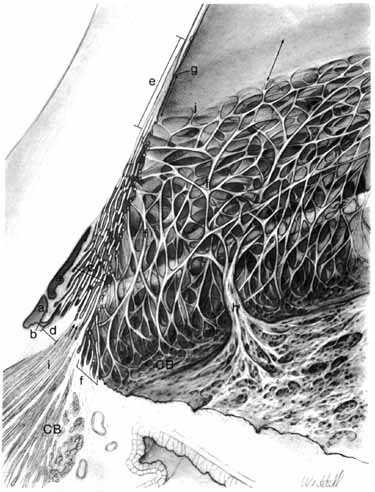
| Fig. 12 Drawing of the aqueous outflow apparatus and adjacent tissues. Schlemm's canal (a) is divided into two portions. An internal collector channel (Sondermann) (b) opens into the posterior part of the canal. The sheets of the corneoscleral meshwork (c) extend from the corneolimbus (e) anteriorly to the scleral spur (d). The rope-like components of the uveal meshwork (f) occupy the inner portion of the trabecular meshwork; they arise in the ciliary body (CB) near the angle recess and end just posterior to the termination of Descemet's membrane (g). An iris process (h) extends from the root of the iris to merge with the uveal meshwork at approximately the level of the anterior part of the scleral spur. The longitudinal ciliary muscle (i) is attached to the scleral spur but has a portion that joins the corneoscleral meshwork (arrows). Descemet's membrane terminates within the deep corneolimbus. The corneal endothelium becomes continuous with the trabecular endothelium at j. A broad transition zone (double-headed arrows) begins near the termination of Descemet's membrane and ends where the uveal meshwork joins the deep corneolimbus. (Hogan M, Alvarado J, Weddell J: Histology of the Human Eye—An Atlas and Textbook. Philadelphia: WB Saunders, 1971:137) |