|
|
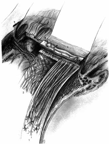
| Fig. 15 The ciliary body showing the ciliary muscle and its components. The cornea and sclera have been dissected away, but the trabecular meshwork (a), Schlemm's canal (b), and two external collectors (c), as well as the scleral spur (d), have been left undisturbed. The three components of the ciliary muscle are shown separately, viewed from the outside and sectioned meridionally. Section 1 shows the longitudinal ciliary muscle; in section 2, the longitudinal ciliary muscle has been dissected away to show the radial ciliary muscle; in section 3, only the innermost circular ciliary muscle is shown. According to Calasans (1953), the ciliary muscle originates in the ciliary tendon, which includes the scleral spur (d) and the adjacent connective tissue. The cells originate as paired V-shaped bundles. The longitudinal muscle forms long V-shaped trellises (e) that terminate in the epichoroidal stars (f). The arms of the V-shaped bundles formed by the radial muscle meet at wide angles (g) and terminate in the ciliary processes. The V-shaped bundles of the circular muscle originate at such distant points in the ciliary tendon that their arms meet at a very wide angle (h). The iridic portion is shown at i joining the circular muscle cells. (Hogan M, Alvarado J, Weddell J: Histology of the Human Eye—An Atlas and Textbook. Philadelphia: WB Saunders, 1971:305) |