|
|
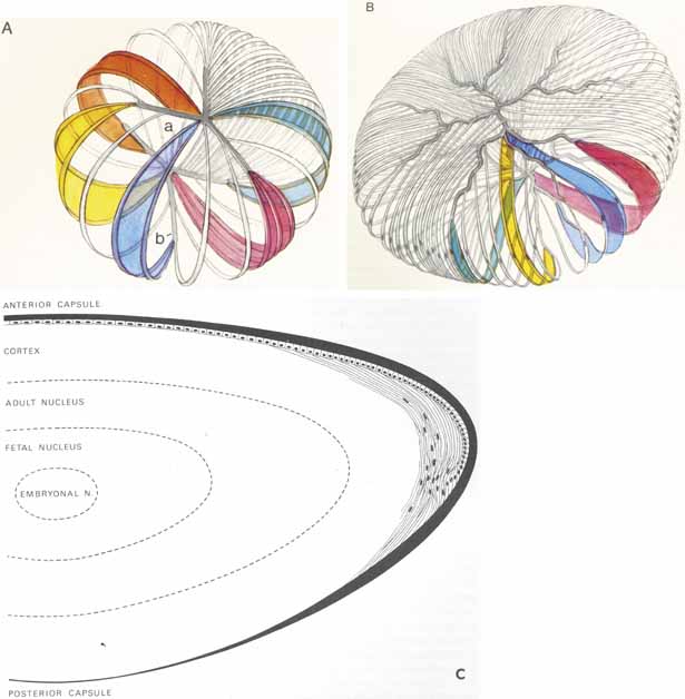
| Fig. 17 Embryonal and adult lens to show the sutures and arrangement of the lens cells. A. Drawing of the embryonal nucleus. The anterior Y suture is at (a) and the posterior at (b). The lens cells are depicted as wide, colored bands. Those cells that attach to the tips of the Y sutures at one pole of the lens attach to the fork of the Y at the opposite pole. It can be seen if the lens cells attaches to the tip of a Y suture anteriorly or its distance from the equator is shorter at that pole of the lens. B. Adult lens cortex. The anterior and posterior organization of the sutures is more complex. Those lens cells that arise from the tip of a branch of the suture insert farther anteriorly or posteriorly into a fork at the posterior pole. This arrangement conserves the shape of the lens. This drawing shows the suture to lie in a single plane for pictorial reasons, but it should be remembered that it extends throughout the thickness of the cortex and nucleus to the level of the Y sutures in the embryonal nucleus. C. Schematic representation of the adult lens, showing the nuclear zones, epithelium and capsule. The thickness of the lens capsule in various zones is shown. (Reprinted with permission from Hogan MJ, Alvarado JA, Weddell JE. Histology of the Human Eye—An Atlas and Textbook. Philadelphia: WB Saunders, 1971) |